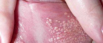
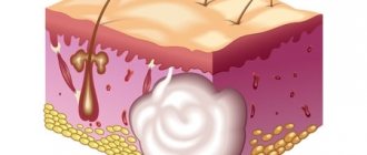
An epidermal cyst (atheroma) is a skin formation that looks like a capsule filled with sebum and particles of keratinized epithelium. Pathology of this type is often diagnosed in patients 25-45 years old. An abnormal manifestation occurs in both men and women, which excludes hormonal prerequisites for development.
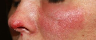
Epidermal cysts occur on any part of the body where hair grows, as the formation forms around the hair follicle. The cyst can grow up to 50 mm in diameter. Outwardly, it is similar to a dense pink ball. On the surface of the formation you can see a capillary network. Enlarged pores are also visible. Inside the capsule the secret is a pasty light yellow substance.
The nature of the neoplasm is benign. But there is a risk of degeneration into a malignant form. Penetration of pathogenic microflora into the capsule provokes inflammation. If the epidermal skin cyst is located in the eye area, vision may be impaired due to its pressure. Formations behind the ear provoke headaches and nervousness. Therefore, the appearance of pathology cannot be ignored; if possible, it should be treated.
Don't forget about the cosmetic component. Cysts on the face, neck, and head can significantly spoil a person’s appearance. Large formations cause increased attention from others.
Modern dermatology offers several methods for treating atheromas. Experts from the Lasersvit clinic tell you more about the methods, as well as the causes and symptoms of the appearance of epidermal cysts. If after reading the article you still have questions, our qualified doctors are ready to answer them during a face-to-face appointment.
Epidermal cyst: causes
Unlike dermoid cysts, which are congenital, epidermal (skin) cysts appear due to blockage of the gland duct with sebaceous masses and dead epidermal cells. Provoking factors for the development of pathology:
- When metabolic processes are disrupted or hormonal imbalance occurs, sebum is secreted with a thicker consistency than that of people in good health. Due to the viscous structure of the substance, the physiologically normal outflow of the substance is disrupted. It, like a plug, closes the duct of the glands.
- When there is inflammation on the skin, for example, acne, folliculitis, tissue swelling occurs. This causes a narrowing of the lumen of the excretory duct of the sebaceous gland.
- Scars and scars consist of dense connective tissue, which causes compression of the ducts of the sebaceous glands.
- Improper skin care and use of inappropriate cosmetics can cause blockage of the sebaceous gland.
Any of these factors can provoke the formation of this type of pathology. And if there are several of them, then with each new potential cause the risk of occurrence increases by 10-15%.
What is a dermoid cyst?
Many patients are diagnosed with a cyst, most of them women. However, cysts are different.
We asked the executive director of Clinic Expert Orenburg LLC, Yuri Andreevich Podlevskikh, to talk about one of its varieties - a dermoid cyst.
- Yuri Andreevich, what is a dermoid cyst and should we be afraid of this diagnosis?
This is one of the types of benign tumors that develops during human embryonic development.
The anxiety of patients who have been diagnosed with this is understandable. But there is no need to panic. This is a benign formation in nature, which is already reassuring. But, nevertheless, it should not be normal.
-Can a dermoid cyst occur in any organ?
It can be almost anywhere. However, there are areas where dermoid cysts are more common. For example, this is the small pelvis in women (in particular, dermoid ovarian cyst).
-What are the causes of dermoid cysts?
These are disturbances in the process of embryonic development of the body. There are also isolated reports of their appearance after injury.
- Is a dermoid cyst more often an accidental finding or is something bothering the patient?
Some patients do not present specific complaints: the formation is detected by chance (for example, during a preventive, screening study).
In a number of patients it can be detected during routine examination (particularly under the skin).
The symptoms of a dermoid cyst also depend on the area of the body where it is located.
If it is, for example, a dermoid cyst of the brain, then nonspecific symptoms may be observed - for example, headaches, dizziness. When it is located in the small pelvis, pain syndrome is also possible (in some cases, intense - in particular when the cyst ruptures), impaired urination, signs of intestinal obstruction, and with inflammation - increased body temperature. In most cases, it is impossible to make an unambiguous conclusion based on such signs that this is a dermoid cyst. The diagnosis can be clarified only after additional research.
- How are dermoid cysts diagnosed?
In addition to clinical methods in the form of questioning, examination, and palpation, instrumental diagnostic methods are widely used. The most commonly used are ultrasound, magnetic resonance imaging and computed tomography.
In practice, statistically more often, people undergo screening ultrasound, which detects this formation for the first time, after which they are sent to MRI or CT to clarify the diagnosis. Dermoid cysts are well detected by both methods, but MRI is somewhat more preferable - of course, if there are no contraindications to it. The main advantage in this case is the absence of radiation exposure, which to a certain extent is inherent in CT.
- Yuri Andreevich, if an MRI reveals a dermoid cyst, what should such a patient do?
Contact a doctor whose specialty depends on the area where the education is located. If it is intracranial (inside the skull), then consultation with a neurosurgeon is necessary; if in the pelvis - a gynecologist; in the abdominal cavity - the surgeon.
- How are dermoid cysts treated?
Usually treatment is surgical. Depending on the specific situation, the dermoid cyst is either excised or first drained and freed of contents, and then removed.
Depending on the doctor's decision, they are sometimes monitored dynamically.
- What complications can occur if a dermoid cyst is not treated?
This is, for example, inflammation, rupture of a cyst. Its malignancy is also described: according to various sources, the dermoid cyst degenerates in approximately 8% of patients.
For reference:
Podlevskikh Yuri Andreevich
Graduate of the Orenburg State Medical Academy in 2012 with a degree in Pediatrics.
In 2013, on the basis of the above-mentioned educational institution, he completed an internship in the specialty “Radiology”. In the same year, he underwent advanced training in magnetic resonance imaging.
Currently he holds the position of executive director of Clinic Expert Orenburg LLC.
Other interviews with Yuri Podlevskikh:
Do's and Don'ts for MRI
How can MRI help a patient with a traumatic brain injury and its consequences?
What is neurovascular conflict?
What are men silent about...What does an MRI of the abdominal cavity show and what is included?
Hit below the belt
Why undergo an MRI of the brain when nothing hurts?
Diagnostics
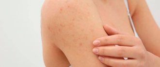
Diagnosis of the disease is based on visual examination, hardware and laboratory tests. Externally, as already mentioned, atheroma looks like a painless bulge on the skin. The capsule is mobile and can move under the skin under mechanical stress. Atheroma can be detected in areas of the skin rich in sebaceous glands. A cyst often appears on the face, scalp, behind the ears, and on the back. Education is characterized by slow growth.
In a calm state, a cyst on the body does not cause physical discomfort to patients. However, in the presence of an inflammatory process, the tumor can look quite scary - redness and swelling of nearby tissues, thinning of the skin in the area around the tumor. Touching the subcutaneous lump can be extremely painful. The size, condition of the cyst and its location influence the choice of treatment tactics.
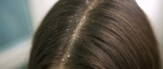
Symptoms and diagnosis of atheroma on the head.
It is not difficult to identify atheroma on the head, however, during examination you need to be extremely careful to exclude a cerebral hernia. If the doctor has doubts, then a craniography (head x-ray) is prescribed, which will clearly confirm the diagnosis. The following signs are characteristic of an inflamed atheroma on the head:
- pain on palpation;
- release of liquid with a specific odor;
- a reddened, inflamed area of the scalp around the tumor;
- intensive growth of atheroma;
- elevated body temperature.
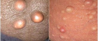
Rice. 2. Atheroma of the scalp, scalp.
Treatment methods
Since the formation takes a long time to develop and is benign, it is not necessary to remove it. The decision to undergo surgical intervention is made for cosmetic reasons or in the event of an unsuccessful pathology location or frequent mechanical damage to a neoplasm protruding above the surface of the skin. This increases the risk of bacterial infection with subsequent transition to purulent processes. In this case, radical treatment in the form of removal is the only way to prevent more serious diseases and complications.
Epidermal cysts are treated in several ways:
- If the formation is large, it is removed with a scalpel. A scar remains at the site of the incision.
- Laser removal of atheroma is considered the preferred method of choice due to the minimal list of contraindications and excellent results.
- Burning with liquid nitrogen is affordable, but has its drawbacks - inaccuracy of the effect, prolonged healing of the burn.
Important: Do not try to treat epidermal cysts or other skin formations with folk remedies. This is fraught with serious damage and the appearance of a scar at the site of exposure. In addition, without histology it is impossible to accurately determine the nature of the neoplasm, therefore it is impossible to talk about its benign quality. Any unprofessional interventions can cause serious health problems.
Types of cystic formations
Based on the time of appearance, all cysts are classified into 2 categories:
- Congenital, or dysontogenetic, arise due to disruptions in the process of intrauterine development.
- Acquired ones appear as life progresses.
According to the mechanism of development of cysts, there are:
- Retention - the most common. They are formed if the excretory duct of the gland is compressed or blocked from the inside. The secretion has no outlet, accumulates and stretches the surrounding tissue.
- Ramolitic - arise at the site of dead cells, for example, in the brain after a stroke.
- Parasitic is a capsule within which the parasite is located. In this way, the helminth protects itself from the body’s immune attacks.
- Traumatic ones form at the site of bruises due to the accumulation of tissue fluid.
- Tumor - lined with epithelium with signs of metaplasia; inside there are seals and places where fluid accumulates.
Epidermal cyst: laser removal
Removal of a subcutaneous cyst with a laser is a simple procedure that is performed on an outpatient basis and does not require hospitalization or long-term rehabilitation. After conducting an examination and determining the exact location and size of the capsule, the doctor sets the intensity and depth of the laser beam, taking into account the data obtained. The laser acts in a targeted manner without affecting healthy tissue.
During manipulation, the wound is disinfected and cauterized, which eliminates the risk of infection. Laser pulses are short, pain receptors do not have time to react to the impact. Therefore, during the operation, light local anesthesia is used to prevent discomfort.
The whole procedure takes 15-20 minutes. Almost immediately after it, the patient can return to his usual lifestyle. The recovery period involves wearing a sterile bandage for several days until the wound is completely healed.
After laser surgery, there are no scars or scars, which is important if the pathology has formed on the face. And if the epidermal cyst is on the head (pilar), then the laser method is the only way to preserve the hair without the risk of developing a bald spot at the site of the operation.
How much does it cost to treat atheroma?
The cost of treating epidermal cysts depends on the size and location (availability). You will also have to pay for the necessary package of studies, without which the operation is impossible. The advantages of the Lasersvit clinic are that we offer a comprehensive service - from seeing a dermatologist to monitoring the patient during the recovery period after surgery. A comprehensive service is always cheaper, and all manipulations and procedures are carried out in one building. This increases the safety and comfort of treatment.