Original title of the article: “The effectiveness of using the Stop Demodex series of drugs in the complex treatment of patients with demodicosis”
Pritulo O.A., Ngema M.V., Prokhorov D.V., Smolienko V.N., Vintserskaya G.A., Ravlyuk D.A. State Institution “Crimean State Medical University named after.
S.I. Georgievsky" Demodicosis is a chronic parasitic disease of the skin of humans and animals, widespread in all climatic zones of the globe. Its causative agents are demodicid mites, representatives of the group of Arachnids, the family Demodecidae, most adapted to the parasitic lifestyle [3,6,8,11,13,15].
It has now been proven that the causative agents of human demodicosis are mites Demodex folliculorum and Demodex brevis, belonging to the phylum Arthropoda, the class Arachnida, and the genus Acarina. They are representatives of an independent family Demodecidae from the order Trombicliformes [1,2].
Pets also suffer from demodicosis. Infection of cats and dogs by Demodex cati (a parasite of cats) and Demodex canis (a parasite of dogs) mites causes a disease known as glandular red mites, which can be accompanied by severe inflammation of the hair follicles and sebaceous glands, hair loss, peeling, thickening of the skin, the formation of pustules, sometimes boils, generalization of the pathological process and even end in death.
According to the concept of the life pattern of species, mites Demodex folliculorum and Demodex brevis belong to permanent monoxenic skin parasites; in addition, mites are community-hospital, that is, they parasitize together on the same host [5]. In accordance with the high level of topical and trophic specificity, the causative agents of demodicosis predominantly affect the skin in the area of nasolabial folds, chin and eyelids [7,9,14].
Demodex folliculorum is most often found in the area of the nose and outer ear in 38% of cases, in the forehead in 30%, and in the perioral area in 29% [10]. As for Demodex brevis, its primary localization is the skin of the upper half of the chest and neck.
Depending on the predominance of certain symptoms, several clinical forms of demodicosis . Some researchers distinguish erythematous-squamous (or seborrheic), papular, papulovesicular, pustular, rosacea-like, combined and asymptomatic forms.
Others note low-symptomatic, erythematous-squamous, acneiform, rosaceomorphic, sycosiform, small-nodular forms, while others note erythematous, pustular, papular, and combined forms. Depending on the prevalence of the A.A. process. Antonev et al [1]. The following forms of demodicosis are distinguished: limited (with localization of rashes mainly in the folds and near the corners of the eyes), diffuse, in which the entire skin of the face is affected; common when the inflammatory process extends beyond the face.
According to the course of the disease, acute, subacute and chronic recurrent demodicosis are distinguished. According to the clinical picture, the following forms of skin demodicosis have been identified: low-symptomatic erythematous-squamous, papular, pustular, combined and cystic [4].
In addition, there are several types of arrangement of efflorescences during demodicosis :
- central type , in which the rashes are localized mainly in the T-zone (the central part of the forehead, the glabella, the back and wings of the nose, the nasolabial zone, the central part of the chin) - the zone of greatest density of the sebaceous glands;
- medial type - the rashes are located mainly in the area of the frontal tubercles, the central part of the cheeks and the area of the chin protuberance;
- asymmetrical type - rashes are found only on one side of the face; lateral type - rashes are located on the sides of the face;
- total type - rashes are distributed evenly over the entire surface of the face.
The clinical picture of human demodicosis may resemble the clinical picture of rosacea, acne vulgaris, photodermatosis, and seboroid. Often, on a congestive-hyperemic, slightly swollen, flaky background with symptoms of oily seborrhea, follicular rashes of the small-papular and small-pustular type are located. Also, some researchers note that the clinical picture of demodicosis resembles rosacea, identifying demodicosis with rosacea.
The cause of demodicosis is the acne iron mite
The demodex mite (D. follicullorum) belongs to the genus Demodex, family Demodicidae, suborder Trombidiformes, order Acariformes.
A little history
- The parasite was first discovered in 1841 by two independent researchers Henle (identified the parasite on human skin) and F. Berger (identified the parasite in earwax).
- In 1842, C. Simon identified mites in hair follicles and described the parasite for the first time. Between 1917 and 1923, acarologist Hirst identified 21 species and several subspecies of demodex mites in animals.
- In 1963, L. Kh. Akbulatova described a new species of iron mite, Demodex folliculorum brevis.
Spreading
Demodex mites are the most common parasites living on the surface of the body of humans and animals. They live in the mouths of hair follicles and the excretory ducts of the sebaceous and meibomian glands. Of the 100 known species of Demodex mites, 2 species live on human skin: Demodex folliculorum longus (long) and Demodex folliculorum brevis (short). Contamination of the skin occurs in childhood through contact. The maximum incidence occurs in females aged 20–40 years. At older ages, tick parasitism is registered in 80–100% of the population.
Acne iron mites live in places with high humidity; the parasites do not live long outside the host, are distributed everywhere, and are recorded in all races and in all geographical zones.
General characteristics of parasites
Acne gland mites live in the mouths of hair follicles and excretory ducts of the sebaceous and meibomian glands of the skin of the face (T-shaped zone), in the ears, and less often on the back and chest. There is an atypical localization of parasites.
Demodex mites feed on epidermal cells and sebum.
At night, ticks crawl to the surface to mate. Ticks move along the surface of the skin at a speed of 8 - 16 mm/hour.
At the head end (gnathosoma) there is a mouth opening equipped with sharp chelicerae that facilitate the absorption of food (epidermal cells and sebum). Digestion of food occurs under the influence of lytic enzymes.
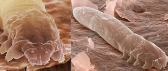
Rice. 2. Photo of demodex mites under an electron microscope.
Reproduction and development
Ticks crawl onto the surface of the skin at night to mate. That's when they end up with other hosts. The spring-summer period is the most favorable for parasites.
The life cycle of parasites is 14 - 18 days and consists of 5 developmental phases. After fertilization, the females return to the hair follicles and sebaceous gland ducts and lay diamond-shaped eggs, from which larvae emerge after 60 hours. In the developmental cycle, larvae transform from eggs, protonymphs and nymphs into adults. The appearance of a large number of parasites coincides with the first rashes on the skin.
Rice. 3. The photo shows (from left to right) the developmental stages of the demodex mite from egg, larva, protonymph and nymph to adult.
Features of Demodex folliculorum longus (long mite)
The tick of this species is long (0.3 - 0.4 mm), females and males are of the same size. The body is worm-shaped. The head end (gnatosoma), thorax (subsoma) and abdomen (opisthosoma) are well differentiated. The chelicerae of the oral opening are sharper.
The subsoma has setae. The covering (cuticle) is transparent. There are very short three-membered legs. Transverse striations of the posterior part of the body are noted. They live in hair follicles in groups.
Features of Demodex folliculorum brevis (short mite)
This species of tick has a short body (0.18 - 0.19 mm). The opisthosoma is short with a pointed cone-shaped end. The head end is short and flattened. Males are smaller than females and die after fertilization. The breast is wide and lacks setae. The cuticle is less transparent. They live in the mouths of the excretory ducts of the sebaceous and meibomian glands singly, which makes them difficult to detect. They rarely rise to the surface, making treatment difficult.
Demodex folliculorum longus is found 4 times more often in men and 10 times more often in women than Demodex folliculorum brevis.
Rice. 4. On the left are long demodex mites (Demodex folliculorum longus), on the right are short mites (Demodex folliculorum brevis).
Rice. 5. In the photo there is a short and a long demodex mite.
Indications for examination
Analysis for demodex must be carried out if there is:
The doctor's or patient's assumption of infection.- Clinical manifestations in the form of:
- affected ciliary margin;
- redness and inflammation;
- gluing eyelashes;
- severe scabies at the affected area;
- heaviness in the eyes;
- eyelash loss, which usually falls out without a root;
- the appearance of plaque or white scales on the eyelashes.
The danger is not the tick itself, but the products of its vital activity; it does not live long (up to 18 days in the larval stage and 5 days as an adult). When the tick dies, it begins to disintegrate, releasing toxins that subsequently lead to the appearance of unpleasant symptoms.
Epidemiology of demodicosis
Source of infection
The main source of demodex mites are sick people or carriers.
Distribution routes
- Iron mites can be transmitted through bedding, underwear, and personal items. The parasites were found in house dust, the source of which was bedding.
- Some researchers suggest that ticks can get on a child during breastfeeding from a sick mother.
Sustainability
Constant humidity, darkness and air temperature within 30-40°C are optimal conditions for iron mites. Outside the host, at room temperature, constant humidity and in the dark, ticks live up to 9 days.
According to the observation of some researchers, parasites remain viable in water for 7 days, and up to 2 weeks in immersion oil.
Rice. 6. Demodex mite under a microscope.
3.Diagnosis of the disease
After a visual examination and conversation with the patient, the dermatologist prescribes a microscopic examination
.
Material for analysis is taken by scraping the epidermis from the affected areas of the skin. It is also recommended to analyze eyelashes
taken from different parts of the eyes. Express analysis makes it possible to get instant results. During the study, adult ticks, their eggs and shed chitinous shells are discovered. In order for the dermatologist to see a complete picture of the density of mite colonization of the skin, it is recommended not to wash your face for 24 hours before the examination. What is important for making a diagnosis in this case is not the fact of detecting a tick in itself, but the number and activity of the parasite, as evidenced by the number of larvae and eggs.
About our clinic Chistye Prudy metro station Medintercom page!
How the disease develops
Demodicosis in humans can develop for the first time and occur as an independent disease, or develop against the background of existing skin diseases (acne, perioral dermatitis, rosacea, seborrheic dermatitis, etc.). From 55 to 100% of the population are carriers of iron ticks and do not have any manifestations of the disease, which proves that the parasites belong to the opportunistic flora. Damage to follicles by parasites increases with age.
Factors in the development of the disease
Acne iron mites are an opportunistic infection. They can parasitize a person for many years without causing him any harm. Their activation and active reproduction depends on a number of factors.
- Changes in microbiocenosis, the function of the sebaceous glands and the composition of sebum are the trigger mechanism of the disease. Changes in the composition of surface lipids lead to the development of dysbiosis.
- Demodex mites are carriers of viruses and microbes into the sebaceous glands and hair follicles. When pathogenic pyococci and fungi Pityrosporum spp are introduced, purulent-necrotic inflammation develops.
- The bacillus Bacilluss oleronius found on the surface of the parasite increases the activity of ticks and stimulates staphylococci, streptococci, acne propionobacteria, and fungi of the genus Malassezia to produce anti-inflammatory proteins, which triggers a cascade of immune reactions.
- The presence of foci of chronic infection, disturbances in the functioning of the liver and gastrointestinal tract, endocrine glands, nervous system, and corticosteroid therapy contribute to the activation of ticks.
- The rise in incidence in the spring-summer period is associated with increased insolation. An increase in the production of vitamin D under the influence of ultraviolet radiation leads to an increase in the synthesis of cathelicidins, which support the inflammatory process.
- The likelihood of infestation by Demodex mites increases in individuals with a sharply reduced functioning of the immune system, which occurs with primary and secondary immunodeficiencies, long-term use of corticosteroids and cytotoxic therapy, HIV infection, leukemia, T-cell lymphoma and other malignant neoplasms.
- The mite infestation maintains the activity of the existing inflammatory process. An imbalance in the cytokine cascade develops when the immune response is too strong.
Mechanisms of action of iron mites on humans
The impact of Demodex mites on humans is multifaceted:
- When feeding, the parasites secrete aggressive saliva and mechanically destroy the cell walls of the sebaceous glands and follicles, inflammatory infiltrates and granulomas form in the dermis, and the processes of keratinization and pigmentation develop.
- Substances formed after the death of ticks have antigenic properties, which leads to allergization and increased immune response.
- When the immune system is suppressed, opportunistic flora is activated, which successfully colonizes the host.
Acne iron mites act as carriers of bacteria and viruses, which complicates the course of demodicosis and complicates treatment.
Rice. 7. Histological preparation of a skin section. Arrows indicate the location of demodex mites.
Patient preparation
It is recommended to perform a demodex test in the morning.
To obtain an error-free and reliable result, it is necessary not to wash your face a day (or better yet, 2-3 days) before taking the test, not to put drops in your eyes and not to use decorative cosmetics, especially mascara.
Try to avoid getting shampoo, soap and other chemicals in your eyes. Previous course of antimicrobial therapy may also influence the outcome.
It is quite difficult to identify demodicosis in a patient, because the mite itself is active at night and is afraid of sunlight, so in order not to create additional obstacles to the detection of demodex, it is necessary to prepare in accordance with the above recommendations.
If these rules are neglected, there is a possibility of incorrect diagnosis.
Clinical variants of demodicosis
Demodicosis in humans can develop as a primary independent disease, or it develops against the background of existing skin diseases (perioral dermatitis, rosacea, acne, seborrheic dermatitis, etc.), aggravating their course.
Primary demodicosis
In primary demodicosis, inflammatory skin elements resolve after antiparasitic therapy. About 40% of cases of primary demodicosis develop in patients suffering from rosacea.
Primary demodicosis is characterized by:
- the onset of the disease over the age of 40 years;
- The areas most often affected are around the mouth, eyes and outer ear;
- inflammatory elements are located asymmetrically and are accompanied by itching;
- acne and rosacea are absent;
- high content of mites on the skin;
- positive effect of the antiparasitic treatment.
Secondary demodicosis
Secondary demodicosis develops against the background of diseases accompanied by a sharp suppression of the immune system (HIV infection, leukemia, etc.), long-term use of corticosteroids and cytotoxic therapy, or develops against the background of acne and rosacea, complicating their course. This form of the disease is registered in 33% of patients with acne.
Secondary demodicosis is characterized by:
- the disease appears at any age;
- lesions are widespread;
- presence of anamnesis and clinical picture of relevant diseases.
Papulopustular rashes on the skin of the face are a reason for diagnosing demodicosis.
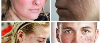
Rice. 8. Demodectic mange on a woman’s face.
conclusions
Taking into account the presence of clinical and laboratory changes in various clinical forms of demodicosis, a scientifically based treatment complex with a series of drugs “Stop Demodex” has been introduced into practice. Its effectiveness can be seen both in complex treatment and in the prevention of demodicosis. There is an elimination of the very cause of the disease - the uncontrolled reproduction of the Demodex mite, as well as a strong anti-inflammatory and restorative effect - rapid elimination of the symptoms of the disease. As a result of the use of drugs in this series, after just a few days there is a significant cleansing of the skin from foci of inflammation and, subsequently, a complete regression of this dermatosis.
Literature
- Akbulatova L.H. Human demodicosis./L.Kh. Akbulatova // Bulletin of Dermatology. – 1964. – No. 3 – pp. 32 – 42.
- Akbulatova L.H. The pathogenic role of the Demodex mite and clinical forms of demodicosis in humans. /L.H. Akbulatova // Bulletin of Dermatology. – 1966. – No. 12. – P. 57–61.
- Valne M.S. Populations of iron mites in perioral dermatitis and rosacea./M.S. Valne, A.B. Lange .//All-Russian Congress of Dermatologists and Venereologists: (VI; 1989; Moscow) VI All-Union Congress of Dermatologists and Venereologists: Abstracts. report – M., 1989. – Part 2. – P. 135 – 138. 04. Kogan B.G. Morphological, biological and functional characteristics of human demodicosis mites – mites Demodex folliculorum and Demodex brevis at the current stage. /B.G. Kogan, V.I. Stepanenko//Dermatovenerology.Cosmetology. Sexopathology. – 2001. – No. 23(4) – P.70 – 75.
- Kogan B.G. Specificity of Demodex folliculorum and Demodex brevis mites – causative agents of human demodicosis / B.G. Kogan, V.T. Gorgol //Ukr. Journal of dermatology, venereology and cosmetology. – 2001. – No. 1. – P.37 – 41.
- Kulaga V.V., Romanenko I.M. Treatment of skin diseases. – Lugansk, 1996. – 414 p.
- Maychuk Yu.N. Parasitic eye diseases./Yu.N. Maychuk – M.: Medicine, 1988. – 288 p.
- Malakhova M.Ya. Methods for recording endogenous intoxication: A manual for doctors./M.Ya Malakhova – St. Petersburg, 1995. – 125 p.
- Malygina E.L. "Ivomek" in the external treatment of demodicosis. // Problems of achievement of biomedical sciences and practical healthcare. Proceedings of KSMU named after S.I. Georgievsky / E.L. Malygina - Simferopol: Publishing center of KSMU, 2001. - 137 T., Part 1. - P. 78 -79.
- Ponomarev B.A. The main problems of ectoparasitic infection /B.A. Ponomarev., V.P. Kulagin, G.D. Selissky.// Vestn. dermatol. – 2000. – No. 1. – P. 39 – 40.
- Potekaev N.N. Rosacea: Etiology, clinic, therapy. / N.N Potekaev - M.: Biom, 2000. - 142 p.
- Endogenous intoxication syndrome in patients with demodicosis / D.A. Slevareva, O.A. Pritulo // Problems, achievements and prospects for the development of biomedical sciences and practical healthcare: Proceedings of KSMU named after. S.I. Georgievsky. – Simferopol: Publishing Center of KSMU, 2007. – T. 143, Part V. – P. 325-327.
- Desch CE Morphology and functional anatomy of Demodex folliculorum (Simon) of man / CE Desch, WB Nutting //Acarology. - 1977. - V.19, No. 3. - P. 422.
- Erbagci Z. High incidence of demodicidosis in eyelid basal cell carcinomas. / Z. Erbagci, I. Erbagci, S. Erkilic. //Int. J. Dermatol. – 2003. – V.42. – P. 567 – 571.
- Katz AM Rosacea: epidemiology and pathogenesis/A. M.Katz.//J. Cutan. Med. Surg. – 1998. – V. 2. – P.5-10.
Signs and symptoms of demodicosis
The disease always occurs gradually and tends to spread and progress.
The main localization of demodex mites
Short mites live in the mouths of the excretory ducts and the secretion of the sebaceous glands of the skin of the face, ear canal, chest and back, sometimes on the scalp, as well as the meibomian glands, which are modified sebaceous glands that open at the edges of the eyelids. Long mites live in hair follicles. Mites were found on the penis, buttocks, and in the area of ectopic sebaceous glands.
Rice. 9. Demodectic mange on the face in the area of the nasolabial folds.
Rice. 10. Demodectic mange on a woman’s face in the chin area.
Rice. 11. Demodectic mange on the skin of the cheeks, nose and around the mouth.
Rice. 12. Demodectic mange on the face of women on the skin of the forehead, cheeks and nasolabial folds.
Rice. 13. Demodectic mange on the face of women.
Rice. 14. Demodectic mange of the eyelids.
Characteristics of the rash
- Initially, erythematous (red) spots appear around the mouths of the follicles. The rash is located asymmetrically and is accompanied by lamellar peeling.
- The appearance of the rash is accompanied by a feeling of heat, burning, crawling and distension.
- Very quickly, pink or red papules with a cone-shaped top and gray scales appear in the reddened area. Papulovesicles (a vesicle is a blister filled with clear fluid) or papulopustula (a pustule is a blister filled with purulent fluid) often appear.
- Inflammatory infiltrates and granulomas form in the dermis. The skin over the affected areas thickens and its elasticity decreases. A feeling of tightness appears. The affected areas are covered with serous, serous-sanguineous or purulent crusts. Over time, hyperpigmentation develops: the skin becomes brownish or yellowish-brownish in color.
- With the massive development of a pyogenic bacterial infection, large follicular pustules (purulent elements) appear at the site of inflammatory infiltrates. Over time, they spread into the deeper layers of the dermis, which leads to facial disfigurement.
- The clinical picture of demodicosis for different types of mites is somewhat different from each other. Thus, when Demodex folliculorum is affected, erythema and peeling (desquamation of the epithelium) are more often recorded; when Demodex brevis is affected, papulopustular elements are often recorded, which are often located symmetrically.
- With long-term (chronic) demodicosis, thickening of the skin, loss of elasticity, and a feeling of tightness appear. Bloody-purulent crusts are located against the background of hyperemic skin. When a pyogenic infection is attached, pustules and microabscesses appear. Dense infiltrates form in the deep layers of the dermis, disfiguring the face.
Rice. 15. Demodectic mange on a woman’s face, severe.
Deterioration in appearance creates aesthetic discomfort for the patient and leads to the development of neuroses and depression.
2. Symptoms of the disease
In low concentrations, the demodex mite does not manifest itself in any way. The skin's own immunity does not allow the parasite to have a negative impact on it. But under certain unfavorable conditions, the protection weakens, and the tick begins to actively feed and reproduce. Sebum contributes to this. Signs appear on the skin, collectively called “problem skin”: acne and pimples, enlarged pores, inflammation of the sebaceous glands. Most often, the parasite is localized on the chin, nose, cheeks, and forehead. The skin becomes unhealthy, oily shine appears, firmness and elasticity are lost. Mite damage to the eyelids also leads to inflammation, sometimes even leading to eyelash loss. The skin becomes lumpy, painful, itching and a tickling sensation on the skin often occur - these are parasites moving along the surface. Demodex is especially active in the evenings and at night.
Visit our Dermatology page
Forms of demodicosis in humans
Depending on the nature of the manifestations on the skin, several forms of demodicosis are distinguished, the symptoms of which are characteristic of the corresponding diseases. Not to be confused with these diseases!
Acneform form
The acneiform type of demodicosis is characterized by the appearance of papules and pustules on the facial skin, which are similar to acne.
Rice. 16. Demodectic mange of the face in women. Acneform form of the disease: papular (photo on the left) and papulopustular (photo on the right).
Rosacea-like form
The rosacea-like type of demodicosis is characterized by the appearance of papules against a background of diffuse erythema. Demodex brevis (short mite) causes the appearance of inflammatory infiltrates and granulomas in the deep layers of the dermis, which are similar to the granulomatous form of rosacea.
Rice. 17. Rosacea-like form of demodicosis: in the form of diffuse erythema (photo on the left), granulomatous form (photo on the right).
Seborrheic (erythemo-squamous) form
The seborrheic form of demodicosis is characterized by the appearance of redness and rash on the facial skin, accompanied by lamellar peeling.
Ophthalmic form
The ophthalmic form of demodicosis occurs with damage to the eyeballs and eyelids. Redness, dryness, irritation, sensation of a foreign body in the eye, fatigue are the main symptoms of eye demodicosis. Itching of the eyelids, redness and swelling of their edges, the appearance of scales or crusts on the eyelids are the main symptoms of demodicosis of the eyelids.
Read in detail in the article “Demodicosis of the eyelids and eyes”
Rice. 18. Demodectic mange of the eyelids. Lamellar peeling is clearly visible (photo on the left). On the ciliated edge of the eyelid, scales form a “collar” around the eyes (photo on the right).
The role of demodex mites in the formation of androgenetic alopecia
There is an assumption that the presence of mites in the hair follicles aggravates the development of androgenetic alopecia. During inflammation, T-lymphocytes are activated, which induce collagen synthesis, resulting in fibrous degeneration of hair follicles.
Rice. 19. Demodicosis of the scalp.
Important information
Compound
The powder is created on the basis of natural ingredients. It includes:
Dimethicone – promotes moisture retention.
Silica gel – provides increased water resistance.
Hydrogenated lecithin - restores skin elasticity and helps eliminate flaking.
Titanium dioxide is an effective UV filter that protects the skin from solar radiation and preserves its youth.
There are no parabens and other harmful components, as well as oils in the powder. The packaging is a stylish mirror case, which has a separate section for a sponge.
Demodectic mange against the background of rosacea
Demodicosis complicates the course of rosacea in 88.7%. The disease is more common in women over 30 years of age. The phymatous form of the disease is more common in men. The pathological process is characterized by redness of the skin of the face, associated with the expansion of small superficial vessels, the appearance of papules and pustules. Papules increase in size over time and merge, forming dense infiltrates. The sebaceous glands become hyperplastic. Fibrosis develops. Persistent redness and fibrous lumps on the nose are called rhinophyma.
There are many reasons for the development of diseases. Factors contributing to the development of the disease can be divided into endogenous and exogenous.
Rice. 20. Erythematotelangiectatic form of rosacea. Multiple telangiectasias (dilated subcutaneous arterioles) are visible on the skin.
Rice. 21. The photo shows the papulopustular form of rosacea. Against the background of erythema (redness), multiple rashes are visible, which are papules with thin scales on the surface. The affected areas are infiltrated and swollen.
Rice. 22. Demodex brevis (short mite) causes the appearance of inflammatory infiltrates and granulomas in the deep layers of the dermis, which are similar to the granulomatous (phymatous) form of rosacea. The photo shows the phymatous (papular-nodular) form of rosacea.
Rice. 23. Severe form of rosacea.
Rice. 24. The ocular form of rosacea is registered in 50% of patients. Inflammation of the eyelids, redness of the conjunctiva, iritis and keratitis are the main manifestations of the disease.
Types of tests for demodex
Laboratory diagnostics is of paramount importance in identifying mites in human hair.
The test procedure is absolutely painless and does not cause discomfort to the person.
The main methods for conducting analysis to identify parasites in eyelashes are:
- Eyelash research. The material for analysis is the removed eyelashes. From each eye, 4-6 hairs, completely extracted from the hair follicles, are selected and collected in a test tube, then 1-2 drops of glycerin are added, after which a microscopic examination is carried out.
- Scraping from the surface of the ciliary edge of the eyelids. To ensure that demodex leaves the eyelash follicle, the laboratory technician massages it with a blunt scalpel or an eye spoon and collects part of the damaged skin. Next, the collected material is evenly distributed onto a glass slide, glycerin or a 10-20% solution of caustic alkali is dripped, covered with a cover glass and examined under a microscope for 5-10 minutes.
Using these techniques, it is possible to determine the number of mites in the cells and their type, which will further help the doctor in choosing treatment methods and prescribing medications.
Demodicosis on the background of acne
Among acneiform dermatoses, demodicosis accounts for 10.5%. Acne (acne vulgaris) is a chronic, relapsing disease that affects the sebaceous glands and hair follicles. Most often, acne occurs in males during puberty between the ages of 14 and 16 years. The cause of juvenile acne is propionobacteria acne, epidermal staphylococci, pityrosporum ovale and orbitale, which constantly live on the skin of the face. Many factors contribute to the development of acne.
Rice. 25. Demodicosis is registered in 10.5% of patients with acne. The photo on the left is papular acne. When a staphylococcal infection is attached, pustules (ulcers) and microabscesses develop - pustular and abscess acne (photo on the right).
Rice. 26. With the indurative form of acne, inflammatory infiltrates appear in the deep layers of the dermis. The skin has a bumpy appearance.