Cryodestruction is a modern method of treating superficial benign neoplasms, based on cooling tissues to extremely low temperatures and their subsequent destruction . In fact, cryodestruction belongs to the field of cryosurgery and is considered a surgical treatment method.
Cryodestruction with liquid nitrogen is widely used in cosmetology, dermatology and medicine. For example, cryodestruction of warts, condylomas, papillomas and other neoplasms is popular. The procedure is widely used to treat vascular formations and cervical erosion. In many cases, it can successfully replace surgery.
Indications for cryodestruction
Cryodestruction with liquid nitrogen is indicated for various pathologies that are infectious in nature or related to skin defects. The most common is cryodestruction of warts. This method is widely used to remove various neoplasms of pigmented or vascular type: nevi, hemangiomas, papillomas, condylomas, pigmentary granulomas, hyperkeratosis, squamous cell carcinoma, epidermal cyst, melanoma, mastocytoma.
Cryodestruction with liquid nitrogen is widely used in gynecology for cervical erosion, condylomas and papillomas of the vagina and vulva, ectopia of columnar epithelium, cervical dysplasia, cervical leukoplakia and other gynecological diseases.
Cost of the procedure
| Name | Price |
| Removal of molluscum contagiosum (as part of a consultation with a dermatologist) 1-3 units | 550 rub. |
| Removal of molluscum contagiosum (as part of a consultation with a dermatologist) 4-5 units | 700 rub. |
The information and prices presented on the website are for reference only and do not constitute a public offer. The services indicated in the Price List may be provided in other medical institutions. We ask you to clarify the address and cost of services in advance at the 24-hour call center by phone +7 (812) 331 17 04
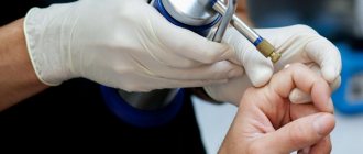
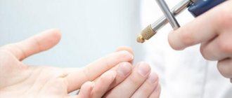
Operating principle of cryodestruction
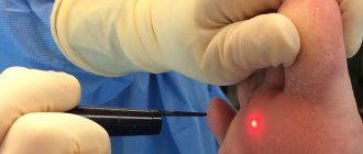
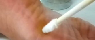
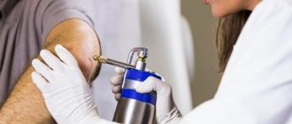
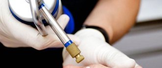
The procedure is performed using special equipment (cryodestructor) or liquid nitrogen. When using the hardware method, the tip of the applicator is pressed against the required area, after which it is cooled to a temperature of -180-190 degrees Celsius. When using liquid nitrogen, a cotton swab is soaked in it, and then simply applied to the damaged tissue for a short period of time (no more than 2-3 minutes).
The procedure involves instant cooling of tissues due to exposure to ultra-low temperatures. As a result, the intracellular and intercellular fluid freezes, the vital activity of cells is disrupted, protoplasm stops and microcirculation in the pathological tissue is disrupted. Tissue destruction occurs. After cryodestruction, the destroyed tissues are gradually replaced with healthy ones.
What complications can there be during the procedure?
- Immediate complications: pain, fainting, headache, swelling (especially periorbital), blistering.
- Delayed: infection, bleeding, slow healing.
- Long-term but temporary: hyperpigmentation, milia.
- Constantly: hypopigmentation, ectropion, atrophy, alopecia.
The procedure is usually safe if done carefully, although there are sometimes exceptions. For example, cryotherapy treatments near the eyes may cause the eyelid to swell for a day or two, although the swelling will soon subside.
Stages of cryodestruction
For different purposes, different tips are used, which are selected depending on the size of the surface. In this case, the planes of contact must be parallel.
The cryodestruction process is divided into the following stages:
- Freezing tissue using liquid nitrogen. In this case, the tissue becomes dense, cold, white and insensitive. This is often accompanied by a tingling sensation, a slight burning sensation and mild pain.
- After cryodestruction, collateral edema and hyperemia occurs, lasting from 1 to 3 hours.
- The appearance of epidermal blisters, which disappear after 6-24 hours.
- Necrosis is completely rejected within 2-6 weeks. After cryodestruction, an inconspicuous spot remains, and the process of epithelization of the defect and surrounding tissue begins.
- Complete tissue regeneration occurs in about six months.
Removal of benign tumors with liquid nitrogen
Fibroepithelial polyps
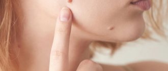
Fibroepithelial polyp (papillomas) are benign neoplasms on the skin in the form of wart-like growths, rising above the level of the skin, with a wide base or on a stalk, grayish or brown in color, with a diameter of 0.3 - 1.5 cm or more, round or irregular in shape. Most often they are located on the neck (along the lateral surface) and in large folds. The surface of papillomas is villous or covered with easily detachable horny masses.
Fibroepithelial polyps occur in 50% of the world's population, and in women during pregnancy they increase in number and size. In pregnant women, these tumors often appear on the nipples, which is dangerous for breastfeeding for the unborn child.
Seborrheic keratosis
Seborrheic keratosis (syn.: seborrheic wart, senile wart, basal cell papilloma) is the most common benign skin tumor. Usually develops in middle and old age from the epidermis. Its pathogenesis is unclear, but it is believed that this lesion develops from cells of the basal layer of the epidermis or keratinocytes of the most superficial part of the hair follicle and is not associated with human papillomavirus. In accordance with modern ideas, there is no connection with solar radiation. It occurs predominantly in elderly and senile people, but is often observed in young people, equally often in men and women.
The tumor develops very slowly, sometimes over several decades. Initially, a limited spot of yellowish or brown color appears, which gradually increases, sometimes reaching 4-6 cm or more in diameter. The surface of the stain is gradually covered with greasy crusts, which are easily removed. Over time, the crusts become denser, but often retain a greasy surface, riddled with cracks. The thickness of the crusts can reach 1-2 cm. The tumor becomes yellow-brown, dark brown or black. Localization can be very diverse. Lesions can be single or multiple. In rare cases, keratoses can become malignant.
Removing warts with liquid nitrogen
Warts are small, usually painless growths on the skin caused by the human papillomavirus (HPV).
More than 100 types of HPV are known. HPV affects the top layer of skin and usually enters the body through damaged skin. The virus causes the top layer of skin to grow rapidly, forming warts.
Most warts, but not all, are usually harmless and will disappear on their own within a few months or years. Warts can grow on any part of the body. They are most common among children and young adults - so liquid nitrogen removal is especially effective for hard-to-reach wart habitats.
Sometimes warts can be disfiguring, especially if they grow on the face or hands and cause psychological discomfort to their owner, and some of them also cause itching and pain.
Warts can appear at any age. Infection occurs through the shoes of an infected patient (plantar warts), in swimming pools, baths, gyms with exercise equipment, and in hairdressing salons through manicure accessories (periungual warts).
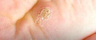
Vulgar warts
Warts vulgaris are the most common type of wart in humans. Other names are simple or common warts.
Remember: the cause of the appearance of vulgar warts is the already known Human Papilloma Virus (type 2).
Infection occurs in childhood. The virus gets onto injured skin (abrasions and scratches are common in children) and penetrates the skin. While in the body, it gradually multiplies in the basal layer of the skin and after a few months ordinary warts appear.
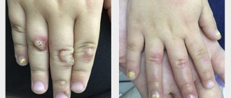
The clinical manifestations of the disease are simple. Rounded elevations ranging in size from 3 to 10 mm, no more, appear on the skin of the fingers and on the back of the hands and feet. A characteristic feature of this disease is the multiple nature of the rashes. That is, next to one simple wart on the hand, a second, daughter wart may soon appear, then another one nearby, etc.
Another sign characteristic of vulgar warts in most cases is the so-called kissing warts. This is their location when two simple warts appear on two adjacent fingers, opposite each other, touching each other when the fingers are closed.
Flat warts
Among all neoplasms caused by the human papillomavirus, flat warts are considered the most harmless. But this is only at first glance, sometimes leaving the problem without proper attention, a person gives the green light to further development, contributing to the spread of flat warts over a much larger area of the body. Flat, or juvenile, warts usually grow on the face, arms, or legs. They are small 1-5 mm, have flat tops in irregular nodules, and can be pink, light brown or light yellow in color. They occur mainly in children and adolescents. During the treatment of warts, the feet can and should be washed, because treatment with liquid nitrogen is not traumatic, does not open tissue, and does not release the virus. It is advisable to use liquid soap with tea tree oil. After warts on the feet are rejected, it is necessary to disinfect shoes, get rid of slippers, socks, and stockings to avoid reinfection.
Genital warts or genital warts
Transmitted sexually in 60% of cases through contact with an infected partner. This is the most common HPV infection (HPV types 6, 11, 16, 18, 42, 44, 54). Externally, genital warts are moist, soft, cauliflower-shaped nodules. They often ulcerate, turning into foul-smelling sores. Favorite localization is the genitals (mucous membranes and skin folds). Removal of warts with nitrogen is a painless removal without anesthesia followed by healing without scars.
During the treatment of benign epithelial tumors of the skin and mucous membranes, after a blood test for HPV, the use of immunomodulators and antiviral drugs is indicated both on the skin and mucous membranes, and, if necessary, orally.
Benefits of cryodestruction
Cryodestruction with liquid nitrogen has a number of undoubted advantages:
- the area of necrosis does not bleed;
- does not leave scars or scars;
- ease of procedure;
- cryosurgery does not require pain relief, since rapid cooling produces an analgesic effect;
- minimal injury;
- speed of the method;
- stable desired effect;
- cryodestruction is tolerated easily and painlessly;
- does not require lengthy preoperative preparation;
- during and after the procedure, metastasis stops;
- no relapses;
- does not require postoperative care or stitches.
What diseases can be treated with cryodestruction?
Dermatological _
Riosurgery allows you to treat a wide range of benign and precancerous skin conditions, such as:
- Angiomas
- Warts, including plantar warts
- Seborrheic and actinic keratosis
- Birthmarks
- Age and sun spots
- Dermatofibromas
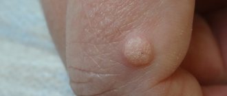
- Molluscum contagiosum
- Condylomas
- Hemangiomas
- Acne
- Keloids
In most cases, a single application of cryodestruction is sufficient. Keloids require three times of treatment in two months, and dermatofibromas and ingrown toenails require two times of treatment in two months.
Rehabilitation: skin care after cryodestruction
- Do not allow the treated area to come into contact with clothing to avoid damaging the protective film.
- Do not cover with adhesive tape so as not to disturb air exchange. It is acceptable to cover with a gauze bandage.
- Do not puncture the blister. If its integrity is compromised, an infection may enter the wound, and a scar will form in its place after healing.
- Do not peel off the crust so that a scar does not appear.
- You can take a shower, but you cannot rub or steam the treatment area.
- Use healing ointments as prescribed by a doctor. They will speed up the healing of the skin after cryodestruction and prevent complications.
Dermatologists
- Ayvazyan Linda Volodyevna
Experience: 5 yearsDermatovenerologist, mycologist, trichologist
Rating: 0/5 — 0 votes
Make an appointment
- Osipova Daria Sergeevna
Experience: 14 years
Deputy chief physician for medical work. Dermatovenerologist
Rating: 0/5 — 0 votes
Make an appointment
The cryodestruction method is widely used in dermatology to remove various skin tumors. This is a modern, effective way to correct cosmetic defects and treat diseases. Cryodestruction can remove warts, moles, basal cell carcinomas, papillomas, condylomas, scars, old calluses and other undesirable phenomena from the skin. There are practically no traces left.
At the Kutuzovsky Children's Center, cryodestruction treatment is performed by experienced dermatologists. The procedure is performed on an outpatient basis in our clinic in Moscow. Prices for cryodestruction are indicated on the website.
Contraindications and complications
Contraindications to cryosurgery include cryofibrinogenemia, cryoglobulinemia, Raynaud's disease, agammaglobulinemia, and multiple myeloma.
Serious complications from cryosurgery are rare. Hypopigmentation often occurs as a result of increased sensitivity of melanocytes to cold injury. This is much more noticeable in patients with darker skin. Deeper freezing can destroy hair follicles, which can lead to alopecia patches. Although rare, prolonged freezing can result in scarring caused by damage to the basement membrane and necrosis of the epidermis. Some degree of paresthesia occurs in a quarter of patients and may persist for one to three months. Permanent sensory loss is rare and is best prevented by avoiding cryosurgery of nerve trunks (eg, preauricular areas). The risk and extent of scarring, alopecia, hypopigmentation, and paresthesia can be reduced by reducing freezing time to less than 30 seconds.
How to prepare and what to do after the procedure
Before you go for the procedure of removing papillomas with liquid nitrogen, you must consult with your doctor. Specialists must study the tumor, exclude oncological aspects, and only then admit the patient to cryodestruction. If the patient is experiencing an acute phase of infection at the time of contacting a specialist, the procedure should be postponed until complete recovery. A week before cryodestruction, you should not take anticoagulants.
After cryodestruction has been carried out, swelling appears at the site of the former papilloma, and the areas underneath harden and darken. After necrosis of the tumor tissue, a scab appears, which is rejected only a few weeks after the procedure. The scab acts as a protective layer that prevents germs from entering the wound. The scab cannot be removed on its own; this can only be done by a doctor if the scab does not fall off on its own.
When very large tumors are removed, blisters with serous or hemorrhagic contents appear in their place, which under no circumstances should be opened. The blister will break on its own within 1 week, and after that you will need to apply a sterile bandage in its place.
During the first week after cryodestruction, the site of the former neoplasm cannot be covered with cosmetics. You can wash the wound with soap, but you cannot rub it with a washcloth. In the future, when everything has healed and a new epithelium has appeared, it is not recommended to expose the skin to direct sunlight, treating it with a cream with a high radiation protection factor.