Microsporia and its prevention
Microsporia (more popularly known as “ringworm”) is a contagious disease transmitted to humans from sick pets. The disease, which belongs to the group of dermatophytosis, is caused by a microscopic fungus of the genus Microsporum (from ancient Greek the name literally translates as “small crop”), the microorganism affects the coda, hair, and in some cases the nail plates of the patient.
Microsporia
The disease is very common, for example, in the Primorsky Territory alone, 600–800 cases are registered annually. An increase in the number of cases is typical for the summer period, which is associated, firstly, with the increase in offspring of cats and dogs, and secondly, with an increase in the possibility of contact with an infected animal. The end of August is a period of increased spread of microsporia, the peak of activity is October/November.
Most often, children (preschoolers and schoolchildren) become ill with microsporia; this category of the population accounts for 90% of all cases. By coming into contact with a sick animal, one child freely spreads the contagious disease among other children with whom he communicates, so the disease becomes widespread. As a rule, the most common source of infection is stray animals, usually dogs and cats; much less often, infection occurs from rodents, rabbits or foxes. Sick people with skin lesions in the scalp area pose the greatest epidemiological danger, because this form of mycosis is not immediately diagnosed and presents certain difficulties in treatment.
Symptoms and clinical picture
The first signs of a fungal infection depend on the location of the infectious focus. Symptoms on smooth skin differ from the manifestations of fungus on hair structures. The clinical picture in children may be similar to the adult course and may differ from it.
Symptoms of smooth skin lesions
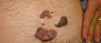
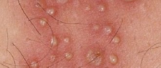
Microsporia of smooth skin is characterized by the formation of a spot with clearly defined boundaries. As the disease progresses, such a spot increases in diameter. The edge of the spot resembles a defined roller, rising above the surface of the skin, having a bubbly structure with crusts and nodules. The center of the spot is a focal inflammatory process with a pink tint and pronounced peeling. The number of ring-shaped lesions when affecting smooth skin is from 1 to 3, and the diameter varies from 0.5 to 3.5 cm. More often, lesions are recorded on the neck, face, shoulders or forearms. Significant symptoms include moderate itching and flaking.
In children during the neonatal period or in young women, peeling may be completely absent, but the inflammatory process will be more significant. With a complicated allergic history, the Microsporum fungus can be mistaken for atopic dermatitis or other chronic skin diseases, so it is not always detected and identified in a timely manner. If you continue hormonal treatment of the underlying disease, fungal colonies begin to grow rapidly. When the pathological process spreads to the nail plate, the outbreak is usually localized along the edge of the nail from the cuticle. The form of the disease is characterized by the appearance of a whitish spot, gradual thinning and destruction of the nail structure.
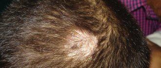
Signs of fungus on the scalp
Microsporia of the scalp is often recorded in children aged 4 to 13 years. The frequency of occurrence in children is explained by the absence of special organic acids in the hair, which prevent the spread of fungal colonies and are found in sufficient quantities in the hair of adults. If a small patient is a carrier of a fungus of the genus Microsporum with its localization in the scalp, then spontaneous recovery is possible when he reaches puberty. An interesting fact is the rarity of the occurrence of lesions on hair structures in red-haired children. Characteristic signs are the following manifestations:
- the formation of 1 or 2 large lesions on the crown, temples or crown;
- the diameter of the lesions varies from 2.5 to 6 cm;
- the shape of the pathological spot can be round or oval;
- the presence of screenings along the edge of the lesion with a diameter of about 1 cm;
- peeling;
- hair fragility;
- slight itching.
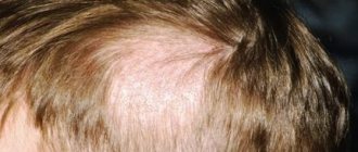
As the disease progresses, the hair in the affected area breaks off and resembles a “hedgehog” after a haircut. Hence the name “ringworm”. The pathological ring exhibits slight swelling and redness.
Symptoms of the suppurative form
The purulent form is characterized by the formation of a focus with visible inflammation, the formation of soft nodules of a bluish tint. Pustules form on the surface of the affected skin area. With slight pressure, purulent exudate is released. The development of a suppurative form is provoked by inadequate treatment of the superficial form of microsporia, the presence of a burdened clinical history and chronic diseases, and advanced forms of microsporia.
Making a diagnosis of microsporia only based on primary signs and the appearance of the pathological formation may be unreliable. If there is any doubt, doctors resort to different methods of differential diagnosis.
The causative agent of microsporia and ways of its spread
Microscopic fungi of the genus Microsporum are the causative agents of this disease. The unique microorganism is very resistant to environmental factors (temperature, moisture, etc.); the virus can persist in the fur of sick animals and fragments of nail plates for several years.
Signs of microsporia in animals:
- Bald patches on the face;
- Areas of baldness in the area of the external ears;
- Bald patches on the paws (usually the front, less often the rear).
Ways of spread of the disease:
- Contact (when interacting with a sick animal or person);
- Contact-household (in contact with items caring for a sick animal or items belonging to an infected person).
Infection can also occur through the common local area (sand in sandboxes, dust on staircases).
From the moment of infection until the first signs of the disease appear, it can take from 5 days to 6 weeks; a narrowing of the interval for the manifestation of microsporia is usually associated with low immunity and poor compliance with personal hygiene requirements. Often the first signs of the disease go unnoticed.
The main symptom of the presence of a microsporia pathogen in the body is a pink-red spot with swelling and clear boundaries. Over time, the spot increases in size and infiltrates. A ridge of small bubbles or crusts forms around the affected area. If the infection is in the scalp, the growing fungus attacks the hair follicles and causes them to weaken, resulting in hair breaking off at the roots.
Microsporia
Microsporia
Potekaev N.N.
Central Research Institute of Dermatovenerology, Ministry of Health of the Russian Federation, Moscow
Microsporia is a fungal disease from the group of dermatophytosis, which affects the skin and hair, and in extremely rare cases, the nail plates . The name of this mycosis comes from the name of its causative agent - a fungus of the genus Microsporum, which belongs to the dermatophytes. The disease is also known as “ringworm” (the term combines microsporia and trichophytosis), which is due to the peculiarities of its clinical picture.
Etiology
The causative agent of microsporia was first described by Gruby in 1843. The scientist discovered a sheath of small spores on the surface of the affected hair and gave the fungus the name Microsporum audouinii in honor of the late Dr. Audouin. However, the author’s discovery was not appreciated, and highly respected dermatologists (in particular, Bazin) identified microsporia with trichophytosis. Sabouraud managed to restore the truth in 1893, who, having carefully studied the biology of the causative agent microsporia , indicated the signs that distinguish this mycosis from trichophytosis. In Russia, microsporia was first described by S.L. Bogrov in 1912.
Currently, more than twenty species of the Microsporum fungus are known. Of these, the following are identified as pathogens:
- M. distorum, M. rivalieri, M. langeronii.
- Zoophilic group —
- M. canis, M. nanum, M. persicolor.
- Geophilic group - M. gypseum, M. cookeii, Keratynomyces ajelloii.
Of the listed species, only M.canis (seu lanosum) has become practically the only pathogen of microsporia in recent years. It is no coincidence that it is called a cosmopolitan mushroom.
Once on the skin, the pathogen penetrates it and begins to multiply. When located near the mouths of hair follicles, fungal spores germinate, leading to hair damage. Quite quickly spreading over the surface of the latter, the mycelial hyphae destroy the cuticle, between the scales of which spores accumulate. Thus, the fungus surrounds the hair, forming a sheath, and tightly fills the follicular apparatus.
Epidemiology
Microsporia is the most common mycotic infection among dermatophytoses, not counting mycoses of the feet. The disease occurs everywhere. In Russia, up to 100 thousand patients with microsporia are registered annually. Mycosis is highly contagious, children are more often affected. In the last two decades, there has been an increase in the incidence of microsporia in newborns [1]. Adults rarely get sick—mostly young women. The rarity of the disease with microsporia in adults, especially with damage to the scalp, and the usually occurring spontaneous recovery at the beginning of puberty is explained by the presence of fungistatic organic acids (in particular, undicylenic acid) in the hair of adults. Patients with lesions of the scalp pose a particular danger in epidemiological terms. This is due to the fact that this form of mycosis, firstly, is most often diagnosed untimely, and, secondly, its therapy is associated with certain difficulties. Unfortunately, data from recent epidemiological studies conducted in Russia indicate an increase in the number of patients with hair damage [2].
As already indicated, the most common pathogen of microsporia is Microsporum canis , a zoophilic fungus that is found in 90-97% of patients. The main source of the disease is cats (usually kittens), less often dogs. Infection occurs through direct contact with a sick animal or objects contaminated with fur or scales. Once in the soil with an affected hair or scale, M.canis remains viable only for 1-3 months. Thus, soil is only a factor in the transmission of infection and does not serve as its natural reservoir [3]. Intrafamily spread of infection is common. In this case, infection usually occurs from one animal. Transmission of zoonotic microsporia from sick family members is possible, but this is quite rare. There are isolated observations of families in which three generations were sick with this mycosis. It should be emphasized that in such situations, women and children of younger age groups, including newborns, are at greatest risk of infection.
Clinical manifestations in animals are characterized by areas of baldness on the face, the outer surfaces of the ears, as well as on the front, less often the hind, legs. Under Wood's lamp a green glow is detected. Often, clinically healthy cats can be mycocarriers, and then only a luminescent study helps to identify the fungus. However, situations are possible when the fact of carriage cannot be confirmed either by clinical or luminescent examination. In such cases, and they are observed in 2-3% of carriers, wool is sown from various areas [4].
The incidence of zoonotic microsporia varies throughout the year. Seasonal fluctuations are associated with litters in cats, as well as more frequent contact of children with animals in the summer. The rise in incidence begins at the end of summer, the peak occurs in October-November, and the decrease to a minimum occurs in March-April. The emergence of epizootics of microsporia in cats and kittens in a number of regions and cities leads to the formation of epidemic foci among children.
Clinic
Since the main causative agent of microsporia in our time is Microsporum canis, when describing the clinical picture of the disease, more attention will be paid to the zoonotic form rather than the anthroponotic one.
The incubation period for zoonotic microsporia is 5-7 days . The nature of the clinical picture of the disease is determined by the localization of the lesions and the depth of penetration of the pathogen. There are microsporia of smooth skin and microsporia of the scalp.
Microsporia of smooth skin
, a swollen, raised erythematous spot with clear boundaries appears . Gradually the spot increases in diameter and infiltrates. A continuous raised ridge is formed along the periphery, represented by small nodules, bubbles and crusts. In the central part, the inflammatory phenomena resolve, as a result of which it acquires a pale pink color, with pityriasis-like peeling on the surface. Thus, the focus has the appearance of a ring. As a result of autoinoculation of the fungus in the central part and repeated development of the inflammatory process, bizarre foci of the “ring within a ring” type are formed. Such iris-like figures are more common with anthroponotic microsporia. Vellus hair is often involved in the process, which complicates the treatment of the disease. The number of foci with microsporia of smooth skin is usually small (1-3). Their diameter ranges from 0.5 to 3 cm. The rash can be located on both open and closed areas of the skin, since a sick animal is often warmed under clothes and taken to bed. However, the most common lesions are located on the skin of the face, neck, forearms and shoulders. There are no subjective sensations or moderate itching.
In persons with a reduced delayed-type hypersensitivity reaction, an abortive course of microsporia is possible. In this case, the lesion has the appearance of a pale pink scaly spot without clear boundaries, along the periphery of which there are no nodular and vesicular elements. In 1957, J. Esteves first described parasitic achromia , a rare variant of microsporia characterized by minimal symptoms [5]. With parasitic achromia, along with damage to the scalp, there are numerous depigmented spots of round shape, with slight peeling on the surface.
In newborns and young children, as well as in young women, due to a hyperergic reaction, an erythematous-edematous form of microsporia is often observed, in which pronounced inflammatory phenomena and minimal peeling are noted.
The papular-squamous form occurs when microsporia is localized in seborrheic areas of the skin - on the face, chest and back. The lesions are characterized by infiltration and lichenification, accompanied by significant peeling and itching. Since this form of microsporia is usually observed in individuals with signs of atopy (in particular, in patients with atopic dermatitis), mycosis is often masked by manifestations of the underlying process and is not always diagnosed in a timely manner. The use of local corticosteroid drugs only increases the spread of mycotic infection.
In young women with hypertrichosis, follicular-nodular elements with a diameter of 2-3 cm may appear in the lower leg area - the so-called deep form of microsporia of smooth skin.
Localization of single foci of microsporia in places atypical for it can sometimes lead to difficulties in diagnosing the disease. T.I. Meerzon, in particular, described an isolated focus of zoonotic microsporia on the skin of the shaft of the penis in an 18-year-old patient [6].
A rare type of microsporia includes damage to the skin of the palms, soles and nail plates . On the palms, and less often on the soles, dyshidrotic and/or squamous-keratotic rashes are observed. Microsporic onychomycosis is characterized by isolated lesions of the nail, usually its proximal part [7]. Initially, a dull spot is formed, which becomes white over time. The nail in the area of leukonychia becomes softer and more fragile, and subsequently can collapse as onycholysis. When examining the affected nail under a Wood's lamp, a bright green glow is detected. Microsporic onychomycosis not diagnosed in time can cause reinfection and further spread of the disease among others.
Microsporia of the scalp
Damage to the scalp occurs mainly in children 5-12 years old . Over the past 20 years, there has been a 20-fold increase in the incidence of this form of microsporia in newborns [1]. It is generally accepted that the rarity of this form in adults is explained by the presence of fungistatic organic acids in their hair and water-lipid mantle of the skin. This fact indirectly confirms the spontaneous recovery of children during puberty, when the composition of sebum changes. Perhaps the difference in hair thickness between children and adults matters. It is noteworthy that microsporia of the scalp practically does not occur in children with red hair [8].
Foci of microsporia of the scalp are located mainly on the crown, in the parietal and temporal regions (Fig. 2). Usually there are 1-2 large lesions ranging from 2 to 5 cm in size, with round or oval outlines and clear boundaries. Along the periphery of large lesions there may be screenings - small lesions with a diameter of 0.5-1.5 cm. At the beginning of the disease, a peeling area forms at the site of infection. In the first days, the fungus is localized only at the mouth of the hair follicle. Upon closer inspection, you will notice a whitish ring-shaped scale surrounding the hair like a cuff. On the 6-7th day, the process spreads to the hair itself, which becomes fragile, breaks off above the level of the surrounding skin by 4-6 mm and looks as if it has been trimmed (hence the name “ringworm”). The remaining stumps look dull and are covered with a grayish-white sheath, which is the spores of a fungus. If you “stroke” the stumps, they deviate in one direction and, unlike intact hair, do not restore their original position. The skin in the lesion, as a rule, is slightly hyperemic, edematous and moderately infiltrated, its surface is covered with grayish-white small scales.
With microsporia of the scalp caused by anthropophilic fungi , numerous small foci with minimal inflammation and unclear boundaries are observed. A characteristic feature of anthropophilic microsporia is its localization in the marginal zone of hair growth, when one part of the lesion is located on the scalp, and the other on smooth skin.
Atypical, rare variants of microsporia of the scalp include infiltrative, suppurative (deep), exudative, trichophytoid and seborrheic forms.
In the infiltrative form of microsporia, the lesion on the scalp rises somewhat above the surrounding skin, is hyperemic, and the hair is often broken off at a level of 3-4 mm. It should be especially emphasized that with this type of microsporia, the sheath of fungal spores at the root of broken hair is weakly expressed.
In the suppurative form, against the background of significant inflammation and infiltration, soft bluish-red nodes form, the surface of which is covered with pustules. When pressed, pus is released through the follicular openings. Such clinical manifestations correspond to the picture of kerion Celsi (Celsius honeycomb) - infiltrative-suppurative trichophytosis. The formation of infiltrative and suppurative forms of microsporia is facilitated by irrational (usually local) therapy, the presence of serious concomitant diseases, and late consultation with a doctor.
Exudative microsporia of the scalp is characterized by severe hyperemia and swelling, with small bubbles located against this background. Due to the constant impregnation of the scales with serous exudate and gluing them together, dense crusts are formed, which, when removed, exposes the moist, eroded surface of the lesion.
The listed three forms of microsporia of the scalp are often complicated by regional lymphadenitis, and patients with suppurative microsporia may also experience symptoms of intoxication.
With the trichophytoid form of microsporia, numerous small foci with weak pityriasis-like peeling are scattered on the scalp. The boundaries of the lesions are unclear, there are no acute inflammatory phenomena, the hair is broken off at a level of 1-2 mm above the surrounding skin. Along with broken hair, there are healthy hairs. Trichophytoid microsporia is more common in older age groups with serious concomitant diseases.
With seborrheic microsporia of the scalp, thinning of the hair is mainly observed. The areas of rarefaction are abundantly covered with yellowish scales, upon removal of which a small amount of broken hair can be found.
Late diagnosis and inadequate treatment of atypical forms of microsporia lead to further changes in clinical symptoms, dissemination of rashes and chronicity of the process, irreversible alopecia in the patient and dissemination of infection in the environment.
Diagnostics
To confirm the clinical diagnosis of microsporia, fluorescent, microscopic and cultural studies are used.
Luminescent study
The method is based on identifying a bright green glow in hair affected by fungi of the genus Microsporum when examined under a Wood's lamp. At the same time, both long and vellus hair glows. The reason for this phenomenon has not yet been established. Luminescence testing must be carried out in a darkened room. The lesions are first cleaned of crusts, ointments, etc. When examining fresh lesions, there may be no glow, which is due to insufficient hair damage. In such situations, the hair should be epilated from the suspected site of fungal penetration, and the glow can be detected in its root part. When the fungus dies, the glow in the hair remains.
The luminescent method is used for:
- pathogen identification;
- identifying affected hair;
- evaluation of therapy results;
- control over persons in contact with the patient;
- determination of infection or mycocarriage in animals.
Microscopic examination
To confirm the fungal etiology of the disease, scales from lesions of smooth skin lesions are subjected to microscopic examination, and if the scalp is involved in the process, hair fragments are subjected to microscopic examination. Immediately before collecting pathological material, the lesion on smooth skin must be treated with 960 alcohol. Then, using a blunt scalpel, carefully scrape off the scales from the periphery of the lesion. On the scalp, using tweezers, hair fragments are also removed from the marginal zones of the lesion. Then the pathological material is placed on a glass slide in a drop of 20% potassium hydroxide solution. Microscopic examination is carried out after 30-40 minutes.
In scales from lesions on smooth skin, twisted threads of septate mycelium are found. Microscopic examination of the affected hair reveals many small spores (2-3 microns) on its surface (ectothrix-type lesion). In this regard, the boundaries of the hair appear blurred. The spores surrounding the hair are arranged chaotically, like a mosaic.
Cultural examination
Carrying out cultural diagnostics with positive results of luminescent and microscopic studies is required to identify the causative fungus. The method allows you to determine the genus and type of pathogen and, therefore, carry out adequate therapy and prevention of the disease. Pathological material (scales, hair) is placed on Sabouraud's medium. The growth of Microsporum canis colonies (the main pathogen of microsporia) is observed on the 3rd day after sowing. By the 10th day, the colony reaches a diameter of 4-5 cm and is represented by a flat disk covered with a whitish, delicate fluff, which spreads like rays along the walls of the test tube. The reverse side of the colony is yellow in color.
Treatment
When treating microsporia of smooth skin without affecting vellus hair, external antimycotic drugs are used. Apply 2-5% iodine tincture to the foci of mycosis in the morning, and apply antifungal ointment in the evening. Use traditional 10-20% sulfur, 10% sulfur-3% salicylic or 10% sulfur-tar ointments. Modern ointments are used twice a day: clotrimazole, ciclopirox, isoconazole, bifonazole, etc. The allylamine drug terbinafine (Lamisil) , produced in the form of 1% cream and spray, has proven itself well.
Terbinafine has a fungicidal effect (i.e., it leads to the death of the fungus) and is the most active antimycotic agent against dermatophyte fungi. The drug inhibits the functions of svalene epoxidase, resulting in disruption of the synthesis of ergosterol, the main component of the fungal cell membrane. At the same time, the amount of squalene, a high molecular weight hydrocarbon, increases inside the cell. These disturbances lead to the death of the fungal cell. The sensitivity of svalene epoxidase in fungi is 10,000 times higher than in humans, which explains the selectivity and specificity of the action of terbinafine in relation to the fungal cell. The drug can be used once a day. It should be emphasized that, having a keratophilic ability, Lamisil accumulates in the stratum corneum of the epidermis and is present here for a long time in fungicidal concentrations. This circumstance explains the persistence of a pronounced antifungal effect even after discontinuation of the drug. The convenient dosage form of terbinafine spray ensures contactless application of the drug to large areas of affected skin. Terbinafine cream and spray are quickly absorbed and do not leave marks on clothing.
In case of a pronounced inflammatory reaction, it is advisable to prescribe combination drugs containing additional corticosteroid hormones. Similar products include mycozolon and travocort .
When a secondary bacterial infection occurs, Triderm cream is useful . With severe infiltration of the lesion, as well as with deep forms of microsporia, preparations containing dimexide , which is known to have conductive properties, are indicated. In particular, in such situations, a 10% solution of quinosol is widely used (quinosole and salicylic acid 10.0 each, dimexide 72.0, distilled water 8.0). The solution should be applied 2 times a day until clinical manifestations resolve and the fungi disappear.
When vellus hair, and especially long hair, is affected, systemic antimycotic therapy for microsporia is necessary.
In the treatment of microsporia of the scalp, griseofulvin , a chlorine-containing antibiotic produced by the mold Penicillium nigricans, remains the drug of choice. Griseofulvin, available in the form of 125 mg tablets, is prescribed at the rate of 22 mg per 1 kg of patient body weight. The drug is taken daily in 3-4 doses during meals with a teaspoon of vegetable oil, which is necessary to increase the solubility of griseofulvin and increase the duration of its action (a-tocopherol contained in oils delays the metabolism of griseofulvin in the liver). For children under 3 years of age, it is preferable to prescribe griseofulvin in the form of a suspension, 8.3 ml of which corresponds to 1 tablet (125 mg) of the drug. Continuous therapy is carried out until the first negative test result for fungi, after which griseofulvin is taken at the same dose every other day for 2 weeks, and then for another 2 weeks, 2 times a week. The general course of treatment is 1.5-2 months. During therapy, it is necessary to shave your hair weekly and wash your hair 2 times a week . It is recommended to simultaneously rub any antifungal ointment into the affected area. In parallel with oral administration of the antimycotic, manual hair removal can be performed with preliminary application of a 5% griseofulvin patch to the lesion.
Side effects of griseofulvin include headache, allergic rashes, and discomfort in the epigastrium; Granulocytopenia and leukopenia are less common. Unfortunately, due to hepatotoxicity, griseofulvin is contraindicated in children who have had hepatitis or suffer from liver disease. The drug is also not prescribed for kidney diseases, gastric and duodenal ulcers, neuritis, blood diseases, and photodermatoses.
In recent years, terbinafine (Lamisil) . Local forms of the drug have already been mentioned earlier. In the treatment of microsporia of the scalp, terbinafine is used in the form of tablets, available in doses of 125 and 250 mg. The drug has a high safety profile, which is largely due to the peculiarities of its mechanism of action. Squalene epoxidase, which is inhibited by terbinafine, is not associated with the cytochrome P-450 system, so the drug does not affect the metabolism of hormones and other drugs. Since terbinofine is lipophilic, after oral administration it quickly reaches the dermal layer of the skin, overcomes it and accumulates in the lipids of the stratum corneum of the epidermis, hair follicles and hair.
When treating microsporia of the scalp in children, the dose of terbinafine is determined depending on body weight. The manufacturer recommends prescribing the drug for a child weighing less than 20 kg at a dose of 62.5 mg per day; children weighing from 20 to 40 kg - 125 mg; more than 40 kg - 250 mg. However, our experience shows that these doses are often insufficient, since we obtained the maximum therapeutic effect by changing the officially recommended treatment regimens [9]. In this regard, the doses of terbinafine we offer are 50% higher than those recommended by the manufacturer: 94 mg/day (3/4 tablets of 125 mg) for children weighing 10-20 kg and 187 mg/day (1.5 tablets in 125 mg) - 20-40 kg. For body weight over 40 kg, terbinafine is prescribed at a dose of 250 mg/day. For adults, terbinafine is prescribed at a dose of 7 mg per 1 kg, but not more than 500 mg per day.
Terbinafine is taken once a day. The drug is well tolerated. Patients may be bothered by a feeling of fullness in the stomach, minor abdominal pain. Following a diet aimed at relieving flatulence relieves patients from unpleasant sensations.
Prevention
Prevention of microsporia consists of timely identification, isolation and treatment of patients with microsporia. In children's institutions, periodic medical examinations should be carried out. A child diagnosed with microsporia must be isolated from other children and sent for treatment to a specialized mycological hospital. For each sick person, a notification is filled out according to registration form 281. Things belonging to a patient with microsporia are subject to disinfection. Relatives and people in contact with the patient must be examined. Particular attention should be paid to pets, since they are often the source of infection. Animals with microsporia are either destroyed or given full antifungal treatment. An important role in the fight against microsporia is assigned to health education authorities, as well as veterinary supervision of stray animals.
Literature
1. Mohammad Yusuf. Clinical and epidemiological features of microsporia in modern conditions and the development of treatment with new medications. Author's abstract. diss...candidate of sciences. M., 1996
2. Fakhretdinova Kh.S. Clinical and epidemiological features of modern microsporia. Author's abstract. diss... doc. med. sc. M., 1999.
3. Sheklakov N.D., Andriasyan S.G. Some ecological features of Microsporum canis and the incidence of zooanthroponotic microsporia. Vestn dermatol. 1979; 2: 18-23.
4. Stepanova Zh.V., Davydov V.I. On the carriage of fluffy microsporum by clinically healthy animals. Vestn dermatol. 1970; 3:42-6.
5. Esteves J. Acromia parasitaria devida ao M. Felineum. Trab. Soc. Derm. Vener. 1957; 15:43.
6. Meerson T.I. Atypical localization of smooth skin microsporia caused by Microsporum canis. Vestn dermatol. 1985; 5:70.
7. Stepanova Zh.V., Klimova I.Ya., Shapovalova F.S. Onychomycosis caused by fluffy microsporum. Vestn dermatol. 1997; 4:37-9.
8. Feyer E., Olah D., Szatmari S. et al. Medical mycology and fungal diseases. Budapest. 1966.
9. Potekaev N.S., Kurdina M.I., Potekaev N.N. Lamisil for microsporia. Vestn. Dermatol. 1997; 5: 69-71.
Diagnosis of microsporia
The clinical picture and history of contact with animals provide the doctor with grounds for making a preliminary diagnosis. To confirm it, the following diagnostic studies are prescribed:
- Inspection using a fluorescent filter. This method is based on the ability of fungi of the genus Microsporum to fluoresce when using ultraviolet radiation. For this purpose, a device known in medicine as a Wood's lamp is used. This lamp emits ultraviolet light, which, when passed through a darkened filter, helps identify pathogenic fungi: when examined, they glow pale yellow or greenish.
- Histological examination. An area of skin is scraped for microscopic examination, which makes it possible to identify the fact of infection and the degree of inflammation. However, microscopy does not determine the type of pathogen.
- Cultural research. To determine the species of the pathogen and its sensitivity to antifungal drugs, the fungus is sown and grown on a nutrient medium. The disadvantage of the cultural method is that you have to wait at least a week to get a reliable result.
- Additionally, clinical tests of blood and urine are performed. If the results deviate from the norm, the tests are repeated once every 10 days. A biochemical study of blood serum is done before the start of the therapeutic course and 3 weeks after its completion.
Sources and routes of infection
The most common source of infection is the fungi Microsporum canis, which parasitizes mainly dogs and cats. To a lesser extent, rabbits, sheep, rodents, poultry, pigs and large wild animals are susceptible to microsporia.
Infection can occur in any environment. However, the combination of heat and high humidity is most favorable for the fungus, so outbreaks of microsporia are characterized by seasonality - most often they occur in late spring and early autumn. Microsporum spores can live in soil for up to several months, so frequent contact with the soil may pose a certain risk. An additional predisposing factor may be increased sweating and disruption of the secretion of the sebaceous glands. Timely washing of hands with soap reduces the risk of infection to a minimum, even if fungal spores have already reached the skin.
Important!
For infection, the so-called entry gate of infection is necessary, which can be abrasions, scratches, calluses and other microtraumas of the skin. If the integrity of the skin is not compromised, the fungus has virtually no chance.
Treatment
Treatment of microsporia in most cases is carried out on an outpatient basis, that is, the patient is not hospitalized. A hospital stay is advisable only in cases where there are concomitant pathologies that require diagnosis and treatment. Due to the high resistance of the pathogen to antifungal drugs, the course of treatment can last several weeks or even months. Treatment methods are divided into general, systemic and local.
General methods
If microsporia spreads to the scalp, once a week you should carefully shave the hair at a distance of approximately 1 cm from the lesion to ensure normal access to it. Regardless of where the source of inflammation is located (on the scalp or on smooth skin), you should adhere to the rules of hygiene, washing the skin around it 2-3 times a day.
Important!
Sports and physical activity should be temporarily limited, since sweat on the areas affected by ringworm is undesirable, as is frequent contact with water. This is why it is better to take a shower instead of a bath. It is advisable to change washcloths every 8-10 days, and towels every 2-3 days.
Systemic treatment
To eliminate the external manifestations of microsporia and avoid its further relapses, it is necessary to take antifungal drugs in tablets and capsules. The following medications have proven themselves to be the best:
- Griseofulvin. Used orally at the rate of 12 mg per 1 kg of body weight per day. It is recommended to take the drug with a teaspoon of olive or sunflower oil.
- Terbinafine. Adults are recommended to take 250 mg of the drug once a day (for children, depending on age and weight, the daily dosage varies between 60-120 mg). The average course of treatment is 10 weeks. The drug is taken after meals.
- Itraconazole For adults, the dosage is 200 mg once daily. The drug is taken after meals. Typically, the course of treatment varies between 4-6 weeks.
Prevention of microsporia in children
“You need to teach your child how to communicate properly with animals:
- The child should know not to touch sick animals. After petting a cat or dog, you need to wash your hands.
- You should not let animals, especially cats and dogs, into your bed or warm them under your shirt. After playing with them, you should immediately wash your hands with warm water and soap.
- Contact with stray animals should be avoided; domestic animals should be examined, including those that do not come into contact with wild animals.
- It is unacceptable to use other people's objects - such as a comb, handkerchief, etc. You cannot wear someone else's clothes.
- Examinations of children in organized groups (kindergartens, children's camps) should be carried out at least 2 times a year using a Wood's lamp.
- If a disease is detected, the child should be isolated from other children.”
Kuldibaeva Alina Talgatovna
expert
Pulmonologist, Allergist-immunologist, Pediatrician
Local treatment
For local treatment of microsporia, ointments, gels, alcohol tinctures and creams are used. They have an antifungal effect, cleanse the skin and promote the regeneration of damaged tissue. The following drugs are recommended:
- ciclopirox (cream, course of treatment 4-5 weeks);
- ketoconazole (cream, ointment, course of treatment 5-6 weeks);
- salicylic acid 3% + sulfur 10%;
- 2% alcohol tincture of iodine;
- 10% solution of ichthammol (course of treatment up to 3 days);
- potassium permanganate, solution 1:6000 (course of treatment 1-2 days);
- furatsilin, solution 1:5000 (course of treatment 1-2 days);
- rivanol, solution 1: 1000 (course of treatment 1-2 days).
Important!
During pregnancy and lactation, the use of systemic antifungal drugs is contraindicated. Despite their high effectiveness, during this period women are advised to limit themselves to local treatment only.
Criteria for successful treatment:
- elimination of all symptoms of microsporia;
- three consecutive negative results of a control microscopic examination;
- lack of glow under ultraviolet radiation using a Wood's lamp.
Since even with successful treatment the possibility of relapse of the disease cannot be ruled out, the patient needs clinical observation for 1-3 months. Control microscopic examinations are carried out on average once every 10 days.
Damage to the scalp by fungi of the genus Microsporum. Photo: Surg Cosmet Dermatol / ResearchGate (CC BY-NC 4.0)
A patient with microsporia should be hospitalized in the following situations:
- low efficiency of outpatient treatment;
- a large number of lesions on the body;
- deep suppurative form of the disease;
- presence of severe concomitant diseases;
- isolation from healthy people is necessary (applies to children from boarding schools and orphanages, as well as from large or dysfunctional families).
How to distinguish microsporia from other diseases
Separately, differential diagnosis is carried out with diseases such as psoriasis, trichophytosis and pityriasis rosea. The clinical picture of these diseases is in many ways similar to microsporia, but there are several important differences:
- With the superficial form of trichophytosis, small foci of peeling of a round or irregular shape are observed, while hair thinning and inflammation are weakly expressed. When conducting a differential diagnosis, the specialist pays attention to broken hair: its base is covered with muff-like layers, and peeling is observed around it. Fluorescence testing plays a key role.
- With pityriasis rosea, attention is drawn to such a characteristic feature as the absence of clear boundaries of the source of inflammation. The flaky skin resembles crumpled tissue paper, and the lesion itself has a characteristic pinkish color. During microscopic examination, the characteristic emerald glow of pathogenic microorganisms is not observed.
- Psoriasis is characterized by dry lesions and clearly defined boundaries. There are no muff-like layers on the affected hair, and the scales have a silvery tint.
To diagnose microsporia, a Wood's lamp is used. Under ultraviolet light, pathogenic fungi glow a certain color. Photo: freepik.com