Symptoms
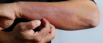
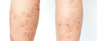
Externally, eczema appears as irregular rashes on the skin, usually in the form of blisters and papules. Microbial eczema often appears as tiny blisters containing serous fluid; when they are opened, the liquid pours out, and a slight erosion remains in place of the bubble. Sometimes instead of bubbles there may be simply tubercles not filled with liquid. The skin at the site of the rash turns red. At the initial stages of development, the redness around the rash has a fairly clear outline, which blurs over time and loses its outline.
A feature of microbial eczema is small but numerous foci with a tendency to merge. The diameter of the rashes rarely exceeds 3 cm, but there are quite a lot of them. Most often, microbial eczema first appears on the hands, in particular on the fingers. Then it moves closer to the elbows; lesions may begin to appear on the torso. The more the disease develops, the more the localization of microbial eczema shifts to the body; lesions appear on the chest, back, and lower back.
The rashes cause moderate itching, but even if you don't scratch them, they don't go away.
Treatment of seborrheic dermatitis in adults and children
Seborrheic dermatitis may resolve without treatment. But it often occurs in waves, with remissions and exacerbations.
Several repeated courses of therapy or ongoing maintenance therapy may be required until the symptoms go away completely. After some time, exacerbation may occur again.
You should consult a doctor if repeating the usual regimens of care and relieving acute symptoms does not help, as well as if there are signs of a secondary infection.
The basis of therapy is the use of medicinal shampoos, creams and lotions.
For children, it is recommended to use special children's shampoos; before washing, you can apply oil or cream to help soften the crusts.
In children, it is important to stop itching, since scratching can cause secondary infection of the scabs.
Features of the treatment method
When selecting external skin care products, choose products containing the following ingredients that reduce skin inflammation:
- Keratolytics: salicylic, lactic acid, urea, propylene glycol.
- Topical antifungal agents: shampoos and creams containing ketoconazole (ciclopirox).
- Products with selenium disulfide or zinc pyrithione.
- Local low-strength corticosteroids can only be used as prescribed by a doctor to relieve the acute phase of the disease, except for the facial skin.
- In case of resistant forms, patients may be offered oral antifungal drugs.
- In severe cases, isotretinoin or phototherapy may be considered.
How is seborrheic dermatitis treated at the Rassvet clinic?
We evaluate the patient's skin type, severity and location of the rash.
The diagnosis of seborrheic dermatitis is based on the clinical picture. The detection of Malacesia has no diagnostic value, since this is a normal microflora of human skin.
A dermatologist evaluates the condition and makes a differential diagnosis with psoriasis, rosacea (on the face), pityriasis versicolor (on the body), and atopic eczema in children. At first, we usually recommend shampoo and only if the selection of permanent skin care products fails, do we move on to active drug therapy.
Etiology of microbial eczema
Microbial eczema appears during secondary inflammation of the skin affected by fungus or pathogenic bacteria. The causes of microbial eczema can be both pathogenic bacteria (for example, Neisser's gonococcus or fungi from the genus Candida) and an immune reaction to autoantigens.
Varieties
Based on the various causes of the rash, doctors divide microbial eczema into:
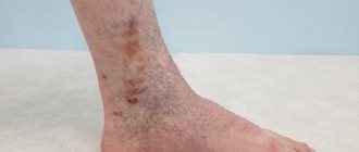
- varicose. It appears in connection with venous insufficiency and is usually localized in areas of ulceration;
- post-traumatic. These are rashes that appear in the area of healing wounds, abrasions and postoperative sutures;
- eczema of the nipples. Breastfeeding mothers most often suffer from these rashes. Eczema appears due to regular trauma to the skin of the nipples during feeding.
Causes of the disease
Today, the main cause of seborrheic eczema is considered to be an allergic reaction to an infection. Another reason is fungus.
There are certain risk factors that contribute to the formation of eczema
:
- excessive production of fat by the sebaceous glands;
- pathologies of the gastrointestinal tract;
- liver disorders;
- hormonal imbalances;
- VSD (vegetative-vascular dystonia);
- chronic infectious pathologies;
- weak immunity.
Treatment of microbial eczema
The essence of the treatment course is to eliminate the source of infection and restore the skin in the damaged area. For this purpose, topical medications, oral medications and physical therapy are used.
Medicines
For topical use use:
- antibacterial (if eczema is caused by bacteria) or antimycotic (if affected by fungus) ointments;
- lotions from a solution of lead water (during exacerbations);
- non-steroidal ointments;
- hormonal ointments (if there are a lot of rashes).
In addition, the doctor may include antibiotics, antihistamines and vitamins B and A in the course of treatment of microbial eczema.
Diagnostics
The identification and treatment of seborrheic type eczema is carried out by a dermatological specialist. Basically, an external examination of the affected areas of the patient is sufficient to make a diagnosis.
Studies may be ordered to confirm
:
- dermatoscopy;
- analysis of scrapings of the affected area for fungus;
- histological examination, etc.
During diagnostic measures to detect side pathologies and infections, patients may be prescribed consultations with specialists in other fields: gynecologist, ENT specialist, endocrinologist, etc. In addition, additional examinations may be required: ultrasound of the abdominal cavity and pelvic organs, gastroscopy, blood tests, rhinoscopy, etc. As part of the diagnosis, it is necessary to distinguish seborrheic eczema from occupational eczema, from skin diseases such as microsporia, psoriasis, etc. All these pathologies have outwardly similar symptoms.
Bibliography
- Ado A.D. General allergology. M.; "Medicine", 1970.-543 p.
- Results of a study of the effectiveness of non-halogenated corticosteroids in the treatment of chronic eczema / I.M. Korsunskaya, N.A. Lukashova, Z.A. Nevozinskaya, E.E. Agafonova // Clinical. dermatology, venereology. 2008. - No. 4. — P. 101-105.
- Ryzhko, P.P. The use of antihistamines in the treatment of various dermatoses / P.P. Ryzhko // Ukr. magazine dermatology, venereology, cosmetology. 2002. - No. 1. — P. 39-41.
- Skripkin, Yu.K. Allergic dermatoses / Yu.K. Skripkin, B.A. Somov, Yu.S. Butov. M.: Medicine, 1975. - 245 p.
- Tishchenko, A.L. Optimal therapeutic doses of vitamin A in patients with eczema and psoriasis / A.L. Tishchenko // Vestn. dermatology and venereology. 1998. - No. 4. — P. 36-38.
- Turchina, I.P. Complex treatment of patients with eczema and neurodermatitis localized on the lower extremities / I.P. Turchin // Dermatovenerology, cosmetology, sexopathology. 2002. - No. 1-2 (5). - S. 98101.
- Yusupova, L.A. Treatment of patients with eczema / L.A. Yusupova, R.Kh. Kha-fizyanova // Ross. magazine skin diseases. 2005. - No. 6. — P. 20-22.
- Brazzini, B. New and established topical corticosteroids in dermatology: Clinical pharmacology and therapeutic use / B. Brazzini, N. Pimpinelli // Am. J. Clin. Dermatol. 2002. - N3. — P. 47-58.
- Buznikov, GA Serotonin and serotonin-like substances as regulators of early embryogenesis and / GA Buznikov, HW Lanbert, JM Lauder //Cell Tissue Res. 2001.-N 39.-P. 177-186.
- Catania, A. Inhibition of acute inflammation in the periphery by central action of salicylates / A. Catania, J. Arnold, A. Macaluso et al. //Proc. Natl Acad Sci USA. 1991. -Vol.88, N19. -P. 8544-85
Seek help from a dermatologist
It is important to confirm the diagnosis before prescribing either treatment or diet.
If you find suspicious rashes on your skin, be sure to consult a dermatologist. The effectiveness of treatment directly depends on the correctness of the diagnosis. Therefore, it is important not to delay or self-medicate. We invite you to a consultation at the PsorMak Institute for Healthy Skin. We have extensive experience in successfully treating various types of dermatitis, and we guarantee the achievement of long-term remission. April 28, 2021
Author of the article: dermatologist Mak Vladimir Fedorovich
results
Eczema is an acute or chronic allergic recurrent inflammatory skin disease caused by various exogenous and endogenous factors, characterized by a polymorphic rash caused by serous inflammation of the skin, polyvalent sensitization and severe itching [9, 10].
Classification of eczema
. There is no generally accepted classification of eczema. Based on clinical manifestations, domestic dermatologists distinguish the following forms of eczema: true (pruriginous, microbial, dyshidrotic), seborrheic, horny (tilotic), childhood and occupational. Varieties of M.E. are coin-shaped (nummular or plaque), paratraumatic, mycotic, varicose, sycosiform and eczema of the nipples and pigment circle in women [9, 10]. In accordance with the Federal Clinical Guidelines for the Management of Patients with Eczema (2016), all of them, regardless of the form, are encrypted as L.30 [10]. However, this section contains only L.30.0 - coin-shaped eczema; L.30.1 - dyshidrosis; L.30.2 - skin autosensitization; L.30.3 - infectious dermatitis; L.30.4 - erythematous intertrigo; L.30.5 - pityriasis white; L.30.8 - other specified dermatitis; L.30.9 - dermatitis, unspecified. This significantly complicates the statistical accounting of the occurrence of various clinical variants of eczema. Some authors designate ME as “infectious eczema” [11], which according to ICD-10 probably corresponds to infectious dermatitis (L.30.3). But there are significant differences in the pathogenesis of eczema and dermatitis. Depending on the duration of the disease, acute (up to 3 months), subacute (3-6 months) and chronic (more than 6 months) stages of the disease are distinguished [12] (Fig. 1).
Rice.
1. Stages of microbial eczema. a - acute; b - subacute; c - chronic. Foreign authors identify true eczema with atopic dermatitis (atopic eczema), although these nosological forms have clear differential diagnostic criteria. Eczema also includes nummular, vesicular palmoplantar eczema and autosensitizing dermatitis [13]. The first two types of eczema actually correspond to microbial and dyshidrotic eczema. Returning to the traditions of domestic dermatology, in Russia all scientific publications on ME are based on the clinical forms accepted in our country.
Pathogenesis of microbial eczema
. The etiopathogenetic aspects of ME, according to scientific research, are multifaceted. However, the mechanisms of development of immune abnormalities in the body as a whole and directly in the skin of patients have not been fully established, so an integrated approach to studying the pathogenesis of eczema is necessary [14-16]. Eczema, including ME, is formed under the influence of immunological, neuroendocrine, metabolic, infectious-allergic, vegetative-vascular, hereditary and other factors [9, 17-21]. The predominant significance of certain endogenous and exogenous influences is ambiguous, therefore it is generally accepted to consider ME as a polyetiological disease.
The role of the skin barrier in the pathogenesis of microbial eczema.
Intact skin protects the human body from the penetration of infectious pathogens. This is due to the tight adhesion of horny scales, acidic pH (4.5-5.3), fatty acids of sebum, the synthesis of some interleukins in the epidermis, as well as the biological balance and antagonistic interaction of pathogenic, conditionally pathogenic and non-pathogenic microflora [4] . Changes in the structure of the stratum corneum lead to increased skin permeability to fungi and bacteria [22]. A shift in the pH of the epidermis to the alkaline side leads to increased desquamation of corneocytes, disruption of the formation of lamellar bodies, causes thinning of the skin and slows down the restoration of the skin barrier. In general, post-inflammatory epidermal barrier deficiency may be due to involucrin deficiency leading to impaired corneocyte cohesion; lack of natural moisturizing factors that promote dehydration and lipids of the stratum corneum; genetically determined filaggrin deficiency [23]. Microbial allergens have pronounced antigenic activity and cause and maintain immune inflammation. Violation of the integrity of the skin when scratching it due to itching forms an entry point for infection. Exudation that accompanies eczema promotes the concentration of proteins on the surface of the skin and creates favorable conditions for the proliferation of secondary infections [4].
The state of the skin microbiota in patients with microbial eczema.
In recent years, more and more attention has been paid to studying the state of the skin microbiota in ME patients [21, 24—26]. In a healthy person, the microflora takes part in the implementation of the protective function of the skin by suppressing pathogenic microorganisms with non-pathogenic ones [27]. Species and quantitative changes in the composition of normal skin microflora can be accompanied by both the development of the disease and the manifestation of diseases that occur subclinically [28, 29].
, Staphylococcus
aureus
is sown in 80% of cases
S. haemolyticus
in 14% , and non-lipophilic yeast fungi, mainly of the genus
Candida
spp., in 40.7%.
[24]. A study carried out 10 years later in Moscow also established the predominance of gram-positive coccal microbiota, represented mainly by Staphylococcus
spp.
(64.3%) and Streptococcus
spp
.
(33.3%) [21].
The authors also established the presence of S. aureus
(33.3%),
Klebsiella pneumoniae
(19.8%),
and Proteus vulgaris
(16.7%) in the nasopharynx.
Infectious pathogens with a lower frequency were also sown in the discharge of the urogenital tract and when examining stool for dysbacteriosis. The predominance of coccal microflora in lesions in ME is evidenced by the results of studies performed in Samara [25]. Staphylococci were cultured in 96.7% of patients. species composition was represented by: S. aureus
(65%),
S. epidermidis
) ,
S. saprophytes
(3.3%).
was cultured in 13.4% of patients .
In isolated cases,
Propionibacterium ,
Corinebacterium
(
5% each) and
E. coli , Proteus
,
Klebsiella
(3.3% each).
It is significant that in 2/3 (66.7%) of cases the growth of a monoculture was registered, and in 1/3 (33.3%) - an association of 2-3 microorganisms. , S. aureus
predominated in the lesions 1535.6±41.8 and 1682.2±52.6 CFU/ml, respectively). On externally unchanged skin, its population was significantly smaller (403.0±36.4 and 515.8±61.0 CFU/ml) [11].
Quantitative differences in the species composition of skin microbiota pathogens in ME patients with acute and chronic disease were identified when compared with healthy people [26]. The level of total density of microorganisms on inflamed areas of the skin during the acute course of the process was 38 times higher than on externally unchanged skin (1562±14.9 CFU/cm2 versus 41.2±6.1 CFU/cm2); in the chronic course of the disease, this indicator differed by 21 times (1562±14.9 CFU/cm2 versus 75.2±8.2 CFU/cm2). Compared to healthy individuals (3.6±0.5 CFU/cm2), the differences were already 434 and 20.9 times, respectively. The structure of the microbiota was sharply dominated by gram-positive microorganisms, the density of which in the lesions during the acute process was 10 times higher than during the chronic process (221.2±2.0 CFU/cm2 versus 22.0±4.1 CFU/cm2) and significantly higher than in the same patients on unchanged skin (132.1±1.4 and 14.7±0.2 CFU/cm2, respectively). And when compared with healthy people (0.7±0.1 CFU/cm2), this indicator differed by 315.7 and 34.4 times, respectively. Total density of pathogens of the genus Staphylococcus
spp.
during an acute process on the affected areas of the skin was 37.1±2.6, which was significantly higher than on externally unchanged skin (24.8±2.9 and 16.1±1.8 CFU/cm2, respectively). Compared to healthy individuals, with ME in lesions these indicators were 17.7 and 11.2 times higher. At the same time, the density of yeast-like fungi of the genus Candida
spp
.
was higher in chronic ME compared to acute ME (5.7±0.4 CFU/cm2 versus 3.1±0.2 CFU/cm2) and differed by 28.5 and 15.5 times compared with that in healthy individuals (0.2±0.01 CFU/cm2).
In a chronic process, the contamination of affected and apparently unchanged areas of the skin with Candida
spp. was almost identical.
Immunoallergic concept of the development of microbial eczema.
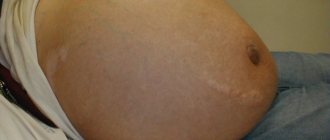
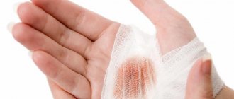
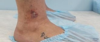
Leading in the pathogenesis of ME are infectious allergens - bacterial, viral, fungal, protozoal, etc. [12, 30-33]. The disease can also be provoked by exogenous stimuli - physical, mechanical and biological. The immunoallergic theory is clearly confirmed by the empirically identified stages of the course of M.E. Initially, the disease develops in the form of eczematization at the site of non-allergic banal pyodermitis, and then through eczematids and localized forms it is transformed into a generalized process [34, 35]. Foci of M.E. often occur in places of long-term persistent pyoderma (Fig. 2) and around purulent wounds (Fig. 3, a, b, c), when scratching scabious lymphoplasia of the skin (Fig. 4) [34, 36-38]. ME is a complication of dermatophytosis of large folds and feet (Fig. 5), superficial skin candidiasis (Fig. 6), and develops against the background of a varicose symptom complex [9, 39-41] (Fig. 7). Initially, sensitization may be monovalent in nature, but over time it becomes polyvalent [3]. The pathogenesis of ME is shown in Fig. 8.
Rice. 2. Microbial eczema at the site of persistent staphylococcal impetigo.
Rice. 3. Paratraumatic microbial eczema. a - at the site of intramuscular injection of calcium chloride; b - at the site of a thermal burn; c - around the fistula opening in osteomyelitis.
Rice. 4. Microbial eczema at the site of scabious lymphoplasia of the skin in the elbow area.
Rice. 5. Mycotic eczema, complicating dermatophytosis of large folds and feet.
Rice. 6. Mycotic eczema, complicating superficial skin candidiasis (absence of eponychium is a diagnostically significant symptom).
Rice. 7. Varicose eczema (a, b), a combination of varicose and mycotic eczema (c).
Rice.
8. Scheme of the pathogenesis of microbial eczema. In the pathogenesis of ME, a decisive role is played by bacterial sensitization with the leading role of S. and ureus
and
St. _ hemolyticus
[30, 41, 42]. Sensitization to these allergens is characterized by an increase in the frequency and severity of an immediate-type hyperergic reaction (IHT) and a decrease in these indicators for a delayed-type hyperergic reaction (DTH). On the one hand, the possibility of activation of ME due to the persistence of bacterial microflora in foci of chronic infection in other organs and systems of the body (nasopharynx, tonsils, gastrointestinal tract, hepatobiliary system, reproductive system, etc.) cannot be ruled out. Foci of chronic infection occur in 65.3% of ME patients [30]. On the other hand, the presence of a deficiency of cellular and humoral immunity in ME patients contributes to the formation of foci of chronic infection and persistence of predominantly pathogenic microorganisms on the skin [43].
The data obtained are clear evidence of the need to use topical drugs with a wide spectrum of antimicrobial activity for ME.
A comprehensive assessment of immunological disorders in ME revealed changes in many indicators of the cellular, humoral and cytokine components of the immune response (Table 1).
Table 1. Changes in indicators of immune status in patients with ME
Patients with ME develop an immunologically determined, latent syndrome of endogenous intoxication, which manifests itself clinically as a complex of nonspecific symptoms. An increase in the concentration of circulating immune complexes (CIC) and the value of the leukocyte intoxication index (LII) is directly proportional to the intensity of immunopathological processes. The clinical form of ME, the prevalence of rashes, and the duration of exacerbation are directly dependent on the severity of immunological changes and endotoxemia. The key indicator of endotoxemia is the content of IgG, IgG2 in the bloodstream. A decrease in the expression of TLR2, TLR4 and TLR9 on blood cells is observed as the severity of the disease increases, which is often accompanied by the development of clinically severe forms of dermatosis. A decrease in the level of antimicrobial peptides in the blood serum of patients correlates with the duration and severity of the eczematous process. During an acute process, the level of CD14+DR+ monocytes increases, and during an exacerbation of the chronic process, their decrease occurs. An increase in IL-2 concentration indicates an active acute inflammatory process. Increased levels of IL-17 and interferon-γ (IFN-γ) are associated with the development of allergic inflammation and protection against infection. IFN-γ ensures the launch of a cytokine cascade aimed at the formation of a focus of inflammation with the involvement of effector cells. A decrease in the level of lactoferrin in the blood serum indicates the activation of a bacterial infection.
These studies indicate that for topical treatment of ME, drugs with a pronounced anti-inflammatory effect should be used. These drugs include topical corticosteroids (TCS), which have taken one of the leading positions in the treatment of steroid-sensitive skin diseases. Currently, dermatologists prescribe them 3.9 times more often than doctors of other specialties. ME belongs to the group of dermatoses for which TCS therapy is pathogenetically justified and used in combination with drugs from other groups. TCS is an etiotropic and pathogenetic therapy in cases where the basis of the disease is inflammation in the skin, especially immune-mediated. TCS have anti-inflammatory, antiallergic, vasoconstrictor and immunosuppressive effects. They regulate the immune response by reducing the number of lymphocytes, inhibiting T lymphocytes, increasing apoptosis of T and B lymphocytes, inhibiting the formation of immune complexes and reducing the activity of the complement system. This leads to inhibition of the hyperreaction of the immune system upon contact with the antigen. At the same time, without local and externally controlled immunosuppression, it is impossible to achieve the required effectiveness of the drug against hyperimmune allergic dermatoses. On the other hand, the presence of an immunosuppressive effect excludes the possibility of using single-component TCS in the treatment of dermatoses combined with infectious pathological processes. Compared to traditional means for external therapy, TCS have significant advantages and suppress all the main components of allergic inflammation. The infectious process should be stopped by prescribing combined TCS. Taking into account the biorhythm of cortisol production in the body and the rhythm of epidermal proliferation, TCS should be used in the morning to enhance the anti-inflammatory effect, and in the evening for the antiproliferative effect [23].
The role of yeast-like fungi of the genus
Candida spp .
in the pathogenesis of microbial eczema.
Candida
can be causative allergens . [3, 45-47]. The widespread use of antibiotics, corticosteroids, and hormonal contraceptives has contributed to the interpretation of candidiasis as one of the serious infectious complications of drug therapy [48, 49]. With M.E. Antibiotics form the basis of traditional treatment regimens, often in the form of repeated courses [10, 50]. The reasons for prescribing antibiotics to patients with ME are traditional treatment regimens, sanitation of foci of chronic bacterial infection in various organs and secondary pyoderma on the skin. Candidiasis of the skin and mucous membranes as infectious complications of antibacterial therapy in our practice was registered in 85% of patients with ME [42]. This high frequency is due to the fact that patients who had been receiving traditional therapy for a long time, including repeated courses of antibiotic therapy with various drugs without antimycotic support, came for consultation with the professor. Complications from the use of antibiotics are numerous and affect the course of ME [1].
Yeast-like fungi of the genus Candida
spp.
belong to opportunistic microorganisms and can induce the formation of HNT in 62.3–78% of patients with ME [30, 33]. skin testing has shown that in patients with ME there is a change in skin reactivity to S. aureus , St. _ hemolyticus , C. albicans
.
The frequency and size of the wheal increases with HNT and the frequency and average size of papules decreases with HRT [30, 41]. Micromycetes Candida
spp., being typical pathogens of opportunistic infections, exhibit their pathogenic potential under the condition of disturbances in the host's antimicrobial resistance system. They complicate the underlying pathological process, causing resistance to therapy and drug intolerance [48].
Allergenic effect of S. and ureus
and
C. _ albicans
in ME are significantly enhanced under conditions of mixed infection.
The synergism of bacterial and yeast microflora in various pathological conditions and in ME has been clinically and experimentally proven [41, 51, 52]. Pathogens of bacterial microflora during associated infection are often characterized by antibiotic resistance, lysozyme, adhesive, hemolytic, lecitellase, and DNA activities [52]. Colonization of the pharynx and nose simultaneously with S. aureus and Candida
spp.
with ME leads to a more severe course of the disease and chronicity of the process. The index for assessing the severity of ME in the presence of candidiasis is significantly higher than in its absence (23.4±7.7 points versus 13.1±4.4 points). The continuous course of the disease is recorded 2.2 times more often, and candidamycids often appear (23%) [42]. response in the form of specific IgM antibodies and IgG antibodies to C. albicans
is registered in 81.2% of patients.
The absence of mannan antigen, the main protein of the cellular machine C, . albicans
is direct evidence of the presence of non-invasive candidiasis and justifies the advisability of using topical antimycotics [33].
The data obtained indicate the advisability of examining patients for the presence of yeast-like fungi of the genus Candida
spp., using clinical and laboratory data for this purpose (Fig. 9).
If Candida
spp. is detected. Specific therapy should be carried out using combination drugs with antimycotics, and in their absence, a course of preventive therapy.
Rice.
9. Scheme of examination of a patient with microbial eczema to identify superficial candidiasis of the skin and/or mucous membranes. Diagnosis of ME is based on the history and clinical picture of the disease. According to indications and for the purpose of differential diagnosis, laboratory tests are prescribed. The most significant are a clinical blood test and an enzyme-linked immunosorbent assay (ELISA) for determining antibodies to antigens of yeast-like fungi of the genus Candida
spp.
,
Giardia, Ascaris, Opisthorchia, Toxocara and others. If candidiasis is suspected, a bacterioscopic examination of the throat, nose and genitals for the presence of pseudomycelium is initially carried out. Its presence is a key mechanism in the pathogenesis of candidiasis. It is advisable to conduct a cultural study of scrapings from lesions on the skin, throat, nose, and genitals to identify bacterial microflora, yeast microflora and determine its sensitivity to antibacterial and antimycotic drugs.
Prevention of seborrheic eczema
Preventive measures for seborrheic eczema are aimed at preventing exacerbation. These include:
- diet – it is necessary to minimize the consumption of potentially allergenic foods;
- personal hygiene – excessive sebum production makes it necessary to shower 1-2 times a day. The water should be warm, for hygiene it is necessary to use products specially designed for sensitive skin;
- limiting external irritants - chemicals can irritate and injure the skin. Therefore, it is necessary to do work that involves contact with chemicals, wearing gloves, and dressing in natural, well-ventilated fabrics;
- timely treatment of chronic diseases;
- regular examinations by a dermatologist and tests.