Erysipelas
There are 20 types (serogroups) of streptococci. The most significant of them are streptococci of serogroups A, B, C, D and G. Beta-hemolytic streptococci of group A (Streptococcus pyogenes) are the cause of many dangerous diseases in humans - pustular diseases of the skin and soft tissues (abscesses, cellulitis, boils and osteomyelitis) , tonsillitis and pharyngitis, bronchitis, rheumatism, scarlet fever and toxic shock. Erysipelas can be caused by any type of group A streptococcus.
Bacteria have a round shape. They are often arranged in chains, less often in pairs. They reproduce by dividing in two.
- In the external environment, including sputum and pus, the bacteria persist for months and survive at low temperatures and freezing.
- High temperature, sunlight and disinfectant solutions have a detrimental effect on microbes.
- Streptococci show high sensitivity to antibiotics, resistance to which they develop slowly.
Streptococci secrete a number of endo- and exotoxins and enzymes that cause their damaging effects.
Rice. 2. Streptococci have a round shape. They are often arranged in chains, less often in pairs.
Rice. 3. Beta-hemolytic streptococci of group A, when growing on blood agar, form zones of hemolysis (light halos) that are 2 to 4 times larger than the diameter of the colonies themselves.
Rice. 4. When growing on nutrient media, colonies of streptococci are shiny, drop-shaped, or gray, matte and granular with uneven edges, or convex and transparent.
Epidemiology of the disease
The reservoir and source of beta-hemolytic streptococci are sick and “healthy” bacteria carriers. Bacteria penetrate the skin from the outside or from foci of chronic infection. Erysipelas in persons with manifestations of streptococcal infection (chronic tonsillitis, caries, diseases of the ENT organs, etc.) occurs 5-6 times more often. Long-term use of steroid hormones is a predisposing factor in the development of the disease.
Minor injuries, cracks, abrasions, abrasions and wounds on the skin and mucous membranes of the nose, genitals, etc. are entry points for infection. Contact and airborne droplets are the main routes of infection.
Group A streptococci often live on human skin and mucous membranes and do not cause disease. Such persons are called bacteria carriers. Erysipelas are more often registered in women during the decline of reproductive function. In some patients, erysipelas is recurrent in nature, which is apparently associated with a genetic predisposition.
The disease often develops with lymphostasis and venous insufficiency, edema of various origins, trophic ulcers and fungal infections of the feet.
Rice. 5. Cellulitis and gangrene are dangerous complications of erysipelas.
Causes of the disease
Streptococcus lives in the body of almost every person, and many people are its carriers. But the development of erysipelas and other streptococcal diseases does not occur if provoking factors are absent.
The occurrence of pathology is possible as a result of:
- damage to the dermis due to purulent, viral infection;
- circulatory disorders due to post-traumatic scars after surgery;
- decreased immunity;
- taking a number of drugs - cytostatics, steroids;
- the presence of pathology in metabolic processes;
- diseases of the immune system;
- AIDS;
- presence of bad habits.
Erysipelas is transmitted by airborne droplets or through direct contact with patients. It quickly begins to develop against the background of diabetes mellitus, sudden changes in temperature, poor nutrition, viral infectious diseases, and chronic ailments in the body.
Another well-known skin disease, streptoderma, is also caused by streptococci. Here you will find information about the treatment of streptoderma in adults.
Provoking factors
Inflammation in the hand can be caused by:
- surgery to remove mammary glands in women;
- excessive insolation;
- overheating or hypothermia;
- infection of abrasions, scratches, bruises, cuts with sharp objects.
In addition, the risk group includes people with pathologies such as:
- diabetes;
- alcoholism;
- obesity;
- varicose veins;
- lymphostasis;
- tonsillitis;
- caries;
- periodontitis;
- fungal infection of the feet;
- thrombophlebitis.
How does erysipelas occur (pathogenesis of erysipelas)
Inflammation in erysipelas is most often localized on the face and legs, less often on the arms, torso, scrotum, perineal area and mucous membranes. The inflammatory process during the disease affects the main layer of the skin, its framework - the dermis. It performs supporting and trophic functions. The dermis contains many capillaries and fibers.
Inflammation in erysipelas is infectious and allergic in nature.
- Waste products and substances released when bacteria die cause the development of toxicosis and fever.
- The cause of the development of the inflammatory process is the effect on tissue of toxins, enzymes and antigens of hemolytic streptococci, as well as biologically active substances. Small arteries, veins and lymphatic vessels are damaged. The inflammation is serous or serous-hemorrhagic in nature.
- Human skin antigens are similar in structure to streptococcal polysaccharides, which leads to the development of autoimmune processes when the patient’s antibodies begin to attack their tissues. Immune and autoimmune complexes cause damage to the skin and blood vessels. Intravascular blood coagulation develops, the integrity of the capillary walls is disrupted, and local hemorrhagic syndrome is formed. As a result of vasodilation, a focus of hyperemia and vesicles appear on the skin, the contents of which are serous or hemorrhagic in nature.
- Biologically active substances enter the bloodstream in large quantities, including histamine, which is involved in the development of hemorrhagic forms of erysipelas.
- Insufficiency of lymph circulation is manifested by edema of the lower extremities. Over time, damaged lymphatic vessels are replaced by fibrous tissue, which leads to the development of elephantiasis.
- The focus of infectious-allergic inflammation consumes large amounts of glucocorticoids. This leads to the development of extra-adrenal insufficiency. Protein and water-salt metabolism are disrupted.
Rice. 6. The inflammatory process during the disease affects the main layer of the skin, its frame - the dermis.
Modern treatment of erysipelas
To avoid the transition from mild forms of the disease to more complex ones, as well as the possible complication of erysipelas in the lower extremities resulting from varicose veins, it must be treated promptly and adequately. Of course, this disease, especially its initial stages, is not a reason for hospitalization, but if erysipelas is advanced, the doctor will suggest hospital treatment as the best solution.
A number of antibiotics are prescribed as drug therapy, since staphylococcus is a bacterium. In addition, various physical procedures are used to dry out the inflamed and non-healing area of the skin, for example, ultraviolet irradiation. Problem areas can also be treated using laser therapy. Modern therapy for erysipelas includes the local application of antiseptic dressings and solutions. This is a very important and even leading component of treatment, as it gives a very good medical effect.
Very often, after the first erysipelas, a relapse occurs. The first time, erysipelas mainly affects only one, specific area of the soft tissues of the face or lower extremities. But for the second time it can affect a large area, causing a significant inflammatory reaction. To protect yourself from this disease, it is necessary to treat varicose veins, which are often the root cause of erysipelas on the legs. If normal blood circulation is restored, the skin will receive enough nutrition, therefore, necrosis and various ulcers will not appear. Of course, good hygiene is also the key to avoiding erysipelas.
Leading modern European studies speak about the polyetiological nature of erysipelas. That is, the disease is caused by a complex of factors. These include disorders of the immune status, diseases of the lymphatic and venous systems. Residents of Moscow and the Moscow region are also significantly susceptible to erysipelas. A good, competent doctor who knows innovative techniques, using modern medications, will be able to stop erysipelas without much difficulty. It is much more difficult to prevent relapse. Here it is necessary to understand the reason that caused the erysipelas. In Moscow and the Moscow region, innovative technologies for comprehensive examination of patients are more accessible. Therefore, it is usually easier to find out the cause of erysipelas in a metropolis like Moscow. Various public and private city medical centers today are engaged in the diagnosis and treatment of erysipelas, diseases of the veins and lymphatic system. However, you must always contact good doctors, European-level professionals who know and apply modern diagnostic and treatment methods, even in Moscow.
Factors influencing the development of erysipelas
The development of erysipelas is influenced by the following factors:
- Individual predisposition to the disease, which is caused by genetic predisposition or increased sensitivity to allergens of streptococci and staphylococci.
- Reduced activity of the body's defense reactions - nonspecific factors, humoral, cellular and local immunity.
- Disorders of the neuroendocrine system and imbalance of biologically active substances.
Characteristic symptoms of erysipelas on the hand
Microorganisms, penetrating into the pores of the skin on the hand, first remain for some time in the incubation period of up to 2-3 days . Infection may not occur if you immediately treat the area with an antiseptic in the event of an unexpected cut or if your immune system is fairly stable.
Otherwise, the primary and characteristic signs of the development of erysipelas are as follows:
- a sharp increase in body temperature;
- nausea;
- dizziness;
- increased fatigue;
- body aches;
- chills;
- loss of appetite;
- redness appears on the hand in the form of a pink-red spot or a roller with uneven edges with tongues of flame;
- further – peeling, burning sensation, swelling at the site of the lesion;
- in some cases, hemorrhages or blisters with serous or bloody fluid appear within the lesion.
Classification of erysipelas
- There are erythematous, erythematous-bullous, erythematous-hemorrhagic and bullous-hemorrhagic (uncomplicated) and abscessing, phlegmonous and necrotic (complicated) forms of erysipelas. This classification of erysipelas is based on the nature of local lesions.
- According to the severity of the course, erysipelas is divided into mild, moderate and severe.
- According to the frequency of manifestation, erysipelas is divided into primary, repeated and recurrent.
- There are localized, widespread, migratory and metastatic forms of erysipelas.
By prevalence
- When a limited area of lesion appears on the skin, they speak of a localized form of erysipelas.
- Extension of the lesion beyond the anatomical region is regarded as a common form.
- When one or more new areas appearing near the primary lesion, connected by “bridges,” they speak of a migrating form of erysipelas.
- When new foci of inflammation appear far from the primary focus, they speak of a metastatic form of the disease. Streptococci spread by hematogenous route. The disease is severe and long-lasting, often complicated by the development of sepsis.
By frequency of occurrence
- Erysipelas that occurs for the first time is called primary.
- If a repeated case of the disease occurs in the same place, but not earlier than 2 years after the first case, or if a repeated disease occurs that arose in another place earlier than this period, they speak of recurrent erysipelas.
- Erysipelas that occurs repeatedly in the same place is recurrent in nature.
By severity
- Mild severity of the disease is characterized by short-term fever and mild symptoms of intoxication, which is characteristic of the erythematous form of erysipelas.
- Moderate severity is characterized by longer-lasting (up to 5 days) fever and more pronounced symptoms of intoxication, which is typical for the erythematous and erythematous-bullous forms of the disease.
- The severe course of erysipelas is characteristic of hemorrhagic and complicated forms of the disease, which occur with high (up to 400C) body temperature, severe intoxication, and the development in some cases of infectious-toxic shock and sepsis. Severe course is observed in migratory and metastatic forms of the disease.
Erased or abortive forms of the disease are observed with adequate, timely treatment. They are rare.
Rice. 7. The photo shows erysipelas of the skin.
1.General information
Erysipelas, erysipelas - these and similar terms are used mainly in Slavic languages; in medical Latin and most Western European languages, the original ancient Greek name erysipelas was fixed, i.e. "red skin" In the old days there were also synonyms “holy fire”, “Saint Anthony’s fire” or, in Russian, “Antonov’s fire”, although some dictionaries define this term as one of the designations for gangrene.
Erysipelas is an acute skin infection (or recurrent exacerbation of a chronic one), the hallmarks of which are swelling and erythema - a bright red, “fiery” hue of the affected area. In general, erysipelas is one of the most common human infectious diseases; the localization of inflammation can be very different, however, due to the peculiarities of the anatomical structure, the disease often manifests itself on the skin of the face, primarily in the soft tissues of the external nose (vestibule, wings, nasal walls): a high concentration of sebaceous glands, capillaries and nerve endings facilitates the penetration and activation of the pathogen .
A must read! Help with treatment and hospitalization!
Signs and symptoms of erysipelas in different forms of the disease
Signs and symptoms of erysipelas during the incubation period
The incubation period for erysipelas in the event of infection from the outside is from 3 to 5 days. As a rule, the disease begins acutely, with an exact indication of the hour of onset of the first symptoms and signs. Headache, general weakness, increased body temperature to 39 - 40°C, chills, muscle and joint pain, often nausea and vomiting, less often convulsions and disturbances of consciousness are the main signs and symptoms of erysipelas during this period. Intoxication with erysipelas develops as a result of the release of streptococcal toxins into the bloodstream.
At the same time, the first signs of local damage appear. Sometimes local symptoms develop 6-10 hours after the onset of the disease.
Streptococci have a tropism for the lymphatic system, where they quickly multiply and spread to regional lymph nodes, which enlarge as a result of developed inflammation. Fever and toxicosis persist for up to 7 days, rarely longer.
All forms of erysipelas are accompanied by inflammation of the lymphatic vessels and lymph nodes.
Rice. 8. The photo shows erysipelas (erysipelas) in children (erysipelas of the face).
Signs and symptoms of erysipelas of the skin in the erythematous form of the disease
A burning sensation and bursting pain at the site of the lesion are the first symptoms of erysipelas. Redness and swelling are the first signs of the disease. In the affected area, the skin is hot to the touch and tense. The inflammatory focus quickly increases in size. The erysipelatous plaque is delimited from the surrounding tissues by a roller, has jagged edges and resembles flames. In the tissues and capillaries of the affected area there are many streptococci, which can be detected by simple smear microscopy. The process lasts up to 1 - 2 weeks. Redness disappears gradually, the edges of the erythema blur, and swelling subsides. The upper layer of the epidermis exfoliates and thickens, sometimes pigment spots appear. Persistent swelling indicates the development of lymphostasis.
Rice. 9. The photo shows an erythematous form of erysipelas on the leg.
Signs and symptoms of erysipelas of the skin in the erythematous-bullous form of the disease
The erythematous-bullous form of the disease is characterized by the appearance of blisters and blisters on the affected area of the skin. Bullous elements contain a light transparent liquid (exudate). Sometimes the exudate becomes cloudy, and the bubbles turn into pustules. Over time, the bubbles subside, and in their place brown crusts form, dense to the touch. After 2 - 3 weeks, the crusts are torn away, exposing the erosive surface. Some patients develop trophic ulcers. Epithelization of the affected surface occurs slowly.
Rice. 10. In the erythematous-bullous form of erysipelas, brown or black crusts form in place of collapsed blisters.
Signs and symptoms of erysipelas in the erythematous-hemorrhagic form of the disease
This form of erysipelas has become more common in recent years, and in some regions of our country it ranks first among all forms of this disease.
A burning sensation and bursting pain, redness, swelling and pinpoint (up to 3 mm) hemorrhages (petechiae) are the main signs and symptoms of the erythematous-hemorrhagic form of the disease. Hemorrhages at the affected area are a consequence of the release of blood from damaged small blood vessels into the intercellular space.
The disease is characterized by a longer (up to 2 weeks) fever and slow regression. Among the complications, skin necrosis is sometimes noted.
Rice. 11. Erysipelas of the hand. Pinpoint hemorrhages (petechiae) are the main symptom of the erythematous-hemorrhagic form of erysipelas.
Signs and symptoms of erysipelas in the bullous-hemorrhagic form of the disease
The bullous-hemorrhagic form of erysipelas of the skin is characterized by the appearance of blisters with serous-hemorrhagic contents against the background of hyperemia. Bleeding is associated with deep damage to the capillaries. After the bubbles subside, an erosive surface is exposed, on which black crusts are located. Healing is slow. The disease is often complicated by skin necrosis and inflammation of subcutaneous fat. After healing, scars and pigmentation remain.
Rice. 12. The photo shows gangrene of the lower limb, as a result of complications of the bullous-hemorrhagic form of erysipelas.
Bullous and hemorrhagic forms of the disease lead to the development of lymphostasis.
Diagnosis and treatment of bacterial skin infections
What is the best way to manage a patient with a staphylococcal skin infection? How can a general practitioner diagnose and treat erysipelas? What treatment is effective for erythrasma?
Normally, human skin is populated by a huge number of bacteria that peacefully coexist on its surface or in the hair follicles.
However, the skin has certain properties that protect it from infection by pathogens. These include a dense and dry stratum corneum, practically impenetrable to microorganisms, and an adhesive intercellular substance - a complex mixture of lipids that tightly connects the cells of the malpighian layer and also protects the skin by clogging the entrance to the hair follicles.
Other factors that stop the penetration of pathogenic microorganisms include the constant turnover of skin cells, acidic pH, the presence of immunoglobulins in sweat and various types of skin flora.
Skin infections typically develop only when injury, overhydration, or inflammatory skin conditions compromise these protective properties. Organisms that cause skin infections may be part of the resident flora of the skin or nearby mucous membranes, or may come from external sources such as another person, the environment, or contaminated objects.
Impetigo is the most superficial skin infection caused by S. aulreuls and S. pyogenes. There are two main clinical variants: bullous impetigo, considered a staphyloccal disease, and nonbullous impetigo, caused by S. aulreuls or S. pyogenes or both organisms.
The disease occurs in children much more often than in adults, developing on exposed parts of the body, face and limbs, in places of scratches, abrasions and insect bites.
Initially, red spots appear, which turn into blisters and pustules, which open easily and form thick, adherent yellowish-brown scales on an erythematous base (see Fig. 1). They are often numerous and may be itchy, but are usually painless.
| Figure 1. Thick yellow crusts at the base of erythema and superficial erosions in a patient with streptococcal impetigo. |
With the bullous form, large vesicles and blisters with a diameter of 1-2 cm can develop. They open more slowly and persist for two to three days. Pathogens are usually identified by culture, but this is not necessary in clinically obvious cases.
The most serious complication of impetigo is post-streptococcal acute glomerulonephritis, the overall incidence of which has decreased in recent years.
Bullous impetigo is caused exclusively by S. aulreuls, which secretes the toxin exfoliatin, which causes breakdown of the intercellular substance in the superficial layers of the epidermis. Absorbed in large quantities into the bloodstream, this toxin causes staphylococcal scalded skin syndrome, which is fatal in 5% of cases.
For moderate and localized infections, a topical antibiotic such as mupirocin or fusidic acid is used, and topical neomycin and bacitracin are also effective. The use of licacin gel is very effective.
For severe and widespread forms, a systemic antibiotic is prescribed. Erythromycin or a first-generation cephalosporin such as cephalexin is usually sufficient.
Ecthyma refers to infections that resemble impetigo, but affect the deeper layers of the skin. It is characterized by the formation of thick, adherent scales (see Fig. 2) covering areas of skin ulceration, preceded by the formation of pustules and blisters. The buttocks, thighs and legs are most often affected. The disease is common in the tropics, where poor hygiene and inadequate nutrition contribute to its development. The causative agents may be S aulreuls or S pyogenes, or both microorganisms, but the ulcerations they cause reach the dermis and heal with scarring, which is not characteristic of impetigo. Treatment is with systemic antibiotics targeting S. aulreuls and S. pyogenes.
| Figure 2. Child with extensive foci of bullous (staphylococcal) impetigo on the body |
Superficial folliculitis, boils and carbuncles. Folliculitis (inflammation of the epithelium of hair follicles) is a common dermatological disease, not always primarily of an infectious nature. Physical or chemical trauma, as well as occupational exposure to tar products also used for medicinal purposes, all cause folliculitis.
When staphylococci penetrate into the deeper layers of hair follicles, inflammation seizes the dermis, causing the formation of boils and carbuncles. An inflammatory vesicle with a purulent head (furuncle) develops or the infection covers several nearby hair follicles and an inflammatory conglomerate is formed, from which pus is released (carbuncle).
Boils are most often found on the face and legs, and the typical location of carbuncles is the back of the neck; as a rule, they accompany diabetes mellitus. Large boils and carbuncles are opened and drained, prescribing a penicillinase-resistant antibiotic.
Recurrent staphylococcal skin infections. Some patients are susceptible to recurrent staph skin infections.
Predisposing factors here are diabetes mellitus, chronic renal failure and some immunodeficiency conditions, but most patients do not have the diseases listed above: these patients are probably chronic carriers of staphylococci, and with the slightest injury to the skin, pathogens cause infection.
They try to prevent recurrences of such infections in various ways: by washing the skin with various antiseptics, treating other family members with antistaphylococcal antibiotics and prolonged therapy with other local or systemic antibacterial drugs. All these methods are aimed at destroying the staphylococcal “trail”.
Unfortunately, these measures are usually nonspecific and ineffective, since bacteria reappear soon after the antimicrobial drug is discontinued. Therefore, long-term use of local antiseptics is preferable.
Erysipelas and cellulitis are acute, rapidly spreading infections of the skin and underlying tissues.
The hallmark of erysipelas is a well-defined, raised edge, reflecting involvement of the more superficial (dermal) layers (see Fig. 3). However, cellulite can be located superficially, and erysipelas deeper, so that in many cases these two processes coexist and it is almost impossible to distinguish them.
| Figure 3. A typical lesion of ecthyma on the dorsum of a boy's foot. He developed multiple lesions during his holidays in Bangladesh. |
It is believed that erysipelas is caused by streptococci, usually group A and sometimes groups G and C. For cellulitis, either S. aulreuls alone or together with streptococcus is cultured. H. influlenzae type b is an important etiological factor for facial cellulitis in children under two years of age.
Erysipelas, which typically affects the face, is a disease of the elderly that develops for no apparent reason or sometimes after facial trauma.
Cellulite affects the lower extremities, especially the calf area. It is often preceded by an injury, ulcer or other damage to the skin, where the infection originates.
As with erysipelas, cellulitis may be accompanied or preceded by fever and chills, but many patients do not develop a fever and do not appear seriously ill.
| Figure 4. Well-defined erythematous swelling of facial erysipelas; more often the lesion is bilateral |
The skin is red, hot and swollen, the edges of the inflamed area are uneven, and bubbles and blisters may develop on the surface (see Fig. 4). In rare cases, lymphangitis and regional lymphadenitis are detected.
Without treatment, complications such as fasciitis, myositis, subcutaneous abscess and septicopyemia may develop. Periorbital cellulitis, usually caused by trauma, may be complicated by cavernous sinus thrombosis, orbital, subperiosteal or cerebral abscess formation, or meningitis.
Patients with these conditions must be hospitalized.
The staphylococcal and streptococcal pyodermas described here make up the majority of skin bacterial infections. You need to be able to distinguish between infectious processes inherent in three clinical situations:
- the infection does not fit into the typical clinical picture of pyoderma or does not respond completely to standard therapy;
- the patient’s body is weakened and cannot withstand the fight against infection;
- There is a history of exposure to unusual skin pathogens.
- Infections caused by resident corynebacteria
Erythrasma is characterized by red-brown scaly areas of skin located in the groin, armpits and interdigital spaces (see Fig. 5).
| Figure 5. Bubbles and blisters developing against the background of erythematous edema of the cellulite area of the foot in a diabetic patient |
Corynebacteriulm minultissimulm is considered the etiological factor of this disease, which is asymptomatic and develops, as a rule, in diabetics, obese and elderly people, as well as in those living in tropical climates.
| Figure 6. Brown, scaly, hyperpigmented area of erythrasma in the axilla of a man from Central Asia. The patient has lesions in the groin and between the toes |
Due to the fact that these microorganisms produce porphyrins, in the ultraviolet light of a Wood lamp, the affected areas fluoresce from coral pink to orange-red, which confirms the diagnosis. Typically, no cultivation is required.
Sometimes vigorous washing with soap is enough to cure. Another approach is topical treatment with erythromycin and clindamycin or topical azoles such as clotrimazole, which are active against some Gram-positive bacteria and fungi. For extensive lesions, erythromycin is probably most effective.
Pockmark keratolysis is a superficial skin infection apparently caused by strains of Corynebacteriulm and characterized by the presence of pockmarks 1–7 mm in diameter on the soles of the feet. Pockmarks, merging, can form surface erosions.
The disease is usually asymptomatic, but sometimes patients complain of itchy, burn-like pain or a cheesy smell.
Pockmark keratolysis appears to be associated with excessive wetness of the feet due to tight shoes, frequent contact with water, or excessive sweating.
Treatment of hyperhidrosis, combined with the methods described for erythrasma, is usually effective.
Axillary trichomycosis is characterized by waxy nodules that form in the hair of the armpit. Yellow, red or black, they are formed by large colonies of coryneform bacteria covering the hair cuticle.
First of all, the disease affects patients who pay little attention to personal hygiene and suffer from excessive sweating.
For successful treatment, as a rule, it is enough to shave your hair and use deodorants for your armpits. Topical application of erythromycin and clindamycin is also effective.
References
1. Noble WC Microbal Skin Disease: its Epidemiology. Arnold, London, 1983. 2. Hoor EW, Hooton TM, Horton CA et al. Mircroscopic evaluation of cultaneouls cellulitis in adults // Arch. Intern. Med. 1986; 146: 295-297.
Note!
- For moderate and localized forms of impetigo, both streptococcal and staphylococcal etiologies, a local antibiotic, such as mupirocin or fusidic acid, is sufficient. Topical forms of neomycin and bacitracin are also effective and are often used in combination. For widespread and severe forms of infection accompanied by lymphadenopathy or if there is reason to suspect nephritogenic streptococcal infection, oral antibacterial drugs that act on both microorganisms, such as erythromycin, are indicated
- Some patients are susceptible to recurrent staphylococcal skin infections. Predisposing factors such as diabetes mellitus, chronic renal failure and some immunodeficiency conditions are absent in most patients. The method of choice is long-term use of local antiseptics
- Erysipelas, usually localized on the cheeks, is streptococcal in nature. It is most common among older patients and develops either for no apparent reason or, in rare cases, sometimes after facial trauma. The drug of choice is penicillin; in more severe cases, benzylpenicillin is prescribed intravenously at 600-1200 mg.
- Erythrasma appears as red-brown patches of skin covered with scales in areas of diaper rash, such as the groin, armpits and interdigital spaces. Intensive washing with soap is sometimes enough to cure. Another approach is topical treatment with erythromycin and clindamycin or topical azoles such as clotrimazole, which are active against some Gram-positive bacteria and fungi. For extensive lesions, erythromycin is probably most effective.
Signs and symptoms of complicated forms of erysipelas
Phlegmonous and necrotic forms of erysipelas of the skin are regarded as complications of the disease.
When inflammation spreads to the subcutaneous fatty tissue and connective tissue, phlegmonous inflammation develops. Blisters filled with pus appear on the affected area of the skin. The disease is severe, with severe intoxication. The affected area of skin is often infected with staphylococci. The phlegmonous form of erysipelas often becomes the cause of sepsis.
The necrotic (gangrenous) form of erysipelas develops in individuals with low immunity. Soft tissues undergo necrosis (complete destruction). The disease begins rapidly, proceeds with severe intoxication, and progresses rapidly. After healing, disinfectant scars remain.
The recovery period for severe and complicated forms of erysipelas is slow. Asthenic syndrome after recovery persists for many months.
Rice. 13. The photo shows erysipelas (erysipelas), a phlegmonous-necrotic form of the disease.
Features of erysipelas in certain areas of the body
Most often, erysipelas is recorded on the skin of the lower extremities, somewhat less often - on the upper extremities and face, rarely - on the torso, mucous membranes, mammary gland, scrotum and perineal area.
Erysipelas on the leg
Erysipelas on the leg develops as a result of a violation of the integrity of the skin, the occurrence of which is associated with injuries and bruises. Often the disease develops in patients with fungal infections of the feet and toenails, circulatory disorders in the lower extremities that develop as a result of diabetes mellitus, varicose veins, smoking and excess weight. The source of infection is also foci of chronic infection in the patient’s body.
A burning sensation, bursting pain at the site of the lesion, redness and swelling are the first signs and symptoms of erysipelas on the legs.
Erysipelas on the legs is often recurrent. Improper treatment and the presence of foci of chronic infection contribute to the development of a recurrent form of the disease.
Frequent relapses lead to the development of fibrous changes in the dermis and subcutaneous tissue, followed by the development of lymphostasis and elephantiasis.
Rice. 14. The photo shows erysipelas of the legs.
Erysipelas on the hand
Erysipelas on the hands often develops in drug addicts due to intravenous drug administration and in women against the background of lymph stagnation as a consequence of a radical mastectomy.
Rice. 15. Erysipelas on the hands.
Rice. 16. The photo shows erysipelas of the hand.
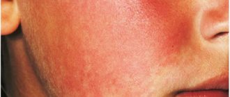
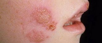
Erysipelas on the face
Most often, the primary erythematous form of erysipelas occurs on the face. Redness often affects the area of the cheeks and nose (like a butterfly) and, in addition to swelling and itching, is often accompanied by severe pain. Sometimes the source of inflammation spreads to the entire face, scalp, back of the head and neck. In some patients, the disease is complicated by the development of abscesses in the thickness of the eyelids and the accumulation of pus under the scalp. When the infection spreads into the subcutaneous fatty tissue, phlegmon develops. Gangrene may develop in weakened individuals and the elderly.
The source of infection for erysipelas on the face is often streptococcal sinus infections and boils. The source of infection for erysipelas of the orbit is streptococcal conjunctivitis.
With streptococcal otitis, erysipelas of the auricle sometimes develops, and the inflammatory process often spreads to the scalp and neck.
Rice. 17. An erythematous form of erysipelas often occurs on the face.
Rice. 18. Erysipelas on the face. Redness often affects the area of the cheeks and nose (like a butterfly).
Rice. 19. Sometimes the source of inflammation spreads to the entire face, scalp, back of the head and neck.
Rice. 20. The photo shows erysipelas of the hand.
Erysipelas of the trunk
Erysipelas sometimes develops in the area of surgical sutures if the rules of asepsis are not observed. Erysipelas develops severely when streptococci penetrate into the umbilical wound of a newborn. Erysipelas of the mammary gland develops against the background of mastitis. The development of gangrene can lead to scarring with subsequent dysfunction of the organ.
Erysipelas of the genitals and perineum
With erysipelas of the scrotum, penis, female genital organs and perineum, an erythematous form of the disease most often develops with pronounced swelling of the underlying tissues. Developed tissue necrosis followed by scarring leads to testicular atrophy. Erysipelas in women giving birth is extremely difficult. The inflammatory process often affects the internal genital organs.
Erysipelas of the mucous membranes
With erysipelas, the pharynx, larynx, oral cavity and nasal mucosa are most often affected. When the mucous membranes are affected, an erythematous form of the disease develops. In the area of inflammation, hyperemia and significant edema develop, often with foci of necrosis.
Rice. 21. The photo shows erysipelas of the oral mucosa.
Relapses of the disease
Erysipelas that occurs repeatedly in the same place is recurrent in nature. Relapses are divided into early and late. Early relapses are considered repeated episodes of the disease that occur before 6 months, late relapses - more than 6 months.
The source of infection is foci of chronic infection, from which streptococci spread through the blood throughout the body, as well as latent (hidden) foci of infection in the dermis, where streptococci turn into the parasitic L-form during a lull period.
Chronic venous insufficiency, lymphostasis, diabetes mellitus and improper treatment of the disease contribute to recurrence. Relapses are often observed in patients working in unfavorable conditions and in the elderly.
When multiplying in the lymphatic capillaries of the skin, streptococci form an inflammatory focus in the dermis. Frequent relapses occur with low body temperature and moderate symptoms of intoxication. Oily erythema and swelling appear on the skin. The demarcation from healthy areas is weakly expressed.
Frequent relapses lead to the development of fibrous changes in the dermis and subcutaneous tissue with the subsequent development of elephantiasis.
Rice. 22. In the photo there is erysipelas (erysipelas) of rare localization.
Treatment
When visiting the clinic, the doctor will first examine the skin, identify the nature, location, degree of damage and shape of the erysipelas. Most likely, the patient with obvious clinical signs will be sent to hospitalization in the infectious diseases department.
For the treatment of erysipelas the following is prescribed:
- antiallergic drugs (Suprastin, Diazolin, Tavegil);
- sulfonamides (Biseptol, Streptocide);
- nitrofurans (Furadonin, Furazolidone) to kill bacteria;
- corticosteroids (Prednisolone) to clear up the infection;
- biostimulants (Pentoxyl, Methyluracil) to stimulate the formation of new healthy immune cells and skin regeneration;
- vitamins (ascorbic acid, Ascorutin) to strengthen vascular walls damaged by bacteria, increase proteolytic enzymes (trypsin, lidase, tactivin).
In addition, treatment is carried out in a hospital with the appointment of:
- benzylpenicillin, as the main antibiotic for streptococcal infections;
- cephalosporins to suppress pathogenic flora in the event of an abscess or phlegmon. The course of treatment is up to 10 days.
Treatment also includes the following:
- Detoxification therapy is carried out in severe cases of the disease by administering intravenously hemodez or saline solution with glucose.
- It is possible to prescribe cardiovascular, antipyretic, diuretic drugs , as well as treat the affected area by applying applications from a dimexide solution, enteroseptol powders to kill bacteria in the affected areas and prevent the addition of another infection.
- Patients are advised to wash their wounds themselves with furatsilin and other solutions with antimicrobial effects to kill bacteria. An oxycyclosol aerosol will help, applying bandages with syntomycin ointment, Vishnevsky's liniment, to relieve inflammation and heal wounds.
You can’t warm up areas - it will only speed up the movement of streptococci through the blood and the spread of bacteria throughout the body. The main treatment is antibiotics, and in no case should you resort to homemade formulations and prescriptions without the knowledge of your doctor.
Those who are sick must strengthen their immune system, take vitamins, multivitamins, and antiallergic medications. Electrophoresis, laser therapy, ultraviolet irradiation, high-frequency magnetic therapy, and physical therapy are indicated for complete suppression of pathogenic microflora.
Erysipelas in children
Erysipelas are rare in children. In older children, the disease is mild. The focus of erysipelas can occur in different places. The erythematous form develops more often. The prognosis is favorable.
In children under one year of age, erysipelas is more severe. Foci of inflammation often appear in areas of diaper rash and on the face, sometimes spreading to other parts of the body. With the phlegmonous form of the disease, sepsis can develop, with erysipelas of the face - meningitis.
Erysipelas develops severely when streptococci penetrate into the umbilical wound in newborns. The process quickly spreads to the child’s back, buttocks and limbs. Intoxication increases, body temperature rises significantly, and convulsions appear. Some patients develop sepsis. Mortality from erysipelas in newborns is extremely high.
Rice. 24. The photo shows erysipelas in children.
Prevention
There are no specific specifics and prevention for erysipelas.
Development can be prevented if:
- do not neglect the rules of personal hygiene, wearing loose clothing and shoes made of natural fabrics;
- use soap containing lactic acid when showering to create a protective layer on the skin;
- immediately treat any damage or abrasions on the skin with antiseptics;
- Avoid ultraviolet irradiation, chapping, frostbite of extremities.
Erysipelas is a common ailment, and it can be treated fairly quickly with timely medication. An advanced disease will ultimately lead to a chronic relapsing course, scarring on the arm, swelling, and lymph stagnation.
Symptoms will recur from time to time, up to the appearance of stiffness in the joints, constant pain, limited mobility and disability. The appearance of a red, itchy and flaky spot on your hand cannot be ignored. It is possible that a streptococcal infection has occurred. The sooner, the better to seek advice from a dermatologist.
Complications of erysipelas
Complications of erysipelas occur in 4 - 8% of cases. A decrease in the activity of the body’s defense reactions and inadequate treatment lead to the development of:
- lymphorrhea - leakage of lymph from damaged lymphatic vessels,
- ulcers - deep skin defects,
- abscess - an abscess surrounded by a dense capsule,
- phlegmon, when inflammation spreads to subcutaneous fatty tissue and connective tissue,
- gangrene - complete destruction of tissues affected by inflammation,
- thrombophlebitis - inflammation of the venous walls with the formation of blood clots,
- pneumonia in elderly people,
- lymphostasis (lymphedema), which developed as a result of impaired lymph outflow and elephantiasis (fibredema),
- infectious psychosis,
- At the site of inflammation, hyperkeratosis, eczema, and pigmentation often develop over a long or recurrent course.
Immunity does not develop after suffering from erysipelas.
Rice. 25. Lymphostasis and elephantiasis during erysipelas often lead the patient to disability.
Rice. 26. A terrible complication of erysipelas is phlegmon.
Rice. 27. In the photo, gangrene of the lower limb is a complication of the bullous-hemorrhagic form of erysipelas.
Complications
With proper treatment, erysipelas on the arm can go away on its own after 2-3 weeks. Redness and swelling will subside and will soon disappear altogether. But pigmentation may remain. Relapses are possible.
New erysipelas can subsequently lead to:
- stagnation of lymph;
- lymph circulation insufficiency;
- pulmonary embolism;
- sepsis;
- dead skin;
- thrombophlebitis.
All this indicates untimely treatment and progression of the disease.
Complications, as a rule, result from untimely consultation with doctors, self-medication, or the addition of a secondary infection. The risk group includes people with diabetes, HIV-infected people, and those who have had meningitis or pneumonia.
Erysipelas with complications can lead to the formation of trophic ulcers on the arm, lymphostasis, abscess, suppuration and thickening of the skin, which will significantly complicate treatment and may even endanger the life of the patient himself.