Erysipelas: what is it?
Erysipelas of the foot is an infectious disease caused by the activity of hemolytic streptococci. Inflammation and deformation usually affect a limited area of the skin. The course of the disease may be accompanied by intoxication. Most often, the disease occurs in men and women over the age of 45. However, it can also appear in children. For a child under the age of 1 year, erysipelas on the leg poses a mortal danger.
The disease is quite common. It ranks fourth after acute respiratory infections, hepatitis and gastrointestinal tract infections. There are several varieties of erysipelas. The list includes:
- Erythematous form. This type of disease is characterized by redness of the affected area. In this case, the erythema rises above the skin and has clear boundaries. It is uniform in color and has an irregular shape. Subsequently, inflammation may be added to the overall picture.
- Erythematous-hemorrhagic form. Almost copies the previous one. The only difference is that hemorrhages occur on the affected area of the skin.
- Erythematous-bullous form. At the first stage, the type of disease is no different from the erythematous form. However, after two to three days, the top layer on the affected area begins to peel off. Then bubbles appear, which are filled with a clear liquid. After they burst, brown crusts appear in their place. What is underneath is directly related to the treatment. If the patient was provided with timely, qualified assistance, after the scabs fall off, smooth pink skin will appear underneath them. If timely treatment is not carried out, there is a possibility of painful lesions. Subsequently, they can turn into trophic ulcers.
- Bullous-hemorrhagic form. Externally, this type of disease is similar to the previous form. The difference is that bubbles appear filled with liquid that contains blood.
Erysipelas can be systematized by symptoms. So, depending on the severity, the lesion can be mild, moderate or severe. There are different types of forms based on the number of times they appear. There are primary, recurrent and repeated erysipelas.
Symptoms of erysipelas of the lower extremities
- quite noticeable headache
- high temperature, which is very difficult to bring down
- chills
- vomiting like food poisoning
- burning sensation on the skin
- severe redness on the skin
- The area of skin affected by erysipelas differs from healthy skin - it is either convex or separated by a small ridge.
If you notice such symptoms, you should immediately consult a doctor so as not to start the disease, because erysipelas can become more complicated.
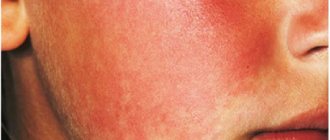
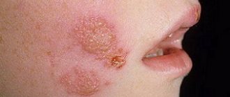
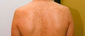
This is what erysipelas looks like
If erysipelas is a consequence of varicose veins, then such a complication may be thrombophlebitis, in which inflammation spreads to the wall of the veins, and then a blood clot forms in the lumen of the vein. Another complication of erysipelas with varicose veins is tissue necrosis, leading to the appearance of trophic ulcers that are difficult to treat.
What does a erysipelas on a leg look like?
Erysipelas on the leg is an affected area of bright red skin. Most often the limbs are affected. In rare cases, inflammation may occur in the genital area or torso. In the initial stage, shiny red spots appear on the leg. They spread quickly, turning into extensive outbreaks. Sometimes blisters form. The fluid in them may be clear or contain blood. In later stages of the disease, the affected area becomes covered with a crust. To better understand what a erysipelas on a leg looks like, it is recommended to study the photo.
General symptoms of pathology
As the situation worsens, erysipelas manifests itself as follows:
- chills and fever in the first hours;
- intoxication of the body;
- cardiopalmus;
- loss of consciousness is rare;
- swollen red spots at the site of the wound;
- rapid increase in edema;
- the affected area turns into a painful cushion, visually standing out against the background of healthy skin;
- formation of bubbles with liquid;
- formation of dense brown crusts;
- the occurrence of trophic ulcers and erosions.
The main complication is damage to the lymphatic system
Therefore, lymphatic tissue swelling, lymphangitis and lymphadenitis occur in the absence of proper therapy.
Reasons for appearance
A streptococcal infection leads to the appearance of erysipelas on the leg. Typically, the pathogen enters the body through:
- diaper rash and cracks;
- skin damage resulting from injuries and bruises;
- insect bites or scratching;
- scratches or burns.
An old streptococcal infection can also provoke the disease. From the main focus, bacteria spread throughout the body, causing problems of varying severity. Having a healthy immune system prevents this from happening. However, if it is weakened for any reason, the risk of erysipelas on the leg increases. Frequently getting cold feet or getting too much tan can also lead to the problem. Stress or a sudden change in temperature are other factors that increase the likelihood of identifying a problem. There are a number of diseases against which erysipelas on the leg can develop. The list includes:
- alcoholism;
- overweight;
- varicose veins;
- fungal infections that are localized on the feet;
- trophic ulcers and thrombophlebitis;
- diabetes.
Most carriers of streptococcal infection do not even suspect it. Therefore, the appearance of erysipelas is often an unpleasant surprise.
Causes of erysipelas on the leg
The main cause of erysipelas is infection by streptococcus bacteria . Streptococci are gram-positive aerobic bacteria that live in the human body. Pathogenic microbes enter through open wounds caused by cuts, scrapes, cracks or burns. Sometimes carriers of streptococci do not even suspect its existence.
Of 100% of carriers, only 15% remain unaware, since their bacteria do not manifest themselves in any way throughout their lives. The remaining 85% of carriers suffer from various diseases that are caused by the proliferation of pathogens.
Erysipelas on the leg can occur at different ages. There is a tendency: in youth, it is mainly men who suffer from erysipelas, and in old age, erysipelas occurs more often in women.
Erysipelas is a deep type of pyoderma. Here you can read about pyoderma in children.
Causes of erysipelas:
- First of all, the disease occurs in people who, due to their profession or lifestyle, are constantly in unsanitary conditions.
- Erysipelas sometimes appears as a consequence of a sedentary lifestyle in older people. Trophic ulcers, bedsores and poor circulation are a favorable environment for the penetration and development of streptococcal bacteria.
- Erysipelas occurs in people with reduced immunity; this may be due to previous illnesses, severe stress and nervous exhaustion.
- Another reason for the appearance of erysipelas on the human body is the systematic exposure of the skin to UV rays, which leads to burns.
- Erysipelas often occurs in patients with diabetes, obesity and varicose veins. And also in people who suffer from alcoholism.
Symptoms of the disease
The symptoms of the disease are quite characteristic. However, they can confuse the doctor, leading to an erroneous diagnosis. The incubation period of the disease lasts from several hours to 3-4 days. Then the patient begins to experience the first symptoms of erysipelas on the leg. The person feels unwell and feels overwhelmed. All this arises against the background of general weakness. Then, quite suddenly, there is a rise in temperature. The person may experience headaches and chills. The temperature persists for the first few hours. The value of the indicator can reach up to 40 degrees. Additionally, there is pain in the muscles in the legs and lower back. A person complains of discomfort in the joints. In some cases, the patient may experience vomiting, diarrhea and nausea. Sometimes anorexia can develop against the background of the disease.
After a day, characteristic local signs are added to the general symptoms. The affected area begins to redden and swell. The person feels a burning sensation and pain. Tension is felt in the area where the erysipelas forms. Further symptoms directly depend on the type of disease.
The lesion has uneven jagged boundaries. Inflamed skin appears slightly shiny. It is dry and hot to the touch. Bright redness may be replaced by bluish, congestive edema. It occurs due to the fact that there is a local disturbance of blood microcirculation. Small hemorrhages often appear. They arise due to damage to the walls of blood vessels, as well as sweating of the formed elements.
On the second or third day of the disease, signs of lymphostasis with the development of dense lymphatic edema may be added to the general symptoms. It is at this moment that bubbles often appear within the lesion. They are filled with clear liquid. Sometimes it may contain blood.
After the blisters open, a dense brown crust appears at the site of the disease. Erysipelas gradually expands. However, if adequate treatment is carried out, the temperature returns to normal within 3-5 days. If the disease manifests itself in an erythematous form, its symptoms will disappear after 8-9 days. In hemorrhagic syndrome, they can persist for 12-16 days.
After the hyperemia and swelling of the skin decreases, its surface begins to itch and peel. During this period, some patients experience dark, congestive hyperemia and uneven hyperpigmentation. Subsequently, they will disappear on their own. If a person has suffered severe bullous hemorrhagic erysipelas, traces of the disease may persist for several years and even decades.
Treatment of erysipelas
If we are talking about the initial stage of the disease, treatment is carried out on an outpatient basis. In case of severe advanced forms, the patient is hospitalized. Usually the doctor prescribes antibiotics. The list includes:
- furazolidone;
- penicillin;
- erythromycin;
- oleandomycin;
- Biseptol.
They are usually prescribed in tablet form. If treatment is carried out inpatiently, the patient is prescribed courses of intravenous and intramuscular injections. In addition to antibiotics, other drugs are also used. So, to improve the general condition of the body, vitamins are prescribed. Inflammation is relieved using anti-inflammatory drugs. If the disease is severe and complicated by intoxication, detoxification drugs are additionally used. They can be used as a glucose solution or Reopoliglucin. Sometimes your doctor may prescribe them together.
To relieve pain and other manifestations of the disease, symptomatic treatment is carried out. In the process, vascular, antipyretic and diuretic drugs can be used. In some cases, agents may be prescribed that reduce the permeability of blood vessels.
To combat erysipelas, local medications are also used. Sometimes erythromycin ointment, furatsilin solution or enteroseptol are used. It may be prescribed in the form of powders or ointments.
If a patient is diagnosed with a bullous form of erysipelas, complex complex treatment is required. When the disease passes the acute stage, the surgeon removes the blisters. The opened bottom is covered with sterile dressings, which are soaked in a solution of furatsilin or rivanol. They must be changed at least once a day. In this case, the bandages should not be tight.
If the bullous-hemorrhagic form is diagnosed, applications are performed with dibunol ointment 5-10%. The procedure is repeated at least twice a day. It is carried out within a week.
In the acute period of the disease, ultrasound irradiation, exposure to electric current discharges, and laser therapy can also be used.
It is strictly forbidden to treat the affected area with ichthyol ointment or Vishnevsky balm. The above agents help to increase the flow of interstitial fluid. All this leads to the healing process slowing down.
Protein C and chronic venous insufficiency in erysipelas of the lower extremities
Erysipelas of the face and erysipelas of the lower extremities as an acute streptococcal disease occurs everywhere, without a tendency to decrease in the foreseeable future. Most often, the inflammatory process develops on the lower extremities (67%), less often on the face (24%). Many authors point out the connection between erysipelas of the lower extremities and the presence of somatic diseases, such as obesity, type 2 diabetes mellitus and underlying fungal infection of the feet and nails. Equally common factors predisposing to the development of erysipelas are chronic venous insufficiency (CVI) and chronic lymphovenous insufficiency (CLVI) [1–6].
In recent years, the frequency of detection of hemorrhagic forms of erysipelas has noticeably increased, which forces researchers to pay special attention to the condition of the vascular wall and the blood coagulation system as a whole [7–10].
Protein C, a vitamin K-dependent protease, is synthesized in the liver, from where it enters the bloodstream. Normally it is in an inactive state. During infectious and inflammatory processes in the blood, binding of protein C to a cofactor (protein C) and its further interaction with thrombomodulin and the endothelial protein C receptor are observed. Activated protein C (APC) inhibits the cascade coagulation mechanism at the early stage of thrombin formation (due to inactivation of Va and VIII clotting factors). Its other important function is the ability to accelerate plasminogen-dependent thrombus lysis, which is a direct manifestation of its angioprotective properties. A decrease in protein C correlates with the level of plasminogen in the blood during infectious and septic conditions [11–19].
Various studies have shown that suppression of protein C activation reactions causes a sharp increase in the production of interleukin-6 and interleukin-8, tumor necrosis factor and other cytokines and reduces the body's tolerance to various endotoxins. Evidence has been obtained that protein C not only blocks the activation of leukocytes, but also regulates the activity of matrix metalloproteinases, which cause degradation of the extracellular matrix and local ulceration [18, 19, 22–26].
Prevention of microvascular endothelial dysfunction is effectively used for the prevention and treatment of many diseases [18, 19]. Against this background, the role of protein C (one of the key endothelium-dependent anticoagulants) in the formation of vascular wall dysfunction in erysipelas has been poorly studied.
We tried to fill the existing gap by studying the dynamics of protein C in patients with erysipelas of the lower extremities (with and without chronic venous insufficiency) in comparison with patients with erysipelas of the face and healthy volunteers to clarify the pathogenesis and determine the feasibility of replacement (antithrombotic) therapy.
Patients and research methods
We examined 60 people aged from 25 to 71 years with a diagnosis of “erysipelas of the lower extremities of II severity” (36) and “erysipelas of the face of II severity” (24). The erythematous form of erysipelas was established in 33% of all cases (in 52% of facial erysipelas), erythematous-bullous in 15%, erythematous-hemorrhagic in 22% and bullous-hemorrhagic in 30% of cases. Erythematous-hemorrhagic (11 cases) and bullous-hemorrhagic (15 cases) erysipelas developed on the lower extremities more often than on the face (2 and 3 cases, respectively). The frequency of hemorrhagic disorders was significantly higher in local inflammation (78%) on the lower extremities compared to the face (20%); odds ratio (OR) = 9.9 [2.8; 34.7].
Primary facial erysipelas (16 women, 8 men) was diagnosed in 92% of cases and predominated in women. In erysipelas of the lower extremities, cases of primary erysipelas were recorded in 50%, repeated in 31% and recurrent in 19%; in contrast to facial erysipelas: primary (92%), repeated (4%) and recurrent (4%). The risk of recurrence of erysipelas was statistically significantly higher when the inflammatory focus was localized on the legs compared to the face (OR) = 5.55 [1; 51.2], p = 0.009.
For erysipelas of the lower extremities, the gender ratio was comparable (17 men, 19 women). Among concomitant diseases, noteworthy was the high (88%) frequency of foot mycoses and onychomycosis. 11 patients had obesity from 2nd to 4th degree, 5 patients had subcompensated type 2 diabetes mellitus.
In patients with erysipelas, the background pathology in 37.5% was represented by skin diseases (behind the ear dermatitis, streptoderma, psoriasis) and in 29% by chronic ENT pathology (otitis media, tonsillitis, rhinitis). Type 2 diabetes mellitus - four people.
The patients were treated as inpatients in the erysipelas department of the ICH No. 2 in Moscow. 32 patients were prescribed antibacterial monotherapy: benzylpenicillin novocaine salt intramuscularly at 0.6 million IU twice a day (7-10 days) and two more patients - cephalosporins (cefazolin) intramuscularly at 1 g 3 times a day (5 days). Combination therapy of two antibiotics (benzylpenicillin novocaine salt intramuscularly at 0.6 million IU twice a day (7–10 days) and Tsiprolet per os at 0.5 g twice a day (10 days)) was carried out in 14 patients. 12 people were treated with a combination of three antibiotics (benzylpenicillin procaine salt intramuscularly at 0.6 million IU twice a day (7 days) + ciprofloxacin intravenously at 800 mg per day (3 days) followed by a transfer to 1 g per day per os (10 days) + cefazolin intramuscularly 1 g 3 times a day (5 days)). Additionally, patients received: antihistamines (Zodak, Diazolin), local physiotherapy and regular treatment of erysipelas in the lower extremities with a tanning solution of potassium permanganate. Patients participating in the study did not receive drugs with a targeted effect on the state of the hemostatic system. The average length of hospital stay for patients with erysipelas of the lower extremities was 11.9 ± 4.1 days; with erysipelas - 8.4 ± 1.6 days.
The study of key indicators of the hemostatic system was carried out at the onset of the disease (days 1-3 - point 1), over time (days 4-6 and 7-10 - points 2, 3, respectively) and during the convalescence period (days 11-15 of illness - 4th research point). Every third patient with erysipelas of the lower extremities underwent a follow-up study (5 months after discharge from the hospital), which made it possible to distinguish changes caused by erysipelas from the background of concomitant diseases.
The control group consisted of 32 healthy individuals aged 24 to 50 years in an equal proportion of men and women.
The content of protein C in blood plasma was determined using an automatic coagulometer SYSMEX CA-500 (Siemens Healthcare, USA, Siemens AG reagents, Germany) in the express laboratory of the Children's City Clinical Hospital No. 13 named after. N. F. Filatova, Moscow Health Department, together with clinical laboratory diagnostics physician N. K. Lagutina. A total of 90 plasma samples were processed.
Statistical processing of the results was carried out using the Statistica 10.0 for Windows 7.0 program. The critical level of significance (reliability) was accepted as p < 0.05. The table shows the results as Me + s (median and standard deviation). In case of statistically significant differences between groups, the odds ratio (OR) and 95% confidence interval (CI) were calculated (OR [-95%CI; +95%CI]).
Results
The initial values of protein C on days 1–3 of illness (at admission) in the group of patients with erysipelas of the lower extremities were lower (81.9 ± 4.9%) than in patients with erysipelas of the face (94.1 ± 6.0%) and significantly below control values (100 ± 5%, p < 0.05) (Table 1).
As the erysipelas faded away, the level of protein C gradually recovered in both groups: 119.6 ± 3.1 on days 4–6 of illness, 129 ± 6.4% on days 7–10 of illness, 153 ± 4.4% on days 11– 15th day of illness - with facial erysipelas (p < 0.05 between 1/4, 2/4 and 3/4 study points) and 103 ± 3.2%, 134.5 ± 4.7%, 139 ± 6.7 % for erysipelas of the lower extremities (p < 0.05 between 1/4, 2/4 study points).
The obtained values of protein C logically fit into the general picture of endothelial dysfunction of the microvasculature, previously described by us in patients with erysipelas [20, 21]. At the same time, the dynamics of protein C in individual patients: from 49.7% (1st point of the study) to 112% (4th point) - an increase of 125%; from 48.9% (1st point) to 110.7% (4th point) - an increase of 126%; from 65.5% (1st point) to 119.7% (4th point) - an increase of 86%, deserves attention as an effective mechanism for the operation of sanogenesis factors in the bloodstream during the period of convalescence (Fig. 2, Table 2) .
In patients with erysipelas of the lower extremities with concomitant chronic venous insufficiency, protein C activity remained below normal throughout the entire observation period (Fig. 1).
Thus, in patients with erysipelas of the lower extremities without CVI (n = 28), the level of protein C was 99.8 ± 4.7% in the acute period of the disease and increased to 140 ± 4.5% in the convalescence stage (p < 0.001) (Table 2). In erysipelas of the lower extremities with CVI, the initially low level of protein C - 69.8 ± 8.1% did not change statistically significantly in the period of convalescence - 79.15 ± 4.0%, p = 0.21 (Table 3). The differences were so expressive that the publication included all absolute values of protein C obtained in patients with erysipelas of the lower extremities with (Table 2) and without signs of CVI (Table 3).
In addition, the period of convalescence in patients with erysipelas of the lower extremities without signs of CVI (and positive dynamics of protein C) proceeded more favorably than in patients with erysipelas of the lower extremities with concomitant CVI, as demonstrated by two clinical observations.
Patient K. (71 years old), diagnosed with bullous hemorrhagic erysipelas of the right lower extremity, II degree of severity, 2nd late relapse, concomitant chronic lymphovenous insufficiency. Protein C level during the study: 37.6% on the 3rd, 39.9% on the 4th, 61.7% on the 9th, 85.8% on the 15th, 93.4% on the 19th sick day. Even after discharge, at follow-up after 5 months, the level of C-protein remained below the level of healthy individuals - 82.1%. Status localis upon admission is shown in Fig. 2, status localis in the dynamics of recovery - in Fig. 3.
A clinical case of erysipelas of similar severity (but without signs of CVI) demonstrates a more favorable course of the disease in combination with restoration of protein C levels on the 11th day of illness.
Patient K. (40 years old), diagnosed with erythematous-hemorrhagic erysipelas of the left lower extremity, grade II, repeated. Protein C level: 74.5% on the 2nd, 74.5% on the 3rd, 119.7% on the 11th, 130.2% on the 12th day of illness. There are no clinical signs of lymphovenous insufficiency (Fig. 4–5).
Restoration of protein C to normal was observed in the third week of illness in erysipelas (108.2 ± 5.1% at admission and 144 ± 4.6% at discharge; 1/4 point - p < 0.001); in the 4th week of illness - with erysipelas of the lower extremities without CVI (99.8 ± 4.7% at admission and 140 ± 4.4% at discharge, 1/4 point - p < 0.001) and was absent in patients with erysipelas of the lower extremities limbs and CVI (69.8 ± 8.1% at admission, 79.15 ± 4.07% at discharge; 1/4 point - p = 0.21) (Fig. 6).
The study made it possible to prove that with normal values of protein C (100.0 ± 5.0%) the chances of a favorable course of erysipelas of the lower extremities are significantly higher (OR = 2.89, [0.15; 55]) than with erysipelas of the lower extremities with chronic venous insufficiency and low protein C levels.
Discussion and conclusion
As our previous studies have shown [20, 21], erysipelas is accompanied by the development of latent intravascular hemolysis and DIC-like syndrome. The study of protein C complemented the overall picture of changes in the hemostatic system in erysipelas, revealing a deficiency of one of the most important natural anticoagulants.
The level of protein C upon admission in the group of patients with erysipelas of the lower extremities was lower than in the group of patients with erysipelas of the face, and lower than the values obtained in the group of healthy individuals. During the period of convalescence, the activity of the endothelium-dependent anticoagulant was restored in patients with erysipelas of the face and erysipelas of the lower extremities, with the exception of patients with chronic venous insufficiency.
It was found that the level of protein C is refractory to standard therapy for erysipelas in such patients, and a documented deficiency of protein C is accompanied by a prolongation of the period of convalescence and an extension of the length of hospital treatment of patients with erysipelas of the lower extremities with signs of CVI. Protein C values in only 7 out of 8 patients with CVI returned to normal 5 months after erysipelas.
The lack of positive dynamics of protein C during the treatment of erysipelas and the protracted period of convalescence in patients with erysipelas of the lower extremities with concomitant CVI require a revision of the existing treatment tactics. Documented protein C deficiency can serve as an indication for replacement therapy, which is advisable to begin in the acute period of the disease [12, 13]. According to the literature, the effectiveness of replacement therapy depends on the level of protein C reduction (not lower than 50–60% of normal) [18, 19, 26].
Compensation of documented protein C deficiency in erysipelas may be a promising therapy aimed at treating chronic venous insufficiency and reducing the systemic inflammatory response.
Literature
- Fazylov V. Kh. State of vascular-platelet hemostasis and correction of its disorders in erysipelas. Author's abstract. dis. ... Ph.D. L., 1990.
- Frolov V. M. Clinic and pathogenetic mechanisms of recurrent erysipelas. Author's abstract. dis. ... Ph.D. Kyiv, 1986.
- Khramtsov M. M. Pathogenetic and prognostic role of factors of intercellular relationships in erysipelas. Author's abstract. dis. ... MD M., 2000.
- Erovichenkov A. A. Clinical and pathogenetic significance of hemostasis disorders and their correction in patients with hemorrhagic erysipelas. Author's abstract. dis. Doctor of Medical Sciences M., 2003.
- Cherkasov V. L. Pathogenesis and treatment of various clinical forms of erysipelas and bicillin prophylaxis of its relapses. Author's abstract. dis. ... MD M., 1977.
- Denis F., Martin C., Ploy MC Erysipelas: microbiological and pathogenic data // Annales de Dermatologie et de Venereologie. 2001, v. 128; No. 3, C2; 317–326.
- Fazylov V. Kh. Impaired hemostasis and immunity during the formation of recurrent erysipelas, their therapeutic correction. Author's abstract. dis. ... MD St. Petersburg, 1996.
- Mitrofanova M. Yu. Disorders of hemostasis and vascular endothelial function in patients with erysipelas. Author's abstract. dis. Ph.D. M., 2010.
- Dubovikova T. A. Nitroxidergic profile and the state of the blood coagulation system in hemorrhagic forms of erysipelas. Author's abstract. dis. Ph.D. M., 2012.
- Ratnikova L. I., Dubovikova T. A. Assessment of the state of vascular-platelet hemostasis in patients with hemorrhagic forms of erysipelas // Journal of Infectology. 2012. T. 4, No. 1, 53–57.
- Egorova V.V., Titova M.I., Demidova V.S. Modern methodological aspects of laboratory diagnostics of the protein C system and the significance of its research in surgery // Medical alphabet. Modern laboratory. 2013. No. 3, 12–17.
- Liaw PCY, Neuennschwander PF, Smirnov MD Mechanisms by wich soluble endothelial cell protein C receptor modulates protein C and activated protein C function // J. Biol. Che. 2000, vol. 275, no. 8; 5447–5452.
- Dahlback B. Resistance to activated protein C as a risk factor for thrombosis: molecular mechanism, laboratory investigation and clinical managements // Semin in hematol. 1997, 34 (No. 3); 217–234.
- Sukhanov V. A. Inflammatory coagulation response as part of the systemic inflammatory response syndrome (SIRS) // Intensive care. 2006, no. 1; 21–23.
- Svetukhin A. M., Amiraslanov Yu. A., Zemlyanoy A. B. et al. Features of disorders of the hemocoagulation system and their correction in patients with purulent-necrotic forms of diabetic foot syndrome // Surgery. 2006. No. 10; 30–34.
- Erin D.N. The role of reducing the level of proteins C, S and antithrombin III in infectious-septic DIC syndrome and correction of their deficiency with cryosupernatant. Author's abstract. dis. Ph.D. Barnaul, 1999.
- Ena Ya. M., Platonova T. N., Sushko E. A. et al. Biological role and clinical significance of protein C // Medical Affairs. 1992. No. 6; 20–25.
- Klimovich L. G., Kozar E. F., Krainyuchenko T. V. et al. Anti-inflammatory and anticoagulant effects of recombinant activated protein C in complex intensive care of sepsis in newborns and infants after heart surgery / 3rd All-Russian Conference Clinical Hemostasiology and hemorheology in cardiovascular surgery: conference materials. 2007. P. 101.
- Bockeria L. A., Lobacheva G. V., Kharkin A. V., Ochakovskaya E. Yu., Samsonova N. N., Klimovich L. G. The first experience of using recombinant activated protein C in complex intensive therapy of sepsis in newborns and infants after cardiac surgery // Children's diseases of the heart and blood vessels. 2006. No. 35. pp. 60–64.
- Fokina E. G., Rosly I. M. Laboratory assessment of erysipelas // Epidemiology and infectious diseases. Current issues. 2014. No. 1; 28–32.
- Fokina E. G. Some features of primary facial erysipelas in modern conditions // Therapeutic archive. 2014. No. 11; 70–77.
- White B., Schmidt M. // Activated protein C inhibits lipopolysacchartde-induced nuclear translocation of NFkB and TNF-alpha production in THP-1 monocytic cell line // Br J Haematol. 2000. V. 110. P. 130–134.
- Esmon CT, Taylor FB Inflammation and coagulation: Linked processes potentialliregulated through a common pathway mediated by protein C // Thromb Haemost. 1991. V. 66. P. 160–165.
- Esmon CT Role of coagulation inhibitors in inflammation // Thromb. 2001. V. 86. P. 51–56.
- Coughlin SR Protease-activated receptors in vascular biology // Thromb Haemost. 2001. V. 86. P. 298–307.
- Bernard GR, Vincent JL Efficacy and safety of recombinant human activated pritein C for severe sepsis // N Engl J Med. 2001. V. 344. P. 699–709.
E. G. Fokina, Candidate of Medical Sciences
FBUN TsNIIE Rospotrebnadzor, Moscow
Contact Information
Possible complications
If erysipelas is not treated, it can lead to serious complications. The disease can provoke:
- abscess;
- the appearance of an ulcer;
- sepsis;
- elephantiasis of the limbs;
- the beginning of necrotic processes;
- thrombophlebitis.
In severe cases, kidney disease and pathology of the cardiovascular system may occur. All of the above diseases pose a serious threat to health and can cause disability.
How to prevent it from appearing?
In order to prevent the occurrence of erysipelas on the leg, it is imperative to treat all skin diseases that appear in this area. The occurrence of a rash and other manifestations of a problem should be a reason for an immediate visit to a specialist.
If a person suffers from excessive sweating, it is necessary to use powder or talcum powder. High humidity is a favorable environment for the proliferation of microorganisms.
Avoid excessive exposure to ultraviolet radiation. The burns that the sun can leave on human skin can cause a number of serious diseases. At the same time, erysipelas is not the worst of them.
It is worth paying special attention to foot hygiene. When washing, you should use warm water, avoiding hypothermia. The use of soap products will prevent the accumulation of large numbers of microorganisms on the skin. When choosing a shower gel, you need to pay attention to the pH level.
Do not ignore the appearance of corns and cracked heels. A minor lesion can cause significant health problems in the future. If a person notices microcracks on the feet, it is necessary to immediately take measures to promote their rapid healing.
The essence of the disease
The main manifestation of an infectious disease is inflammation of the skin, including even the deep layers. In most cases, pathology is localized on the legs, since the lower extremities are located closest to the ground and more often come into contact with dust and dirt - the pathogens of the disease are concentrated there.
Streptococcus infection
from the external environment is considered the main factor influencing the appearance of this skin pathology.
Severe forms of pathology are observed in 80% of cases, whereas in the 70s the percentage of such cases did not exceed 30%.
The disease is contagious, so the main preventive measure is avoiding contact with the patient.
When caring for a patient, additional safety measures are required: only wear gloves when dressing and treating wounds. After any contact with a person susceptible to this disease, you must thoroughly disinfect your hands.