The term “lichen” combines a wide group of fungal and viral skin diseases (dermatoses), different in etiology, differing in external manifestation. According to statistics, lichen in a child most often appears at the age of 3-14 years. Up to 90% of children attending children's groups in a given period of time are affected by various types of it.
Most often, patients present with the following types of lesions:
- with ringworm in the form of microsporia and trichophytosis;
- pityriasis (multi-colored);
- pink (lichen Zhibera);
- red flat;
- encircling.
The disease is highly contagious (infectious). Adults and children surrounding the patient may be at risk of infection. Therefore, in case of any pathological changes in the skin, the child should be shown to a dermatologist, infectious disease specialist or pediatrician.
Causes of illness in a child
Since the disease is bacterial and viral in nature, the etiology of its development is due to exposure to a bacterial or viral pathogen.
- The ringworm appears in two forms - microsporia and trichophytosis. The main causative agent is a fungal infection in humans and animals. Sometimes infection occurs through contact with toys and household items containing fungal spores - hats, combs or washcloths. Ringworm most often develops on the skin of infants.
- The development of pityriasis versicolor in a child is caused by three types of yeast-like fungi - Malassezia furfur, Pityrosporum orbiculare or P. Ovale. These fungi often live on the skin without causing harm to it. The trigger for the development of pathology can be:
- infectious pathologies (AIDS, tuberculosis);
diseases and conditions that disrupt hormonal levels (DM, Cushing's disease, colloid goiter, obesity);
- hyperhidrosis;
- rheumatism;
- leukemia;
- seborrhea;
- lymphogranulomatosis;
- hereditary factor.
- Lichen versicolor (pityriasis versicolor) is low-contagious. More often manifests itself in adolescent children. The favorite location is skin areas with a large number of sweat glands.
- The trigger for the development of pityriasis rosea in children is previous respiratory, intestinal and herpes infections, acute febrile conditions, and vaccinations. Although children from 4 to 10 years of age are mainly affected, pediatric dermatologists sometimes diagnose pityriasis rosea in infants.
- There are no reliable reasons for the manifestation of lichen planus in a child. Only hypothetical versions are considered (neurogenic, immunoallergic, intoxication, viral and hereditary).
- The causative agent of shingles in children are virions - the Varicella zoster viruses, which also cause chickenpox. In children who have had chickenpox, the herpes virus remains in an inert state in the nerve cells. Awakens and activates when exposed to unfavorable, provoking factors:
- decreased immune function;
- overwork;
- ARVI;
- allergies and vegetative neuroses;
- minor skin lesions;
- avitaminosis.
Fungal lichens in children are more often diagnosed in the warm season, viral ones - with the onset of cold weather.
Causes
Skin lesions from pityriasis rosea occur with equal frequency in men and women. Therefore, these skin changes are not hormonal in nature. The highest incidence rate is observed in the autumn-winter period, when a person’s skin is hidden under warm clothing.
The following diseases and pathological conditions contribute to the appearance of plaques:
- weakened immunity due to chronic diseases;
- metabolic diseases;
- helminthic infestation;
- state of chronic stress;
- presence of allergic diseases.
With pityriasis rosea, the surface layer of the skin, the epidermis, is affected, and the middle layer, the dermis itself, becomes thinner. As a result, the skin in the affected area becomes thinner, the blood vessels become visible, giving a pink tint. Then, ridges form along the edges of the plaque, limiting the focus of pathological changes. The epithelium is sloughed off and replaced with new cells. At the site of the lichen, a pale spot remains for some time, which rather acquires a normal flesh color.
Symptoms of lichen in children
The symptoms of the disease differ in external manifestation and clinical course according to the development of a certain form of the disease.
| Ringworm | The most common fungal disease that affects the nail plates, hair and skin of children. The duration of the incubation period (from the moment of fungal infection to the appearance of the first symptoms) ranges from 5 to 40 days. Signs of skin lesions are manifested by the formation of delimited round and oval, highly flaky and scaly red spots. The pathology is manifested by severe itching and burning. When the scalp is affected, large and small, round-shaped foci of baldness are formed. The boundaries of the lesions are framed by hair broken off at the base (as if trimmed). Children with weakened immune systems may experience:
|
| Pityriasis (multi-colored) | The most common localization is the upper body and scalp. The manifestation of the disease begins with the appearance of yellowish dots at the mouth of the hair follicles. The pinpoint formations gradually transform into brown-yellow or pink-yellow spots covered with bran-shaped scales. The gradual growth of spots leads to their merging and the formation of larger, flaky lesions. The affected areas do not tan and form hypopigmented areas on the child's skin. |
| Pink | The primary signs of pityriasis rosea in a child are manifested by a single maternal plaque. This is a medallion-like bright pink oval spot up to 5 cm in diameter. After 1-1.5 weeks, many secondary small rashes (up to 2 cm) appear, accompanied by itching. A distinctive feature of the spots is peeling in the center and the absence of peeling on their red border. The rash can be observed for 1 to 1.5 months, then disappear without a trace on its own. Constant irritation of the affected skin (friction of clothing, water procedures, ultraviolet radiation) can provoke purulent complications - impetigo, folliculitis, hidradenitis. |
| Lichen ruber planus | It is extremely rarely diagnosed by a pediatric dermatologist. It manifests itself as damage to the skin, mucous membranes, and sometimes nails. Characterized by monomorphic nodular rashes. The nodules are flat, shiny, bright red or bluish in color, up to 3 mm in diameter. The fusion of nodules forms small plaques covered with scales. The pathology causes intense itching. The usual localization of lichen planus is the area of the wrist joints, the surface of the forearms in the flexion areas, the axillary and inguinal areas, the inner surface of the thighs, and the oral mucosa. |
| girdling | The appearance of symptoms of herpes zoster in children is preceded by:
A day later, vesicles (up to 0.5 cm in size) filled with a transparent, liquid secretion appear in areas of erythematous-edematous skin. The rash is localized linearly, like a Christmas tree, located to the sides of the nerve trunk along the nerve branches. The period of active rash is accompanied by fever, intercostal pain and lymphadenitis (enlarged lymph nodes). Gradually the bubbles dry up. The crusts formed in their place disappear. Their presence is indicated by light pigmentation of the skin. The duration of the disease is from 1.5 weeks to a month. With the development of herpes zoster on the skin, children may develop neuralgia, neuritis of the oculomotor and optic nerves, iridocyclitis, keratitis, conjunctivitis or stomatitis. With weak immune phagocytosis (protection), this type of lichen can provoke serious complications in the form of serous meningitis, encephalitis, and myelitis. |
Publications in the media
Pityriasis rosea is an acute dermatosis characterized by abundant, finely scaly pink spots on the trunk, neck and proximal extremities, most often occurring in the autumn-winter period. One of the most common skin diseases; usually resolves spontaneously. Risk factors: infectious diseases, prolonged hypothermia. Frequency. 5% of patients who consulted a dermatologist. It is observed mainly in middle-aged women, rarely in children and the elderly.
Clinical picture • The process begins with a primary maternal plaque, which is much larger in size (2–6 cm) than subsequent rashes, and is often located on the trunk, in the area of the collarbone and shoulder blades. After 7–10 days, sometimes later, many spotted or macular-urticarial elements appear, up to 1–2 cm in diameter, pale pink or slightly brownish in color with pronounced pityriasis-like peeling. The long axes of the spots are located along Langer's lines, indicating the direction of maximum extensibility of the skin • The rashes are localized in the axillary and groin areas, on the inner surface of the thighs and the flexor surface of the forearms. In adults, the skin of the face, scalp, hands and feet, and mucous membranes are usually not affected • Itching is mild or absent altogether • Along with typical manifestations, there are other types of pityriasis rosea •• Urticarial form •• Vesicular form •• Papular form •• Lichen annulare bordered Vidal or pityriasis rosea giant is a rare variant of pityriasis rosea, characterized by single large rashes (up to 8 cm in diameter) of red or pink color with small pityriasis-like peeling along the periphery in the form of a ring, as well as a long course; may occur independently or together with typical rashes.
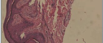
Test methods • Blood test: slight leukocytosis and eosinophilia • Serological test to rule out syphilis • Skin biopsy: inflammation with cytolytic degeneration of keratinocytes adjacent to Langerhans cells. Differential diagnosis • Toxidermia • Trichophytosis • Syphilitic roseola • Psoriasis • Parapsoriasis • Eczema • Lichen planus • Dermatomycosis of the trunk.
TREATMENT General recommendations • Avoid water treatments, hypothermia • Maintain good hygiene to prevent the development of secondary infection • Diet with limited pickles, spices, marinades, concentrated broths • Insolation • Drug therapy is symptomatic only.
Drug therapy • For an inflammatory reaction and itching - topically: ointments with menthol (0.25–1%) • For very severe itching - prednisolone 10 mg 4 times a day orally until the itching disappears (if discontinued, the dose is reduced gradually) • Antihistamines • Anti-inflammatory drugs (salicylates, indomethacin) • Calcium pantothenate. Complications • With irrational therapy, the development of eczema is possible • Secondary infection. Forecast. Recovery is usually within 4–6 weeks. Very rarely, rashes persist for more than 3 months and, as a rule, are due to irrational therapy. Synonyms • Tinea rosacea Giberta • Giberta disease • Roseola exfoliating
ICD-10 • L42 Pityriasis rosea [Gibera]
Zhiber pink lichen: treatment and diagnosis in Kaluga
Despite the fairly accurate symptoms, a full diagnosis is still important. If a spot has formed that looks like lichen , which itches and itches, it is important to determine the type of lichen, as well as differentiate it from other diseases (for example, psoriasis, parapsoriasis, rubella). A dermatologist at our clinic will prescribe a full examination, which includes a number of tests, laboratory tests, and scraping examination. Treatment of pityriasis rosea in an adult is quite easy if the patient applies at the initial stage of the disease. After diagnosis, a treatment plan is prescribed, which must be strictly adhered to:
- multivitamins and immunomodulatory drugs to improve immunity;
- antihistamines to relieve itching and reduce redness;
- ointments to eliminate peeling;
- diet to reduce the allergenic factor.
It is also important to reduce stress. To do this, eliminate all factors that create or contribute to stressful situations. During the treatment period, physical activity and direct sunlight on the affected areas of the skin should be avoided. The disease goes away completely within six to nine weeks. The spots gradually lighten, itching and peeling disappear. For a short period after treatment, barely noticeable marks may remain on the skin: they gradually disappear completely.
We are always happy to help you and invite you to medical! Be beautiful! You can make an appointment by phone: 8 (4842) 27-72-50.
Methods of infection and risk groups
At the moment, there is no reliable evidence of the transmission of this disease from a sick person to a healthy one. There are only infrequent cases when someone in the environment fell ill with lichen upon contact, but in all cases there were provoking factors. These include:
- presence of herpes virus in the body;
- weakened immune system;
- recently suffered viral and colds;
- frequent hypothermia of the body.
The risk group mainly includes women under 40 years of age and children. Men suffer from this disease much less frequently. Almost no cases of infection are detected in infants and people over 40 years of age. In order to reduce the risk of contracting Gibert's disease, it is worth strengthening your immune system. It is reliably known that in most cases the disease appeared against the background of a weakening of the body’s protective functions.
Causes
Despite the fact that to date there is no exact data on the causative agent of pityriasis rosea, many experts are inclined to believe that the cause of the disease is the seventh herpes virus.
However, the disease can develop in people with weakened immune systems, during pregnancy, due to hypothermia and after previous respiratory infections. Science does not know the exact routes of transmission of the infection, but there is an assumption that it is transmitted by airborne droplets. Also, according to experts, pityriasis rosea can be transmitted through household items (combs, towels, etc.).
After recovery, the person gains lasting immunity.
Symptoms
The appearance of pink spots on the skin occurs in strict sequence. Only with Zhiber's lichen does a maternal plaque first appear - an ovoid-shaped spot the size of a quail egg, pink in color, with clear edges. Within two to three days, a rim of darker color forms along the edges of the maternal plaque, and the skin on the surface of the spot becomes flaccid, similar to tissue paper. Then the epithelial cells begin to slough off and be rejected.
After a few days, in places far from the “maternal plaque”, new spots appear, which also have clear outlines, but are smaller in size. The number of fresh plaques is sometimes impossible to count. But the foci of pityriasis rosea never merge, maintaining clear boundaries. The skin in the areas of daughter rashes undergoes the same changes as in the maternal plaque.
The disease is not accompanied by any sensations. No itching, pain, swelling. General changes in the body are limited to an emotional reaction to the appearance of a large number of spots. In the absence of medical care, plaques resolve on their own without dangerous consequences or complications.
The disease is prone to relapse in cases where the background condition of the body does not change or a chronic disease (for example, bronchial asthma) cannot be completely cured.