Ringworm is a dermatological infectious disease with similar symptoms that is caused by viruses or fungi.
They are classified depending on the pathogen, location and degree of damage to the skin. According to WHO statistics, more than 70% of children are affected by this skin disease, both in newborns and older.
The group of lichens includes:
- lichen Zhibera or pink lichen;
- ringworm (the most common form);
- shingles;
- lichen planus or Wilson's lichen;
- ringworm or eczema;
- lichen simplex of the face;
- scaly lichen or psoriasis;
- pityriasis versicolor.
The mechanism of infection has not been reliably identified, since not every child who comes into contact with a carrier of the pathology gets lichen.
There are a number of catalyst factors that increase the likelihood of developing the disease, including:
- genetic predisposition;
- diseases of the digestive tract;
- endocrinological dysfunctions;
- autoimmune pathologies;
- hyperfunction of the sweat and sebaceous glands;
- stress;
- physical stress;
- hypothermia of the body;
- taking certain medications, etc.
Late detection of a lichen spot often leads to an increasingly larger area of skin being affected, causing itching, peeling and discomfort. Often children scratch and injure the affected areas of the skin, which contributes to the addition of a bacterial infection to the underlying disease.
In order to identify and remove lichen from a child’s skin, you need to contact a specialist. A doctor whose competence is the diagnosis and treatment of lichen in children is a pediatric dermatologist.
In addition to treatment, prevention is important in eliminating and preventing relapse of the disease.
Preventive measures for the primary or recurrence of lichen are:
- strengthening the child’s immunity (hardening, etc.);
- compliance with hygiene standards (washing hands, cleaning premises with disinfectants, etc.);
- separate individual items (bed and personal linen, dishes, toys, etc.);
- frequent washing of the child’s clothes, especially after contact with animals or sick people;
- showering rather than bathing to avoid the spread of infection;
- replacing synthetic clothing with natural ones;
- reducing exposure to the sun (heat provokes the growth of fungal colonies), etc.
How is lichen transmitted?
The main sources of the disease are street cats and dogs, or objects containing flakes of their skin affected by the pathogen - a zoonotic form. However, lichen can be transmitted from person to person - an anthroponotic form of transmission, as well as with the help of fungi living in the soil - a geophilic transmission route. Viruses, as a cause of deprivation, can remain in the body of humans and animals for a long time, and only when the child’s immunity decreases can they provoke a disease.
Ringworm is not always contagious. Thus, children's weeping eczema occurs due to infection of microcracks in the skin, dermatoses, or the presence of allergic reactivity in the child's parents. According to the observations of experts, allergic diseases of the mother (asthma, rhinitis or neurodermatitis) increase the likelihood of eczema in the child by 40%. And Zhiber's pityriasis rosea is often a consequence of ARVI and is not transmitted to other people.
The period of high risk of contracting lichen is spring-summer, since the child spends most of his time outdoors and in contact with other children.
Contagious (infectious) types of lichen are pink, ringworm and herpes zoster. They require mandatory isolation of the child to prevent the rapid spread of the disease.
Lichen planus and pityriasis versicolor, as well as psoriasis and weeping eczema are not transmitted.
What does ringworm look like?
The nature of the specific symptoms of lichen depends on the type of pathogen and the characteristics of the body’s response to the pathogen.
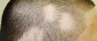
Thus, ringworm or trichophia is caused by the fungus trichophyton and mainly affects children under 14 years of age. The most common locations for ringworm are the scalp and neck, nails and armpits.
Signs of trichophia:
- a clearly defined red or pink spot on the scalp;
- high rate of increase in spot area;
- swelling and rash (blisters) on the affected area of the skin;
- the appearance of white scales on the lichen spot;
- frequent absence of itching;
- hair thinning in the area of inflamed skin, etc.
Lichen Zhibera (pink) is believed to be caused by the herpes virus and is localized primarily on the back, abdomen, face and skin folds.
Signs of pityriasis rosea:
- the presence of one large (maternal) spot, which gradually increases in size;
- a rounded spot with clear boundaries;
- dry flaky skin in the center of the spot;
- itchy skin.
Pityriasis versicolor or versicolor versicolor occurs in children due to the activity of the fungi Malassezia furfur and Pityrpsporum orbiculare, which are localized in the stratum corneum of the skin and gradually cover increasingly larger areas of the skin. This type of disease mainly affects the skin of the chest, abdomen and back, especially in children with hormonal disorders (diabetes, obesity, etc.) and hyperhidrosis (excessive sweating). This form of the disease can also be provoked by: tuberculosis, leukemia or rheumatism.
Signs of pityriasis versicolor:
- at an early stage, spots are greenish in color without clear boundaries, which then transform into yellow, brown or pink;
- tendency to form a large lesion on the skin;
- peeling and itching, especially due to stress and hygiene procedures.
Shingles or herpes zoster is caused by the herpes virus, but in childhood it has the form of chickenpox or chickenpox, to which lifelong immunity is developed.
The etiology of lichen planus has not been reliably established; it is localized both on the skin of the wrists, abdomen, chest, and on the mucous membranes.
Signs of lichen planus:
- flat nodules on the skin of a red or purple color, prone to the formation of large foci of pathology;
- pale pink flat nodules on the mucous membranes;
- itching and sensitivity of the affected skin areas;
- deformation of the nail plate, followed by its destruction.
Lichen simplex of the face or streptodemia sicca occurs due to the activity of streptococcus and is localized mainly on the face, but also on the skin of the buttocks, limbs and back.
Signs of simple facial lichen:
- round pink scaly spots up to 4 cm in diameter;
- no itching;
- disappearance of stains under the influence of ultraviolet radiation;
- the presence of temporary depigmentation at the site of lichen spots.
Your child has microsporia
To children, the world can seem like one big playground full of adventure. But their desire to play in the ground, pet animals, touch other people's things can lead to infection with various infectious and parasitic skin diseases. Children are especially susceptible to this because their immune systems are not yet fully developed. Summer, holidays, children's holidays in camps, outside the city, lead to a seasonal rise in these diseases. Currently, one of the common skin infections is microsporia. Microsporia affects mainly children of preschool and school age, less often adults. Microsporia is caused by a fungus that gets on children's skin called Microsporum. The fungus is very stable in the external environment. In hair, skin flakes, wool, dust, it retains the ability to infect for several years.
How can your child become infected?
Infection occurs through direct contact with a sick animal (cats, kittens, less often dogs), or objects infected with hair or scales (combs, hats). It is possible to transmit microsporia from sick family members, but this happens less frequently. There have been cases of children becoming infected after playing in the sandbox. However, if you follow the rules of personal hygiene, even if the pathogen spores come into contact with the skin, the disease can be avoided.
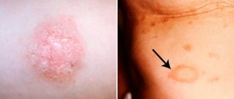
How does the disease manifest in a child?
After an incubation period of 5-7 days on average, foci of infection appear on smooth skin or scalp. Microsporia can appear on any part of the skin, but more often on exposed parts of the body. In mild forms of the disease, round lesions form on the skin, clearly defined, covered with scaly nodules and blisters along the periphery. They often form bizarre shapes, like a “ring within a ring”. At the same time, vellus hair may be affected, which complicates the treatment of the disease. The number of lesions ranges from single to several dozen. Size from 0.5 to 3 cm. When the scalp is affected, 1-2 large lesions are formed, round in shape, with clear boundaries and abundant pityriasis-like peeling. The hair breaks off at a height of 6-8 mm above the skin level (hence the name of the disease - ringworm), because the hair in the lesion appears to be trimmed. There may be slight itching in the affected area. The general condition of children is usually not disturbed. With a decrease in immunity, a child may experience complicated forms of microsporia - infiltrative and suppurative forms. The infiltrative form is characterized by the development of severe inflammation in the lesion. The suppurative form is characterized by the formation of dense foci, round in shape, consisting of deep abscesses (cavities filled with pus). Nearby lymph nodes increase in size. The child's general condition may worsen and the temperature may rise. If a child has suffered microsporia in a mild form, then no traces of the disease remain on the skin or scalp. After a complicated form of microsporia, scars may remain on the skin, and focal baldness may remain on the scalp.
Before treatment, visit a doctor.
If a suspicious rash appears on a child’s skin or scalp, consult a dermatologist without first lubricating the affected areas, as this may complicate diagnosis. Correct diagnosis is half the success in treatment. Skin lesions with microsporia can be confused with other types of lichen, psoriasis, eczema, and hair loss may not be caused by a fungal infection. The diagnosis of microsporia is established on the basis of external signs and must be confirmed by microscopic examination and isolation of the pathogen (growing on a nutrient medium). An important diagnostic criterion will be the greenish glow of the affected hair in the rays of an ultraviolet quartz lamp. A similar study is also used in veterinary medicine, so if you have any doubts about the health of your furry pet, take him to the doctor too, but to a veterinarian. As in children, ringworm in pets (dogs, cats, etc.) can appear as patches of hair loss and skin inflammation. However, many animals infected with microsporia have no symptoms and appear completely healthy and safe. This phenomenon is called colonization.
How to cure microsporia or ringworm in a child?
If the doctor confirms that your child has microsporia, he will be able to prescribe you the correct treatment, without which the disease can take a very long time to develop, can cause complications and can spread to other people. Uncomplicated, single lesions on the skin, without damage to vellus hair, can be treated on an outpatient basis, using only external antifungal agents. During treatment, you should not bathe your child in the bath; wash only in the shower, trying to avoid areas with rashes and using antifungal shampoo for washing. Be sure to provide your child with individual bath and bedding items, and iron clothes with a hot iron after washing. Disinfect all toys with an available antiseptic or boiling water.
Numerous foci of microsporia on smooth skin, with damage to vellus hair, damage to the scalp, and complicated forms are an indication for treating a child in a hospital. In addition to external therapy, systemic antifungal drugs are used for treatment. The dose and frequency of taking systemic antimycotics is prescribed by the doctor, taking into account the child’s weight, his general condition, and the manifestations of the disease. How do you know if treatment for ringworm is helping?
In order to determine how the treatment is going, it is necessary to take control tests from the lesions for fungi.
If the tests show that there are no living fungi in the scrapings, this will mean that the treatment was effective.
Typically, the number of fungal spores in the skin and hair is significantly reduced 2 weeks after the start of treatment. The complete disappearance of fungi occurs later. Sometimes, despite active treatment, the release of fungal spores can continue for several months.
If tests show that, despite treatment, fungi are still present in the skin tissues, the doctor may advise either prolonging treatment, prescribing you other antifungal medications, or increasing the dosage of the drug.
Is there any other way to quickly cure ringworm?
Unfortunately, there are no faster and easier methods for treating ringworm.
Can ringworm go away without treatment?
There are cases when ringworm went away on its own. However, more often than not, it does not go away without treatment and spreads to other areas of the skin.
In a child with ringworm, if left untreated, the disease may persist into adulthood.
Can ringworm reoccur?
The human body is not able to develop immunity against the fungi that cause ringworm, so both adults and children can develop the disease again if they become infected with the fungi again.
Ringworm can also recur if treatment is not completed and if viable fungal spores remain in the skin tissue and hair.
Is there a (vaccine) shot for ringworm?
Unfortunately, an effective vaccine against ringworm in humans has not yet been developed.
Carefully following your doctor’s recommendations will help you quickly cope with the disease, and following preventive measures will prevent infection.
How to prevent infection with microsporia?
Considering that the main source of microsporia infection are stray animals, parents, and caregivers. teachers should explain to children that they should not pet or play with stray cats and dogs. To prevent microsporia, you must follow the following rules:
Keep children's hands, body, and hair clean. Do not allow children to wear other people's things, hats, or use other people's combs. Make sure that children wash their hands thoroughly after playing outside, after contact with pets, especially if they are walking outside, and do not take them to bed. Under no circumstances should you play with homeless animals. If you decide to bring a cat or dog into your home, you should definitely check them with a veterinarian. Do not throw sick cats and dogs outside. This contributes to the spread of the disease among animals and can become a source of infection for children. Be carefull.
How to recognize lichen in a child?
Since there are different types of the disease, differential diagnosis is carried out, usually including:
- examination of skin scrapings under a microscope;
- luminescent tests (characteristic fluorescence of affected areas);
- serological test (blood test for the presence of antibodies to the pathogen);
- bacterial culture for microflora;
- skin biopsy;
- examination under Wood's lamp;
- study of cell morphology, etc.
Under the guidance of a doctor, recovery occurs quite quickly, and compliance with all his recommendations minimizes the likelihood of relapse of the disease.
Various forms of lichen have similar clinical signs. Usually the first symptom of the disease is the appearance of a predominantly pink spot on the child's skin. Subsequently, the next spot appears on an adjacent area of skin, which can merge. In addition to the formation of spots, the infected skin begins to peel and itch; in some cases, the child’s body temperature rises and the lymph nodes become enlarged.
Among the most likely locations of lichen spots are:
- upper limbs;
- scalp;
- stomach;
- localization of sweat glands, etc.
Why is lichen dangerous in children?
The greatest danger for children are the following consequences of deprivation:
- addition of a bacterial infection;
- spread of pathology to internal organs (bones, brain, etc.);
- development of immunodeficiency.
However, the degree and nature of complications after the disease depend on the type of lichen. Thus, pityriasis rosea occurs without side effects and does not cause deterioration in the child’s health. Trichophytosis leads to the formation of bald patches, and pityriasis versicolor, ringworm and red lichen cause a secondary infection, which significantly complicates treatment.
Herpes zoster can cause postherpetic neuralgia, as well as:
- impaired motor activity of the arms and legs;
- reduction and partial loss of sensitivity of the limbs;
- inflammatory process in the structures of the eye (eyeball, cornea, etc.);
- inflammation of the tissues of internal organs (liver, membranes of the spinal cord and brain, etc.).
Contraindications and negative reactions
A contraindication to the use of the product is hypersensitivity to any components in the ointment. Treatment with the drug should not be carried out during pregnancy, lactation and under the age of 6 years.
When using ointment, it is important to avoid getting it into the eyes, mucous membranes of the nose and mouth. If this happens urgently, it is necessary to wash off the ointment with plenty of water to prevent burns. If negative reactions occur, you should seek medical help.
Side effects include allergic reactions. Most often they manifest themselves as redness on the skin and the occurrence of itching and burning. In severe cases, hives may occur. In case of any negative reactions of the body, you should stop using the product. Overdose causes similar symptoms. Against this background, severe burns may occur.
When treating dermatological problems, the product is applied slowly to damaged areas with precise movements. It is important to observe the rules of personal hygiene. After completing the procedure, hands should be washed thoroughly.
Yam ointment can be purchased without a prescription. It is stored in a dry, dark place at a temperature in the range of 0°C - 30°C. When the drug is frozen, its medicinal properties are lost. Shelf life – 2 years. After the expiration date, the product must not be used. In pharmacies you can buy various analogues of yam ointment. But they must be prescribed by a doctor after diagnosis.
Which doctor should I go to if my child has shingles?
As we have already mentioned, a doctor who diagnoses and treats dermatological diseases is a dermatologist. Since the structure and functioning of the skin of a small child is significantly different from the skin of an adult, it is the pediatric dermatologist who should make the diagnosis.
Features of children's skin that determine high sensitivity to external irritants and pathogens are:
- skin thickness (in an adult it is three times thicker);
- loose epidermis;
- lack of elastic fibers;
- weak ability to form skin pigments (low protective function);
- low degree of thermoregulation, etc.
Diseases of the skin and its appendages (hair and nails) can be a consequence of various pathological processes in the child’s body (systemic or autoimmune diseases, etc.), so additional consultation with a pediatric neurologist, immunologist, infectious disease specialist or other specialists may often be necessary.
Indications and use
Yam ointment is characterized by a wide range of applications. It is prescribed for the treatment of various dermatological diseases. The drug is used as monotherapy or the main remedy only when it is necessary to treat minor skin lesions. In advanced cases, the ointment is recommended to be used exclusively as an additional drug.
Main indications for using the ointment:
- Eczema. Its obvious symptom is a rash on the skin in the form of blisters filled with serous fluid.
- Dermatoses. Their sign is severe itching along with the appearance of other symptoms of intoxication.
- Trichophytosis or ringworm. The disease is caused by special types of fungi and is contagious. The carriers of infection are most often animals.
- Demodecosis. This is a seasonal disease caused by microscopic mites. They parasitize on the skin and begin to multiply rapidly when the immune system is weakened. In this case, redness of the skin, itching, irritation, and hair loss occur.
Yam ointment is good for acne on the face caused by dermatitis or other infections. The product reduces discomfort and speeds up recovery. Do not apply ointment to open wounds.
Before use, it is necessary to treat the surface of the skin with an antiseptic. The product is applied in a thin layer to the damaged areas. The procedure is repeated morning and evening. If you experience any discomfort, you should stop using the ointment.
When treating dermatological problems, the product must be left on the skin for no more than 5 minutes for the first three days. Then you can increase the time interval to 10 minutes, and in the absence of unpleasant sensations to a quarter of an hour. If you leave the ointment on the skin for a longer time, the risk of irritation increases.
After the procedure, the product is removed with a cotton swab dipped in vegetable oil, and then the damaged area is washed with warm water. The duration of treatment can be up to 2 months. The first signs of improvement are observed after about a couple of weeks. If there is no positive dynamics, then the use of the product should be abandoned.
When treating demodicosis pits with ointment, it should be understood that the product only reduces the activity of parasites. Therefore, it is important to take measures to eliminate the main cause that provoked the massive proliferation of harmful microorganisms. Additionally, the course of treatment includes the use of drugs that enhance immunity and normalize hormonal levels.
How to cure lichen in a child?
In order to effectively get rid of lichen, you need to consult a doctor. Self-medication and uncontrolled use of medications and traditional medicine can significantly harm the child’s health, and contagious forms of the disease necessarily require ceasing contact with other children and isolating the child.
The duration and composition of the course of therapy depends on the type of pathology and its severity.
Typically, systemic antifungal drugs, local therapy with antimycotic gels/ointments, multivitamin complexes and immunomodulators are used to treat lichen of mycotic origin. After the course of treatment, a fungal test is performed (microscopic analysis for pathogenic fungi); a triple negative test serves as an indicator of complete recovery.
Lichen of viral etiology involves the use of antiviral agents and drugs to detoxify the body.
To reduce itching in pathologies of both fungal and viral origin, antihistamines and anti-inflammatory (corticosteroid ointments) drugs are effective.
Thus, for the treatment of pityriasis rosea, it is often sufficient to use UV therapy, follow a hypoallergenic diet (excluding animal protein) and wear clothes made from natural fabrics. To reduce skin itching, ointments that reduce its sensitivity are recommended. Measures to prevent deprivation of Zhiber include timely treatment of acute respiratory infections, acute respiratory viral infections, as well as comprehensive oral care (treatment of caries, gum and throat diseases). It is important to prevent hypothermia of the child’s body and take vitamin complexes on time.
Therapy for herpes zoster involves taking drugs that activate immunogenesis, antivirals and sedatives. Local exposure to ultrasound, ultraviolet irradiation or laser on the affected areas of the skin is also effective; if necessary, reflexology or novocaine blockade is performed (for intense pain).
To consolidate the effect of treatment, it is recommended to change the diet and include foods containing bioflavonoids and antioxidants (cherries, apples, blueberries, etc.) and enriched with vitamin E (fatty fish, walnuts, etc.). It is also important to prevent scratching of lichen plaques and maintain hygiene.
To eliminate pityriasis versicolor, local and systemic antifungal agents are effective. It is advisable to enrich the diet with foods containing iron (apples, lentils, etc.), as well as dairy and vegetable dishes.
Ringworm responds well to treatment with local antifungal and iodine-containing drugs, but for severe inflammation, oral antimycotics are recommended. Also, to treat this pathology, you need to take vitamin complexes and immunomodulators. Hair on affected areas should be shaved or trimmed.
Lichen planus is treated with PUVA therapy (photosensitizers), corticosteroids, and antimalarial drugs.
There are a number of foods that should be limited if you have any type of lichen, namely:
- tangerines;
- smoked meats;
- canned food;
- sweets (especially cream cakes);
- tea and cocoa.
Among the foods that alleviate the symptoms of the disease and speed up recovery are green vegetables, dairy products and cereals.
However, any actions must be carried out under the supervision and approval of the attending physician. Timely contact with a specialist significantly speeds up recovery and helps prevent the risk of relapse of the pathology.
0
0
7
Article rating:
3.86 out of 5 based on 7 ratings
Author: Mangusheva Victoria Yurievna
Dermatovenerologist, trichologist. Candidate of Medical Sciences, doctor of the highest category. Work experience more than 10 years.
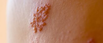
HERPES SIMPLE (HERPESIS SIMPLE)
This paper outlines the present-day concepts of the pathogenesis of herpes simplex, describes its main clinical manifestations, and considers its therapy. V.N. Grebenyuk, doctor med. Sciences, prof., head. Department of Pediatric Dermatology of the Central Research Institute of Dermatovenerology of the Ministry of Health of the Russian Federation. VN Grebenyuk, professor, MD, Head, Department of Pediatric Dermatology, Central Research of Dermatovenereologic Institute, Ministry of Health of the Russian Federation. P
Herpes growth is a serious medical and social problem. This is one of the most common human viral infections, often characterized by a persistent chronic course, affecting various organs, systems and tissues. According to WHO, about 70% of the population of our planet is infected with the herpes simplex virus (HSV) and approximately 10 - 20% of those infected have some clinical manifestations of herpes infection. HSV is a predominantly dermatoneurotropic DNA-containing virus; it also has tropism for other tissues, its size is 150 - 300 nm. The virion, in addition to DNA, consists of an icosahedral capsid and an outer shell containing lipids. It reproduces intracellularly (in the nucleus and cytoplasm) with a 14-hour reproduction cycle. During an acute infectious process, daughter virions are released from decaying cells. HSV infection can cause spontaneous abortions, fetal death and congenital deformities. The herpes virus is associated with the possibility of developing cervical cancer and some cardiovascular diseases. There are two antigenic types HSV-I and HSV-II, which cause lesions of the skin and mucous membranes of various localizations, which is determined by the place of introduction of the virus, usually through contact (coitus, kissing, through household objects). The source of infection can be not only patients with herpes, but also virus carriers who do not have symptoms of herpes.
| Rice. 1. Herpetic lesions of the face. a - forehead, eyelids, bridge of the nose; b - cheeks; c - lips and chin. |
3-4 weeks after infection, antibodies to HSV are formed in the body, the level of which remains relatively constant throughout a person’s life, regardless of the form of infection - manifest or latent. In the vast majority of people, the infection is asymptomatic or subclinical, and only in some infected people does it manifest clinically. Having penetrated the body, the herpes virus reaches a certain regional sensory ganglion (spinal or cranial) through lymphogenous, hematogenous or neurogenic routes, where it constantly persists. The latent state of the virus is based on the biological balance between micro- and macroorganisms. Under the influence of various provoking factors (psycho-emotional arousal, intoxication, overheating, etc.), a relapse of the disease occurs due to the reactivation of latent HSV, which leads to the formation of a recurrent disease. The range of clinical manifestations of the disease - from virus carriage to generalized forms - is determined both by the biological properties of the pathogen and the reactivity of the host. In most people, immune mechanisms, mainly cellular, maintain HSV latency. But in some infected people, antiviral resistance turns out to be untenable and relapses occur. There are two hypotheses that allow for the development of relapses based on both the static and dynamic state of the virus. According to the first hypothesis, the virus is located in the cells of the paravertebral sensory ganglion in an integrated or free non-productive state. Under the influence of the “trigger factor,” the virus, when activated, moves from the ganglion along the axon of the peripheral nerve to the epithelial cells, where it replicates. Cell susceptibility and weakened immune control are thought to contribute to this. According to the dynamic state hypothesis, replication and release of small amounts of virus from the ganglion occur continuously. Reaching the skin nerve, HSV causes microfoci of infection, which are restrained by defense mechanisms, which prevents relapses or weakens their manifestations. The development of relapses is also influenced by the state of local immunity. Its inhibition creates conditions for the replication of the virus that has reached the skin. The immune system plays an important role in containing the spread of herpes infection in the body. Immune protection is determined by the interaction and complex participation of specific and nonspecific factors. The main place in this system belongs to T-cell mechanisms of immunity. Mononuclear phagocytes and neutrophils play a significant role in maintaining local immunity and preventing the dissemination of infection. The protective functions of the body and the preservation of its homeostasis are also greatly influenced by the ability of cells to produce interferon.
Rice. 2. Herpetic felon.
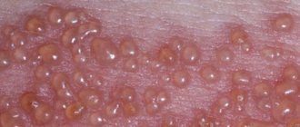
| Rice. 3. Manifestations of herpes. a - on the palmar surface of the hand; b - on the thigh; c - on the buttocks |
Diseases caused by HSV are distinguished by a wide clinical variety of localization, severity, and characteristics of clinical manifestations. Primary herpes usually occurs after the first contact with HSV. More often it is observed in childhood against the background of a reduced immune status, in particular in the absence or low content of specific humoral antibodies. It is distinguished by the high intensity of clinical symptoms. The incubation period lasts several days. Primary herpes in newborns due to hematogenous dissemination becomes systemic, affecting the central nervous system and internal organs. The disease is characterized by herpetic lesions of the oral cavity, eyes, liver, bronchi, lungs, and brain. Usually the disease occurs acutely in the first days after birth and is manifested by anorexia, dyspeptic disorders, convulsions, septic condition, body temperature (39 - 40 ° C), disseminated herpetic rash on the skin and mucous membranes; Deaths are common in the first 2 weeks of illness. Children who have had generalized herpes experience neuropsychic complications. Kaposi's eczema herpetiformis is another severe type of herpes. Occurs mainly in children. It usually occurs in patients with atopic dermatitis, eczema, and other dermatoses in which there are skin lesions. The source of the disease can be patients with herpes in the acute stage. In adults, the disease may be associated with a recurrence of herpes labialis or another clinical form. Kaposi's eczema herpetiformis is characterized by a sudden onset (chills, malaise, body temperature up to 39 - 40 ° C for 1 - 1.5 weeks), a profuse vesicular rash on large areas of the skin, and painful regional lymphadenitis. The rashes appear in paroxysms over 2-3 weeks at intervals of several days. Often, along with skin lesions, the mucous membranes of the oral cavity, pharynx, trachea, and eyes are involved in the infectious process. Grouped and disseminated vesicles soon turn into pustules. In the center of the rash elements there are often umbilical recesses. After the crusts are rejected, secondary erythema remains on the vesiculopustules. Subjectively, the rash is accompanied by itching, burning, and soreness of the skin. Regional lymphadenitis is not uncommon. Patients are subject to hospitalization in an infectious diseases hospital or clinical hospital wards. In severe forms, the pathological process may involve the nervous system, eyes and internal organs. Relapses of Kaposi's eczema herpetiformis are rare, characterized by shorter duration and weakened clinical manifestations.
| Rice. 4. Genital herpes. a — bubble manifestations; b — erosive and ulcerative manifestations. |
The most common clinical form of primary infection is acute herpetic stomatitis.
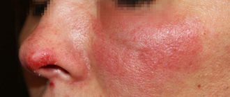
It is more often observed in children in the first years of life; it is rare in adults. In weakened children, dissemination of the virus can lead to visceral pathology (in particular, hepatitis) and death. Acute herpetic stomatitis, occurring after about a week's incubation period, is characterized by a violent clinical picture. Chills, high body temperature (up to 39° C), painful vesicular-erosive rashes in the oral cavity, headache, general malaise, drowsiness - this is a list of the main symptoms of this disease. The rashes are most often located on the mucous membrane of the cheeks, gums, palate, lips, tongue, less often - on the soft and hard palate, palatine arches and tonsils, and spread to the skin around the mouth. The rash initially looks like grouped vesicles against a background of erythematous-edematous islands of the mucous membrane. The transparent contents of the elements become cloudy after 1 - 2 days, the covers of the vesicles are destroyed, and erosions form. In this case, regional lymph nodes are almost always enlarged and painful. Regression of the process usually occurs after 2 - 3 weeks. Recurrences of herpetic stomatitis, as a rule, are milder and resolve earlier. Herpes simplex is more common as a recurrent form. Clinical manifestations compared to primary herpes are less pronounced and not as long lasting. Most often, rashes are located on the face (lips, cheeks, nose), conjunctiva and cornea of the eyes, on the genitals and buttocks. The disease can last for many years and recur with varying frequencies - from several times a year to several times a month. In rare cases, the process becomes permanent when new rashes appear against the background of previous lesions that have not yet resolved. Frequent relapses of genital herpes are especially painful. The localization of herpetic lesions is determined by the site of virus introduction. The appearance of the rash is preceded by prodromal symptoms (burning, itching, tingling and other sensations). Grouped vesicles with a diameter of about 2 mm occur against a background of erythema. The transparent contents soon become cloudy and shrink into lumpy-yellowish crusts. When the vesicles rupture, scalloped erosions form. Their bottom is soft, reddish, the surface is smooth and moist. Regional, slightly painful lymphadenitis with a pasty consistency often occurs. The rash resolves within 1 to 2 weeks, leaving reddish-brown spots. When a microbial infection is added, the duration of relapses increases. Atypical forms of herpes simplex are known: abortive, zosteriform, disseminated, hemorrhagic-necrotic, migratory, elephantiasis-like, ulcerative, rupioid. The abortive form occurs in areas of the skin with a thickened stratum corneum and manifests itself as barely noticeable papules. Abortive manifestations of the disease also include erythematous and pruriginous-neurotic forms, characterized by local subjective disorders without typical rashes. The edematous form is usually located in areas of the skin with loose subcutaneous tissue (eyelids, lips) and is characterized by pronounced tissue swelling. Zosteriform herpes simplex is localized along the course of a nerve on the limbs, trunk, face and is accompanied by neuralgia, headache and general weakness. In the disseminated form of the disease, the rash simultaneously appears on areas of the skin that are distant from each other. The migratory form of recurrent herpes is characterized by a change in the localization of lesions. In hemorrhagic and hemorrhagic-necrotic forms, an admixture of blood is detected in the contents of the vesicles and necrosis develops. The elephantiasis-like form of the disease is characterized by severe swelling followed by the development of persistent elephantiasis in the affected area. Chronic cutaneous herpes simplex is an extremely rare clinical form. It is observed in patients with immunodeficiency and is characterized by permanent active manifestations of infection. Persistent ulcerative lesions up to 2 cm in diameter appear. The ulcerative form of herpes simplex is characterized by the development of ulcerative lesions, which is associated with a weakening of the patient’s immunobiological defense mechanisms and the increased virulence of the virus strain. This clinical type of herpes is characterized by the formation of ulcers at the site of weeping vesicles and fused erosions. The bottom of the ulcers is soft, pink-red in color, sometimes with a grayish-yellowish coating. In the first days of the disease, local pain and burning are expressed. Sometimes the rash is accompanied by inguinal lymphadenitis. The rupoid form of herpes simplex is usually localized on the face. It is caused by pyogenic infection with the development of cracks and layered crusts. Relapses occur several times a year. The rash is often accompanied by tenderness and enlargement of regional lymph nodes. With herpes of the hands, the process is often located on the distal parts of the hands. Limited lesions are represented by single dense blisters, accompanied by severe pain. The most common type of herpes simplex is facial herpes. In most people, these are sporadic focal vesicular eruptions, often resolving within 1 week. In severe cases, the process involves large surfaces of the face - nose, cheeks, forehead, skin and red border of the lips. Genital herpes occupies a significant place in the structure of herpetic diseases. Etiologically, its occurrence is equally often associated with types of HSV-I and/or HSV-II. Infection with one type of virus does not prevent the occurrence of HSV infection of another type, which leads to the formation of intermediate (“double”) antibodies. Mixed infection with HSV-I and HSV-II is quite common. The frequent isolation of HSV-I, which was previously considered the causative agent of non-genital forms of herpes, in genital lesions is due to the prevalence of orogenital contacts. Genital herpes is distinguished by the variability of its clinical picture and its tendency to have a chronic, relapsing course. In men, limited herpetic eruptions are often localized on the inner layer of the foreskin, in the head groove, and less often on the head and shaft of the penis. In women, the labia minora, clitoris, cervix, perineum and thighs are most often affected. Rashes (vesicles, erosions, ulcers, cracks) against a background of erythema and swelling are usually painful and are also accompanied by itching, a feeling of tension and heaviness in the perineum. About a third of patients have inguinal lymphadenitis. When the urethral mucosa is involved in the pathological process, serous discharge from the urethra and pain when urinating appear. The source of infection in the case of genital herpes is usually a patient in the acute stage of the disease; it can also be a virus carrier, given the possibility of asymptomatic persistence of HSV in the genitourinary tract in men and in the cervical canal. The incubation period for primary genital herpes lasts from one to several days. Clinically, primary genital herpes has a more severe and prolonged course. The localization of rashes on the genitals and adjacent areas is determined by the gates of the viral infection. A recurrent course of genital herpes is observed in the majority of infected people. Provoking factors are a variety of influences - psycho-emotional experiences, hypothermia, menstruation, weather and climate fluctuations, and other factors that disrupt the state of biological balance of the body, contributing to a decrease in the immune response and activation of HSV. The clinical picture, the amount of virus secreted by the patient and the associated infectivity are more pronounced with primary herpes than with a recurrent disease. Possible complications of herpes simplex: the addition of a secondary bacterial infection, reinfection with the released virus of other epithelial integuments, neurological manifestations (aseptic meningitis, transverse myelitis), encephalitis, disseminated infection of internal organs, psychosocial consequences (psychological instability). The risk of developing cervical cancer is 2 times higher in women who are seropositive for human papillomavirus types 16/18 and infected with HSV-II.
Diagnostics
The diagnosis of herpes simplex, especially its genital form, in most cases is based on the clinical picture. Difficulties arise with atypical manifestations of herpes. In this case, it is important to carefully collect anamnesis, paying attention to relapses accompanied by itching, burning, and ineffectiveness of antibiotic therapy. In addition, the patient may have a tendency to colds, general weakness, malaise, low-grade fever, and depression. Recurrent herpes is characterized by a wave-like course of the disease - an alternation of relapses and remissions. In women, relapses of herpes may be associated with certain phases of the menstrual cycle. The occurrence of erosions and ulcers on the genitals simulates syphilitic lesions. This similarity is most pronounced when a secondary microbial infection is attached, as well as during irrational therapy. The diagnosis of genital herpes is complicated by the fact that HSV is often associated with some resident autoflora microorganisms: chlamydia, streptococci and staphylococci, gardnerella and others, which can determine the occurrence of mixed infections. In addition, because herpes can be transmitted sexually, the patient must be tested to rule out other sexually transmitted diseases, including syphilis and AIDS. In complex cases, when clinical data is insufficient, laboratory diagnosis is possible. There are a number of specific laboratory tests to recognize HSV infection: isolation of HSV in cell culture, including HSV-I and HSV-II typing, tests to determine HSV antigen or DNA using polymerase chain reaction; serological tests - complement fixation test, ELISA, indirect immunofluorescence reaction, reverse passive hemagglutination reaction, protein-specific immune tests (immunoblotting), cytological examination (detection of multinucleated giant cells in scrapings from the lesion).
Treatment
Treatment of recurrent herpes remains a difficult task, which is not always solved effectively. It is possible to achieve some success if complex etiological and pathogenetic treatment is carried out at different stages of the disease, aimed, on the one hand, at suppressing the infectious agent, and on the other, at increasing the body’s immune reactivity. When choosing treatment, the stage of the disease should be taken into account. For relapses, interferon, antiviral chemotherapy, measles immunoglobulin, human normal immunoglobulin, levamisole, ascorbic acid, deoxyribonuclease, applications of 0.05% zinc sulfite solution are indicated; in the inter-relapse period - herpetic and polio vaccines, pyrogenal. The etiological focus is on antiviral chemotherapy drugs, which are more effective when used in the first hours and days of the appearance of rashes. Among them is the domestic drug Bonafton, which is used orally at 50–150 mg/day for 5–7 days for relapses. Simultaneously with the tablet form, 0.5% bonaftone ointment can be prescribed. It is applied to the lesions in an open manner when signs of relapse appear and is easily rubbed into the skin 2 - 3 times a day for 5 - 7 days. Side effects observed in some patients include malaise, loose stools, and dermatitis. Acyclovar (Zovirax) is effective, characterized by low toxicity and selectivity against HSV. The drug is used intravenously, orally and topically. It gives a pronounced therapeutic effect for Kaposi's eczema herpetiformis. Acyclovir is administered intravenously at the rate of 20 mg per 1 kg of body weight per day. However, the drug does not prevent herpes from recurring, infecting newborns, or infecting other people. Treatment of patients with recurrent herpes with acyclovir 0.1 - 0.2 g 5 times a day for 5 days during relapses shortens the time for resolution of rashes, reduces the severity of subjective sensations, smoothes out clinical manifestations and reduces the degree of virus shedding. Prophylactic administration of the drug 0.1 - 0.2 g 4 times a day for 6 - 12 weeks reduces the duration of relapses and weakens clinical manifestations. Other chemotherapy drugs: famciclovir, alpizarin (2 and 5% liniment), Viru Merz Serol, 1% oxolinic ointment, hevisos, ribavirin (virazol). A certain therapeutic effect is provided by immunocorrective drugs (myelopid, poludanum, arbidol), used both as monotherapy and in complex treatment. Myelopid (0.003 g in 2 ml of saline) is administered intramuscularly once every 3 days (5 injections per course). Treatment is carried out in two courses with an interval of 7 - 10 days. Poludan is administered subcutaneously into the forearm every other day, 100 mcg, for a course of 1000 mcg. Arbidol is prescribed 0.2 (2 tablets) 3 times a day - 5 days with a 2-day break, and then 0.1 g (1 tablet) 1 time per week for 3 weeks. Sodium nucleinate is also used orally at 0.5 - 1 g / day in 2 - 3 doses daily for 2 - 4 weeks. Taktivin is used to stop relapses and for prophylactic purposes. The drug is administered subcutaneously at a dose of 100 mcg every other day, 8 - 10 injections. During the inter-relapse period, 50 mcg is prescribed every other day, a course of 5 injections is repeated every 3-6 months. A course (4 - 5 injections) of treatment with timoptin is also carried out, which is administered subcutaneously at 100 mcg every 3 - 4 days. The courses are repeated after six months.
External treatment
Antiviral ointments, creams, lipsticks accelerate the epithelization of erosions, reduce or reduce subjective sensations in the affected areas. Local use of one or another antiviral drug in the treatment of herpetic lesions for 5 - 7 days shortens the time of regression; use 2 - 3 times a week during the inter-relapse period allows to prolong remission. Interferon has an inhibitory effect on HSV, which is applied to the skin and easily rubbed in for 4 to 7 days. During treatment, it is advisable to alternate antiviral drugs during relapses. Human interferons are effective in the treatment of recurrent herpes in the prodromal period and when the first signs of relapse appear. The ointment is applied to the lesions 2-4 times a day and rubbed in lightly; treatment is continued for a week. The use of interferon ointment during the inter-relapse period prolongs remissions and interrupts the development of relapses. In order to prevent relapses in frequently recurrent forms of herpes, patients for whom treatment is ineffective are prescribed a herpetic vaccine. Contraindications to its administration are lesions of parenchymal organs, diabetes mellitus, stage II and III hypertension, decompensated heart failure, acute infections and allergic diseases. The drug is administered intradermally during the period between relapses, 0.2 - 0.3 ml into the area of the flexor surface of one of the forearms. The first 5 injections are given after 3 - 4 days, the next 5 doses are administered after a 2-week break (once every 5 - 7 days). These 10 injections constitute the main course of treatment, 3–6 months after the end of which 1–2 cycles of revaccination are carried out, each of 5 injections with an interval between injections of 7–14 days and between cycles of 6–8 months. Over the next 2 years, an additional revaccination cycle of 5 injections is carried out every 8 - 12 months. At the injection site, after 18-24 hours, a local reaction develops, manifested by the development of erythema with a diameter of 2-5 cm with a papule in the center and accompanied by a burning sensation. During vaccination, a focal reaction such as abortive relapses may be observed. In this case, a break is taken in the treatment for 2 - 3 days, then it is continued. Specific vaccine therapy leads to an increase in the duration of remissions, a reduction in relapse periods, and the disappearance of subjective sensations. For the purpose of secondary prevention of relapse of herpes, the factors that provoke the disease are controlled. Great importance is attached to the sanitation of the body and health-improving measures in the process of medical examination.
Literature:
1. Barinsky I.F., Shubladze A.K., Kasparov A.A., Grebenyuk V.N.M.: Medicine. 1986, 269 p. 2. Masyukova S. A., Rezaikina A. V., Grebenyuk V. N., Fedorov S. M., Mkhitaryan A. G., Kolieva M. Kh. Immunotherapy of recurrent herpes simplex. Sexually transmitted diseases. Information analytical newsletter. Sanam Association 1995, 3, 27-30. 3. Minde CA. Genital Herpes. A guide to pharmacological therapy. Drugs 1994;47(2):297-304. 4. Whatley JD, Thin RN. Episodic acyclovir therapy to abort recurrent attacks of genital herpes simplex infection. J Antimicrobial Chemotherapy 1991;27:677-81.