Pigmentation on the eyelids are spots of various sizes and colors of various shapes with clear edges. Congenital limited hypermelanosis on the eyelids can manifest as freckles or lentigines. Freckles are small pale yellow or dark brown spots that never rise above the level of the skin. In summer they become a shade darker, and in winter they fade or even disappear. Freckles on the eyelids are quite rare compared to other areas of the face, as well as exposed parts of the arms, neck, and chest. They are intra- or extracellular accumulations of melanin in the spinous epidermis and, to a lesser extent, in the superficial dermis.
Lentigo, or simple nevus, is a pigmented birthmark that can range in size from millet grains to brown lentil grains. They can gradually grow and spread to areas close to the eyelids, for example, to the conjunctiva, or from the conjunctiva to the eyelid area. In old age, nevi often degenerate, forming malignant melanomas and milanocarcinomas. Today, nevi are usually considered as formations of melanocytes, as well as Schwann cells from the sheaths of cutaneous nerves, which are capable of producing melanin - as a viciously developed neuroectodermal part of the ectoderm.
Chloasma
The term is a generalized name for changes in the color of the skin of the eyelids or face, which mainly occurs in pregnant women, as well as with pathologies of the uterus and appendages, diseases of the liver or biliary tract, and helminthiasis. In this case, the color of the skin changes not only on the eyelids and face, but also on other parts of the body - along the white line on the stomach, on the nipples, etc.
According to the condition in which chloasma develops, they are divided into pregnancy chloasma, uterine chloasma, helminthiasis masks or hepatic chloasma.
Such pigmentation is more often observed on the lower eyelids, less often on the upper. Moreover, it tends to continue continuously from the eyelid to the cheek area. To some extent, the appearance of these spots is stimulated by exposure to sunlight. After delivery, chloasma of pregnant women disappears and reappears during a new pregnancy. In liver patients, pigmentation is one of the earliest signs of the disease.
Histologically, in the pigment spots of the chloasma listed above, an excess content of melanin is detected, which is located in the deep epithelium and in the Malpighian layer. The immediate cause of the appearance of chloasma is a variety of toxins that are formed in the body as a result of pregnancy, diseases of the liver, genitourinary organs, as well as the gastrointestinal tract, etc. True, autonomic-endocrine, as well as hormonal disorders play an equally important role in the occurrence of chloasma. In addition, increased pigmentation of the eyelid skin is an early sign of pigment metabolism disorders (with Addison's disease and hyperthyroidism).
In Addison's disease, which is adrenocortical insufficiency of the adrenal glands, the pituitary gland is disinhibited, which leads to increased production of the melanoform hormone. This is expressed in greater deposition of melanin in the epidermis and dermis. Clinically, abnormal pigmentation of the skin of the eyelids is reduced to the appearance of a bronze uniform color.
Changes in the color of the lower, and especially the upper eyelids, can be observed (not often) in the case of Graves' disease. At the same time, it is the change in pigmentation of the skin of the eyelids that should be regarded as one of the early symptoms of thyrotoxicosis. The color of the skin of the eyelids, in this case brownish, is uniform. On the upper eyelid, the area of hyperpigmentation ends at the eyebrow, on the lower eyelid, at the lower edge of the orbit. Pigmentation of the skin of the eyelids with hyperthyroidism is sometimes combined with changes in eyelashes - their loss or graying. It was believed that the change in the color of the skin of the eyelids was associated in this case not with the deposition of melanin, but with hematogenous pigments. This assumption is not true. A significantly large role in the appearance of this symptom belongs to the disruption of the normal relationship in Graves' disease between the thyroid gland and the adrenal glands.
Disease prevention
The best preventative measure to prevent meibomitis is clean eyelids. In other words, maintaining and regular hygiene of the visual organs sharply reduces the likelihood of inflammation of the sebaceous glands. Of course, touching your eyes with dirty hands (for example, to remove a speck of dirt or for self-massage when your eyes are tired) is completely unacceptable - this is fraught not only with meibomitis, but also with a number of serious diseases. In hazardous industries and in unfavorable climates, it is imperative to use personal eye protection.
It is also necessary to avoid hypothermia and, conversely, overheating, monitor the general condition of the visual organs (for example, if necessary, take measures to artificially moisturize the cornea, undergo a course of vitamin therapy and/or immunostimulation). If there is such a possibility, it is advisable from time to time to perform a special massage in the nearest ophthalmology office (not independently) to evacuate excess secretion of the meibomian glands.
Ochronosis
Changes in skin color also occur in the case of so-called ochronosis. This disease is quite rare and is a specific lesion of cartilage, ligaments and tendons, expressed in slowly increasing immobility of the spine with deforming arthritis, otosclerosis, and damage to the cartilage of the larynx. The fabrics are painted in dark colors - ash-gray, brown or almost black. Such changes are also observed on the eyelids. In mild forms of ochronosis, the condition is limited to skin hyperpigmentation and the disease ends in recovery. In severe cases, a change in the color of the skin of the eyelids is accompanied by a spotty dark color of the mucous membrane of the eyes in the area of the palpebral fissure along the limbus. The disease is accompanied by alkaptonuria. The color of the skin during ochronosis is due to the deposition of ochronotic pigment in it. Ochronosis of an exogenous nature can also occur with phenol poisoning or long-term use of carbolic acid for medicinal purposes. The course is chronic.
Treatment: shock doses of vitamin C, measures aimed at reducing alcantonuria.
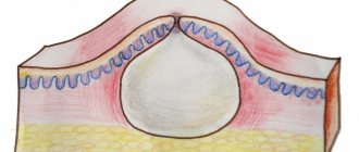
Manifestation of xanthelasma
The following external manifestations are characteristic of xanthelasma:
- appearance: the shape of a plaque protruding above the skin;
- size: from pea to bean;
- location: often close to the bridge of the nose on one or both eyelids;
- consistency: soft, painless;
- color: yellowish;
- quantity: single or multiple, merging into an uneven tuberous strip;
- speed of spread: appear suddenly and grow slowly, becoming a serious cosmetic problem.
Hemochromatosis
This disease is accompanied by bronze coloration of the skin (one of the early symptoms), further development of liver cirrhosis, as well as bronze diabetes mellitus. Skin pigmentation is caused by the deposition of hematogenous pigment - hemosiderin, which is formed due to a violation of iron metabolism, with its accumulation in organs and tissues. Bronze coloration is predominantly localized on the neck and back of the hands. The eyelids turn gray-brown. Hemochromatosis is extremely rare and occurs exclusively in men aged 45-50 years. There are no more than 150 thousand patients in the world.
Treatment: diet including foods with limited iron content, drugs that remove iron from the body, repeated bloodletting in permissibly large doses.
Prognosis for recovery from melanoma of the eyelid
In general, eyelid melanoma is an aggressive tumor and is diagnosed at late stages, when the prognosis for treatment is unfavorable. Five-year survival can be achieved in only half of the patients. But in the initial stages, with a melanoma thickness of less than 0.75 mm, the chances of a complete recovery are much higher.
| Read more about dermatological research at Euroonko | |
| Consultation with a dermatologist-oncologist | from 5,100 rub. |
| Skin examination using the German FotoFinder device | RUB 13,400 |
| Diagnosis of melanoma | from 5,100 rub. |
Book a consultation 24 hours a day
+7+7+78
Dyschromia
This change in skin color, including the skin of the eyelids, can occur with prolonged use of heavy metal salts for medicinal purposes, or with prolonged contact with them in the professional sphere. This kind of hyperpigmentation should be classified as excessive pigmentation of an exogenous order. Among the many varieties of this hyperpigmentation, cases of argyrosis and arsenomelanosis are especially common. With arsenomelanosis, the skin of the eyelids becomes covered with widespread and limited dark brown spots, hyperkeratosis occurs, and scales form. In some cases, the immediate cause of arsenomelanosis was long-term use of salvarsan and Fowler's tincture. The appearance of spots with arsenomelanosis is caused by the deposition of melanin.
Argyrosis is characterized by the appearance of a matte or gray-bluish color of the skin due to impregnation with silver salts, silver hydrochloride salt, in particular. On the eyelids, such impregnation occurs as a result of instillation of the drug into the conjunctival cavity or its introduction into the lacrimal sac. With argyrosis, silver particles are found in the papillary dermal layer, in the walls of the sweat and sebaceous glands, and in the hair follicles. Treatment: potassium iodide is prescribed orally.
The use of picric acid preparations can also lead to dyschromia - the appearance of a light yellow color of the skin of the eyelids. Finally, pigmentation of the skin of the eyelids of an exogenous nature can be caused by the so-called toxic melasma, the former name of which is Riehl melanosis, B-vitaminosis dermatosis. This dyschromia is based on intoxication with heavy hydrocarbons from coal and oil. Often their derivatives are added to cosmetics (vaseline, bergamot oil, etc.). In conditions of hypersensitivity of the body and with intense solar radiation, their use leads to the occurrence of erythema or follicular keratosis (atrophy with telangiectasia). The clinical picture reveals dark melanotic dotted or reticular pigmentation on the skin.
The phenomena of toxomelanoderma are also possible in some individuals when exposed to chlorine and bromine.
By contacting the Moscow Eye Clinic, each patient can be sure that some of the best Russian specialists will be responsible for the results of treatment. The high reputation of the clinic and thousands of grateful patients will certainly add to your confidence in the right choice. The most modern equipment for the diagnosis and treatment of eye diseases and an individual approach to the problems of each patient are a guarantee of high treatment results at the Moscow Eye Clinic. We provide diagnostics and treatment for children over 4 years of age and adults.
Treatment of meibomitis
In most cases, especially with timely treatment, meibomitis can be cured with conservative, non-surgical methods. First of all, disinfection of the ciliary edge of the eyelid is necessary, then local medications are prescribed.
Antibacterial drops and ointments
- Albucid
- Levomycetin
- Tetracycline eye ointment
- Floxal (ointment and drops), etc.
Anti-inflammatory drugs
- Dexamethasone eye drops
- Hydrocortisone eye ointment
Combination drugs
- Maxitrol
- Tobradex
- Sofradex and others.
The duration and frequency of use is determined by the attending ophthalmologist individually, depending on the specific situation.
Our doctors who will solve your vision problems:
Fomenko Natalia Ivanovna
Chief physician of the clinic, ophthalmologist of the highest category, ophthalmic surgeon. Surgical treatment of cataracts, glaucoma and other eye diseases.
Yakovleva Yulia Valerievna Refractive surgeon, specialist in laser vision correction (LASIK, Femto-LASIK) for myopia, farsightedness and astigmatism.
You can find out the cost of a particular procedure or make an appointment at the Moscow Eye Clinic by calling in Moscow 8 (499) 322-36-36 (daily from 9:00 to 21:00) or using the ONLINE REGISTRATION FORM.
How is eyelid melanoma diagnosed?
Although eyelid melanoma is a superficial tumor that is easily accessible for visual inspection, it is not always possible to diagnose the disease at an early stage, since patients do not pay attention to it and do not go to see a doctor. Therefore, much attention is paid to self-examination. You can suspect a malignant process based on the following signs:
- The neoplasm has fuzzy, blurry edges.
- Asymmetrical shape.
- Uneven coloring of the tumor. In melanoma, areas of hyperpigmentation may be replaced by depigmented areas.
- The neoplasm increases in size.
- The skin pattern changes. Melanoma is characterized by a smooth surface; peeling or ulceration is less common.
- There is redness around the tumor.
- The melanoma itself may be itchy, painful, and cause a tingling or burning sensation.
As it grows, eyelid melanoma spreads to the underlying tissues, involving the conjunctiva, sclera and other structures of the eyeball in the process. In order to diagnose melanoma in time and begin treatment, it is necessary to regularly see an ophthalmologist, especially over the age of 50 years.
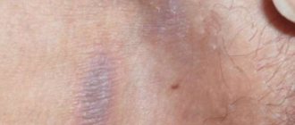
Regime and dark circles under the eyes
In adults, bruises under the eyes sometimes appear due to the most banal reason - violation of the regime.
Overwork, lack of sleep, stress - all this makes the skin of the eyelids thin and dull, and the face pale and haggard. Metabolism is disrupted, the blood vessels of the eyelids appear more prominently, creating characteristic purple or bluish shadows under the eyes. A similar problem arises due to persistent dehydration, when a person constantly stays in rooms with dry air and drinks little water (as well as smokes and abuses alcohol).
A healthy lifestyle is the best friend of cosmetic procedures. In tandem it works flawlessly!
Try a double tactic: sleep 8 hours a day, eat right, spend time in the fresh air more often, and at the same time visit a competent cosmetologist. And you won’t give dark circles under your eyes a single chance.
A consultation with our specialists is the answer to your remaining questions.
Telephone consultation is always free!
Leave your contact, we will tell you everything and offer a discount!
Send
Diagnostics
To exclude diseases of internal organs, you should undergo comprehensive instrumental and laboratory diagnostics, including:
- general and biochemical blood test;
- hormone tests:
- thyroid gland (thyroid-stimulating hormone, free T4);
- produced by the adrenal glands (adrenocorticotropic hormone, cortisol);
- sexual (progesterone, estrogen);
- organs located in the abdominal region;
It is also necessary to consult with different specialists:
- endocrinologist;
- gynecologist;
- urologist;
- ophthalmologist;
- otolaryngologist;
- gastroenterologist;
- cardiologist;
- allergist.