Reasons for appearance
The exact causes of the disease have not yet been established. The main one is regular exposure to ultraviolet radiation. It affects the dermis, epidermal layers, blood vessels, sebaceous glands, and melonocytes.
Gradually, under the influence of sunlight, the disturbances increase, reaching the peak of the disease.
The following factors contribute to the development of pathology:
- genetic predisposition;
- weakened immune system;
- influence on the skin of chemicals (resinous substances, oil, sand, etc.);
- past infections;
- age-related changes (the disease most often affects people over 50 years of age).
Due to weak immunity, AIDS carriers, people with problems with the nervous and endocrine systems, as well as patients who have undergone chemotherapy or complex operations are more prone to the appearance of keratosis.
Some types of keratoses often affect young people. This usually applies to red-haired or fair-haired people with gray, blue or green eyes. Research shows that by the age of 40, 60% of the population has at least one element of keratosis.
Over the age of 80, everyone has some type of this pathology.
MECHANISM OF KERAINIZATION IN NORMAL AND IN HYPERKERATOSIS
Normally, as a result of their vital activity, skin cells gradually move from the lower layer to the upper, while gradually accumulating keratin - a protein that makes them more durable and resistant to external influences. The top layer of skin is nothing more than completely keratinized cells or horny scales, which are peeled off and replaced with new ones during human life. Such cells lack viability, but at the same time, thanks to keratin, they provide protection to living cells located underneath them from negative environmental factors.
With the development of hyperkeratosis as a pathological condition of the skin
- on the one hand, there is an active accelerated division of epidermal cells that produce keratin;
- on the other hand, there is a delay in the process of normal physiological exfoliation of horny scales from the surface layer of the skin.
Types and signs of keratosis
A person may not pay attention to the signs of the initial stage of the disease. It can manifest itself as unnoticeable roughness on any part of the body (cheeks, bridge of the nose, forearms, scalp, ears, etc.). At first the formation is solid, small in size, red or brown in color. The skin around the affected area may peel off, itching begins, and hair loss may also be observed at the site of the keratoma.
There is no unified classification of the varieties of this disease, because it has not been fully studied. According to etiology, the following types of keratosis are distinguished:
- Congenital - appears at birth or at a younger age. Very rare.
- Acquired - appears in adulthood, less often - in adolescence.
- Symptomatic – occurs due to external factors.
According to the affected area, local (single areas of skin are affected) and diffuse (large areas of the skin are affected) keratosis.
There are also several types of clinical manifestations.
Keratosis follicularis
The formation of horny plugs in the hair follicles is observed. These are dead cells that have separated from the skin. By forming nodules, they interfere with hair growth. Most often, keratosis pilaris appears on the abdomen, face, shoulders, buttocks, neck, and armpits. Such manifestations are characteristic of the cold season; closer to summer, symptoms may disappear. This type of pathology is also called pilar keratosis. If the nodule grows more than 3 mm, pain may occur.
Consumption of allergens can cause an exacerbation of the inflammatory process.
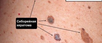
Seborrheic keratosis
This type of disease is characterized by plaque-like or nodular formations with a warty surface. The top of the keratomas is covered with a brown or black keratinized substance. As a rule, it occurs over the age of 50 years, therefore it is also called actinic keratosis.
The most common areas for the appearance of formations are the face, chest, neck, etc. It does not occur on the palms and soles. The development of this form of the disease proceeds slowly and usually becomes chronic. Actinic keratosis does not develop into a cancerous tumor, but a malignant tumor can masquerade as keratosis. In case of inflammation of the affected areas, bleeding and rapid growth of the formation, you should immediately consult a doctor.
Actinic keratosis
Appears on exposed areas of the body. At first it looks like uneven, rough skin. Over time, it develops into a scaly, flaky, compacted spot, ranging in color from skin color to brownish. Such formations may look like growths, rising above the skin. Mostly the face, neck, and chest are affected by keratinization. Such keratosis can transform into cancerous tumors, so it is necessary to be regularly monitored by a dermatologist.
What is follicular hyperkeratosis
Follicular hyperkeratosis is a common dermatitis caused by excessive growth of the stratum corneum of the skin, impaired desquamation of the epidermis and blockage of the mouths of the follicles with epidermal scales.
There is a violation of the release of sebum to the surface of the skin and aseptic inflammation. Visually, this gives the “goose bumps” effect.
Treatment of the disease
Treatment should be carried out by a dermatologist.
You need to visit a specialist after the first appearance of keratomas, since it is important to exclude the malignant nature of the formation. Treatment is long-term and complex, usually including a number of activities.
Conservative therapy
It is carried out with the aim of reducing the number of keratoses before moving on to radical methods of treatment.
Therapeutic agents reduce symptoms and alleviate the course of the disease, but do not cure completely.
Therapeutic agents reduce symptoms and alleviate the course of the disease, but do not cure completely.
To soften keratonic areas, applications using drugs with urea (content - from 12 to 30%) are used: Keratosan, Ureaderm, Ureatop, Akerat..
The following drugs are used in therapeutic treatment: Fluorouracil, Efudex cream, Diclofenac Gel 3%, Imiquimod. Special shampoos are used to treat keratosis of the scalp. Retinoids are taken internally, which help reduce the growth rate of formations, as well as vitamins of groups A, B and C. Additionally, courses of physiotherapy are prescribed.
Radical methods of treating keratosis
Since conservative therapy for keratosis does not guarantee complete cure, it is often necessary to proceed to radical measures - direct removal of the formations. The use of drastic techniques is especially justified if there is a risk of keratosis degenerating into cancer.
The following radical methods of therapy exist:
- Cryodestruction - freezing with liquid nitrogen.
- Radio wave removal. The formation is excised using a radio knife under the influence of radio waves.
- Electrocoagulation - the doctor performs cauterization using high-frequency electric current.
- Laser destruction - a targeted effect of a carbon dioxide laser is applied to the keratoma.
- Photodynamic therapy - methyl aminolevulinate is applied to the affected area with further exposure to a light wave of a certain length. All this leads to necrosis of the affected tissues.
- Surgical removal - the skin is scraped with a curette (special instrument).
- Dermabrasion – removal using an abrasive brush.
HOW TO TREAT HYPERKERATOSIS
Symptoms of hyperkeratosis usually do not cause pain and are considered a cosmetic defect. The exception is calluses and corns on the feet, which cause discomfort when walking. In this case, you should immediately seek qualified help, especially if the manifestations of hyperkeratosis cause pain, discomfort, there are symptoms of infection (redness, swelling, accumulation of pus) or if you have diabetes.
Hyperkeratosis will not go away on its own; in any case, it is necessary to start treatment.
The method of treating hyperkeratosis is chosen by the doctor, taking into account the cause, location, prevalence and shape of the thickenings, as well as compensation for the underlying chronic disease as a result of which the pathological process was formed.
To treat hyperkeratosis, the following are most often used:
- ointments, creams with a softening effect; creams rich in fat;
- taking acitretin;
- Daivonex ointment (calcipotriol);
- medical hardware manicure (if the process is localized on the palms, feet, arms and legs);
- creams with urea;
- peeling, including chemical and laser peeling (face and body);
- radiosurgical removal (keratomas);
- surgical cutting (calluses, corns).
You cannot remove calluses, corns or warts on your own, as there is a risk of infection and other complications.
Forecast and prevention of the disease
The prognosis of the disease depends on the time of initiation of treatment. However, in order to avoid relapses, it is necessary to follow certain recommendations.
The following measures can be taken as prevention:
- use moisturizing creams;
- eat well so that food provides the skin with all the necessary “building” elements;
- limit exposure to direct sunlight to reduce exposure to ultraviolet radiation;
- when in the sun, use sunscreens (ointments and creams) with a high SPF level;
- When working with chemicals, you must use appropriate skin protection.
Keratosis is a skin condition that requires constant monitoring. In addition to an aesthetic defect, sometimes formations can develop into malignant tumors. And to prevent this, it is necessary to undergo regular examinations and monitor changes in the skin.
Clinical picture
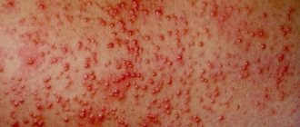
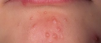
Visually: small reddish rashes, “goose bumps”, dense nodules are localized at the very base of the hair follicle.
to the touch .
Localization : most often on the shoulders, hips, face and buttocks.
In the generalized form of follicular hyperkeratosis, extensive damage to the trunk and extensor surfaces of the extremities is observed.
Diagnostics:
The diagnosis is made by a dermatologist based on a visual examination; specialized techniques are not required.
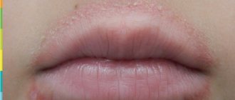
It is necessary to distinguish follicular hyperkeratosis from acne. In the case of hyperkeratosis, the rashes on the face are dry, rough to the touch, small and uniform in size.
Congenital forms of keratosis pilaris
When we talk specifically about follicular keratoses, we mean a large group of congenital diseases manifested by disorders of keratinization processes, the cause of which is hereditary transmission or genetic determination.
The latter is a functional state of the body in which, at one or another period of life and under the influence of certain and often incomprehensible circumstances, activation of pathological genes occurs. The congenital form of keratosis pilaris does not necessarily have to be present at the time of birth or at an early age. It can manifest itself during life at any age, including in adults.
Radical pathogenetic treatment of keratosis pilaris caused by genetically determined or hereditary causes is impossible. It can only be symptomatic, that is, aimed at eliminating or reducing the severity of the manifestations of the disease over a more or less long period of time.
Watch the video about tips for parents about keratosis pilaris:
Classification of congenital follicular keratoses
Hereditary follicular keratoses constitute a fairly large group of diseases, some of which are not casuistic in the practical work of dermatovenerologists.
In the modern classification, follicular keratoses are divided into:
- Papular, which include lichen pilaris, Devergie's disease, persistent lenticular hyperkeratosis of Flegel, Morrow-Brook disease, keratosis squamous of Doha.
- Atrophying - superciliary cicatricial erythema, or superciliary ulerythema, vermiform atrophoderma, Siemens keratosis spinosum decalvans.
- Vegetative, which includes follicular and parafollicular keratosis of Kirle, serpiginous keratosis of Lutz, Darier-White disease.
Diseases belonging to the first two groups are characterized by a tendency to imperceptible or subtle progression and a gradual process of skin atrophy in the affected area.
Keratosis: diagnosis
Keratosis is detected based on examination of the skin using a magnifying glass and a light source. Anamnesis (history of the disease) is important - the doctor clarifies the following nuances of the pathology:
- presence in the family of people with keratosis;
- connection with solar radiation;
- mode of work and rest.
The patient’s age is also taken into account – this applies to the diagnosis of seborrheic (senile) keratosis.
Additional research methods (instrumental and laboratory) are required if it is necessary to differentiate keratosis from other diseases - for example, from skin cancer, when it is necessary to involve a biopsy (sampling of skin fragments followed by examination under a microscope).
List of sources
- Bauman L. Cosmetic dermatology. Principles and practice. Per. from English Potekaeva N. N. M.: MEDpress-inform. 2012. 688 p.
- Afanasyev E.N. Mechanically induced hyperkeratoses of the foot // Plastic surgery and cosmetology. 2012. No. 4. pp. 644–661.
- Kholodilova N.A., Monakhov K.N. The use of basic care products in patients with impaired skin barrier // Russian Journal of Skin and Venereal Diseases. 2009. No. 6. pp. 68–69.
- Lomakina E. A. The role of the barrier function of the skin in the pathogenesis of some dermatoses // Modern problems of dermatovenereology, immunology and medical cosmetology. 2009, no. 2. pp. 87–90.
- Rodionov A. N. Dry skin. Dermatocosmetology. Lesions of the facial skin and mucous membranes. Diagnosis, treatment and prevention. SPb: Science and Technology. 2011. 911 p. pp. 63–69.