Syphilis spots
- I. White spots. With syphilis, the entry of pathogenic bacteria into the skin may be accompanied by a disruption in the functional activity of melanocytes. These are the cells that produce the pigment responsible for the color of the skin. Such spots are not inflammatory in nature. They are characterized by white color, different shapes and sizes.
- II.Atypical localization of the rash, which may indicate syphilis. Spots on the legs develop less frequently, but when they are located on the skin of the flexor surfaces of the joints, oozing (secretion of serous fluid from the rash element) may appear. They are accompanied by pain and the release of a significant number of microorganisms.
White spots with syphilis
White spots on the skin of a patient with syphilis appear 6-8 months later at the second stage of the disease, with secondary prolonged or “recurrent” syphilis.
Let's take a closer look at these elements, how they differ and what to do when stains of this nature appear.
As we have already written, these elements are called syphilitic leukoderma or the “necklace of Venus”.
The spots are limited areas of depigmentation the size of a grain or coin, do not protrude above the surface of the skin and do not cause concern to a person.
Syphilitic leucoderma resembles lace.
The main localization of such formations is the neck.
Leukoderma can also occur on other areas of the skin:
- Back
- Breast
- Stomach
- Small of the back
Often an infected person does not pay much attention to these formations.
Attention! White spots do not cause much concern; they do not hurt or itch.
Depending on whether the spots merge or not, the following types of syphilitic leukoderma are distinguished:
- spotted (without merging)
- mesh (partially merge)
- marble (full drainage)
More often, white spots with syphilis occur in women.
It has been proven that the causative agent of syphilis, Treponema pallidum, is not present in the spots.
The mechanism of formation of leukoderma is not precisely known.
It is believed that the synthesis of melanin is disrupted under the influence of infection.
According to another version, pigmentation disorders occur as a result of damage to the nervous system with disruption of skin nutrition and destruction of pigment.
Syphilitic leukoderma is reversible and disappears with treatment within 2 to 3 months.
Are there brown spots with syphilis?
Rarely, brownish spots on the skin can also be observed.
Let's look at when they might appear.
Brown spots with syphilis, as well as white ones, are a violation of skin pigmentation with syphilitic leukoderma.
Appear with secondary “recurrent” syphilis.
The color of spots with hyperpigmentation can range from yellowish-brown to dark brown.
After 2–3 weeks, light hypopigmented spots, round or oval in shape, appear on these areas of the skin.
Syphilis spots in children
If a child has spots, this should always be a concern.
Let's look at the features of syphilis spots and how to distinguish them from other diseases.
The appearance of a rash with syphilis in children occurs in the second stage of the development of the disease.
This is a sign of secondary syphilis.
Syphilitic rash in children resembles the rash associated with childhood infections (chickenpox, measles, rubella).
Just like with childhood infections, it is accompanied by fever for 1-5 days.
How to distinguish syphilitic rashes from rashes of other etiologies?
The main distinguishing feature is:
- 1.duration of rashes – from 2-3 weeks to several months
- 2. wave-like course - periods of rashes are replaced by periods of deceptive well-being throughout the entire secondary period
The rash in children, like in adults, can be of different colors.
Depending on the nature of the elements, the following types of syphilitic rash are distinguished:
- roseola - spots of 2-3 mm
- papular – nodules from 1mm to 1cm
- pustular - the presence of pustules
After taking antibiotics, the amount of rash may decrease.
Important! If such rashes are detected in a child, you should definitely undergo tests for syphilis.
Pediatricians rarely prescribe this test.
As a result, the child does not receive treatment and the disease progresses.
It is worth understanding that a child can become infected not only from his parents, but also through contact.
For example, if the source of the disease is among his environment or in a preschool institution.
Syphilis can be transmitted:
- Through the dishes
- Through public drinking water
- In close contact (young children)
In the absence of proper treatment, this type of syphilis becomes tertiary!
The infection is extremely dangerous and can lead to damage to internal organs, disability and death.
Treatment of spots due to syphilis in children is carried out only with long-term antibiotic therapy.
If necessary, hospitalization is carried out.
Syphilis without spots
Very rarely, secondary syphilis can occur without the formation of spots.
This significantly worsens the detection of the disease.
And the person remains infectious to others (secondary syphilis is characterized by the possibility of infecting a person through contact and household contact).
Today, for the timely detection of syphilis, a screening laboratory test is carried out for all patients in medical institutions before admission to the hospital.
Spots appear: what to do
If any signs suspicious of syphilis are found, you should consult a specialist.
Remember! You should not self-medicate, as this can lead to the development of complications.
Syphilis spots: which doctor should I contact?
The reason for the development of spots in men and women is almost always the same - infection.
A common cause of infection is unprotected sexual intercourse.
Doctors of several specialties treat such diseases.
This is a dermatovenerologist, gynecologist and urologist.
In case of severe infection, an integrated approach is necessary.
A venereologist is the main doctor in the treatment of syphilis.
In case of inflammation of the bladder, urethra or sexual dysfunction, a urologist is involved.
Women often need to consult a gynecologist.
Infection leads to infertility, menstrual irregularities and other problems.
If syphilitic spots are detected in a child, consultation with a pediatrician may be required.
For treatment and diagnostics for any spots, you can make an appointment at our specialized clinic.
Our own laboratory ensures fast diagnostics.
Timely diagnosis will ensure the appointment of adequate treatment.
Important! We perform all types of diagnostics of syphilis at any stage.
Tests can be taken anonymously, and the result will be ready as quickly as possible.
If you suspect syphilis, contact the author of this article - a venereologist, urologist in Moscow with 15 years of experience.
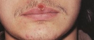
Gumma is a typical manifestation of tertiary syphilis
In 40 - 60% of cases, deep nodular syphilide - gumma - is formed in patients with tertiary syphilis. Syphilide can be single, sometimes it consists of 1 - 3 gummas, rarely more - up to six. Infiltrates appear in the tertiary period of syphilis as a result of local activation of Treponema pallidum. There are a small number of pathogens in the infiltrate. They are located inside the syphiloma and when it disintegrates, they quickly die.
Gumma is formed in the subcutaneous tissue and deep layers of the dermis.
Localization
Most often gummas appear:
- on the mucous membranes of the mouth, nose, larynx and pharynx,
- on the skin of the face, legs, forearms,
- Fibrous gummas (periarticular nodules) may appear around the elbow and knee joints,
- gummous syphilides are found in the bone tissue of the skull.
Gummas in internal organs, including the brain and spinal cord, are rare.
Histology
The cause of the appearance of gummas is the proliferation and transformation of cells capable of phagocytosis. In the affected areas, a pronounced change in the blood vessels is observed - perivascular inflammatory couplings are formed. Proliferation of the endothelium can lead to complete occlusion of the vessel. The edges of the gumma consist of large fibroblasts. In the center of the gumma there is an extensive focus of decay or dense and dry coagulative necrosis.
Development
Initially, a dense mobile nodule appears in the subcutaneous fatty tissue. Gradually, the gummous infiltrate increases in size and adheres to the skin, which becomes thinner and tenser, becoming red-violet in color. The size of gumma becomes the size of a walnut or more.
Decay
Under the thinned skin, a fluctuation begins to appear in the center. When opened, a viscous transparent liquid with an unpleasant odor is released from the granuloma. The resulting ulcer is deep (about 1 cm in diameter). At its bottom there is a “gummy rod” of yellow-green color. After rejection of the necrotic masses, a round, painless ulcer with steep edges, clear boundaries and a dense bottom with grayish granulations is exposed.
Healing
The ulcer heals slowly - weeks and months. In its place remains a pink scar, losing color over time, with a pigment border along the edges, retracted, disfiguring, star-shaped.
Some gummas do not open, but heal “dry” with the formation of an atrophic scar. Very rarely, gummas degenerate fibrously or become petrified and remain unchanged for many years.
When several gummas merge, a gummous infiltration is formed. When gumma grows, it affects nearby tissues, including bone structures, and destroys them. The affected areas are rejected, and scar changes lead to disfigurement and deformities. Such gummas are called mutilating.
Differential diagnosis
Gumma should be distinguished from scrofuloderma, erythema induratum of Bazin, nodular vasculitis, chancroid, atheroma, lipoma, cancerous ulcer, sporotrichosis, chromomycosis, deep blastomycosis, cutaneous leishmaniasis.
Rice. 4. Tertiary syphilis. Gumma on the front of the leg.
Rice. 5. Gumma of the leg and gummatous infiltration of the hand in late syphilis.
Rice. 6. Tertiary period of syphilis. Gummy infiltration of the skin of the back (photo on the left) and gumma of the face (photo on the right).
Rice. 7. The process of scar formation.
Rice. 8. Gummous lesion of the skull bones in late syphilis.
Syphilis - incubation period
The incubation period is a period of illness that lasts from the moment Treponema pallidum enters the body until the appearance of the first signs of syphilis (see photo), including chancre in combination with regional lymphadenitis. During this period, there is a gradual increase in the number of pathogen cells at the site of their introduction into the patient’s body. Treponema reproduces by division on average once every 30-32 hours.
This period of the disease is characterized by the absence of registered clinical and serological changes in the patient’s body; it lasts on average 3-4 weeks, it can be shortened to 8-15 days or lengthened to 108-190 days. A shortening of the incubation period occurs, as a rule, when the body is simultaneously infected from 2 sources; it is lengthened when taking antibiotics after the moment of infection, for example, for a sore throat, although it should be noted that an increase in the incubation period is not always due to the use of antibiotics.
How to treat syphilis?
The treatment of syphilis is approached in a comprehensive manner, taking into account many individual factors (age, gender of the patient, stage of development of the disease, the presence of concomitant diseases, general condition of the body, etc.). In addition, all sexual partners of the suspected patient should also be examined for the presence of syphilis and, if necessary, undergo a course of therapy.
If a patient has primary syphilis, then everyone who has had sexual intercourse with him over the past three months must undergo examination and tests. In the case of secondary syphilis - everyone who had contact with the patient over the past year. The timeliness of the therapy itself, as well as the correct selection of modern medications, is important for achieving success in the treatment of this disease.
The most effective method of treating syphilis is the introduction of water-soluble penicillins into the body. This therapy is carried out in a hospital setting for 24 days with injections every 3 hours. The causative agent of syphilis is quite sensitive to penicillin antibiotics, but there is a possibility of an allergic reaction to these drugs or the ineffectiveness of such therapy. In this case, penicillin is replaced with drugs of the tetracycline, macrolide, and fluoroquinolone groups. In addition to antibiotics, natural immune stimulants, vitamins, and immunostimulants are also indicated for syphilis.
How is it transmitted?
Syphilis is caused by Treponema pallidum, which lives in the external environment for only 3 minutes. Therefore, the main route of transmission of the disease is sexual. Infection of the fetus is possible in utero (vertical route) or intrapartum, when the child passes through the mother’s birth canal.
The household route of transmission is uncommon; infection is possible from persons with the tertiary stage of syphilis, when treponema pallidum gets on dishes, linen, towels, etc. from decaying gums. Hematogenous transmission of syphilis through blood transfusion cannot be ruled out.
Cases of infection of medical workers through contact with the blood of a patient are not that rare. It is possible to become infected through “bloody” objects: a shared toothbrush, razor, manicure set, etc.
Tuberous syphilide
Tubercular primary syphilide is a dense, spherical tubercle (rash) that forms on the skin and mucous membrane simultaneously with gummas. Syphilides of a tubercular nature are located asymmetrically, they are distinguished by a copper-red color with an admixture of cyanosis. The size of the tubercle does not exceed 1 centimeter in diameter, its consistency is dense, and its boundaries are clear.
At least 10 similar tubercles are formed on the human body. It may take several months from the moment they develop until they heal. The process of formation and healing is uneven; the presence of bumps on the skin at different stages of development is a standard phenomenon. Individual tubercles are located in groups, but fusion between them in most cases is not observed.
Healing of the tubercles occurs as dry necrosis, or with the formation of ulcers. The result of the disintegration of syphilide is the formation of atrophic scars. The following types of tubercular syphilide are distinguished:
- Grouped tubercular primary syphilide. The resulting tubercles are homogeneous, never merge, painless, characterized by polymorphism, have a smooth, shiny surface and a red-brown tint. During healing, an atrophic scar or ulcer is formed, which results in the appearance of an unpleasant-looking scar with a pigment spot around it. The shape of the scars is round;
- Creeping primary syphilide. A characteristic feature of this form of the disease is the fusion of tubercles, with their subsequent spread to healthy areas of the skin. At the same time, the nature of the rash at the edges may differ from the quality of the tubercles in the center. It is often possible to identify clear zones of growth, decay and scarring. The healed tissue has a bluish-red tint. Refusal to treat the disease leads to an increase in the affected area;
- Dwarf primary syphilide. A rare type of tubercular syphilide, it mainly appears 10 years after infection. The rash is small, does not exceed 2-3 millimeters in diameter. The color of the pimples depends on the degree of damage; there is a rash of light yellow and dark red, almost brown tint. Pimples do not open, heal dry with subsequent formation of atrophic scars;
- Syphilide platform. This type of syphilis is characterized by the fusion of tubercles and the resulting formation of a plaque-like infiltrate with a diameter of up to 20 centimeters. The color of the rash is brown-red. After the decay is complete, unpleasant-looking scars form on the skin.
- Vegetating syphilide. A group of tubercles forms on the skin, and as a result of their opening, ulcers with lush granulations at the bottom appear.
Regardless of the type of tubercular syphilide, its localization area remains constant. Most often, the rash appears on the face, back, elbows and knees. The patient does not experience any unpleasant sensations.
Diagnosis and treatment
Diagnosis of tertiary syphilis is based on obtaining clinical and laboratory test results. To identify the stage of the disease, a RIF (immunofluorescent reaction) and a RIBT blood test are performed. In order to identify the extent of damage to individual internal organs, patients are referred for cardiac ultrasound, ECG examination, aortography, and bone x-ray.
Treatment of tertiary syphilis begins with taking erythromycin or tetracycline. Upon completion of a two-week course of antibiotics, the patient is prescribed penicillin therapy, combined with intravenous administration of Bismuth.
If there are contraindications to this drug, additional penicillin-containing medications are prescribed. The duration and treatment regimen depend on the form and degree of neglect of the disease. Simultaneously with treatment, blood counts are regularly measured, ECG and ultrasound data are studied,
The tertiary period of syphilis is extremely dangerous, characterized by multiple complications, but fortunately, it is rare; modern diagnostic tools make it possible to identify the first signs of the disease at an early stage.
Forecast
It all depends on the stage of development of the disease and the treatment method. If therapy was started in the early stages of the disease (primary, secondary and early latent syphilis) and is carried out using treponemocidal antibiotics, then in almost all cases without exception, a complete clinical cure occurs, and relapses of early syphilis and the occurrence of late forms of syphilis are prevented.
Treatment of syphilis in pregnant women in the first half of pregnancy in most cases guarantees the birth of a healthy baby. In the case of congenital syphilis, the prognosis is favorable if treatment of the disease was started in a timely manner. Treatment of later forms of the disease is less successful, since it only slows down the progression of the disease, but in all cases it can restore the impaired function of the affected organs and lead to negative serological reactions.
Signs and types
The disease is characterized by long periods of latency. Tertiary syphilides (gummas, tubercles, roseola) develop over many years. The patient does not experience discomfort. Clinical signs are as follows:
- Rashes appear on the skin - a specific type of tubercles (tubercular syphilide);
- A gummous ulcer develops (gummy syphilide);
- The cardiovascular system is affected (myocardial infarction, aortitis);
- The structure of bone tissue changes (osteoporosis, osteomyelitis);
- There are problems with the liver (chronic hepatitis);
- Gastritis and stomach ulcers form;
- There are difficulties in the functioning of the kidneys, intestines, lungs, and nervous system (neurosyphilis).
Ulcers (gummas) and tubercles disfigure the patient’s appearance. Such manifestations of tertiary syphilis are most unpleasant for women.
Most often they appear on the face, hands, and armpits. If the disease is not treated, it becomes irreversible and leads to the death of the patient. Both secondary and tertiary syphilis are successfully treated, the clinical picture of the disease is difficult, but modern drugs cope well with the disease at any stage.