General definition
A nasal bulla is spoken of when a person has a pneumatized middle or (less commonly) superior turbinate.
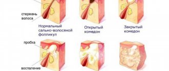
In this case, a bubble is formed at its anterior end, which belongs to the system of air cells of the ethmoid bone. The walls of such a nasal bulla have a bone frame lined with mucous membrane. A secretion produced by glandular cells often accumulates inside the vesicle, which can fester. Other pathological changes in the mucous membrane of the bullous nucleus cannot be excluded. A nasal bulla may be a congenital anatomical feature; in this case, there may be other anomalies in the structure of the facial skeleton. Sometimes the formation and growth of a bubble on the nasal concha is caused by chronic ethmoiditis - a long-term inflammation of the mucous membrane of the ethmoid bone. This process leads to an increase in the volume of its cells and can be complicated by bullous transformation of the upper or middle concha.
Why do polyps appear in the nasopharynx?
The content of the article
Nasal polyps appear to be a reaction of the nasal mucosa to prolonged inflammation and irritation, which in turn may be the result of an allergy, infection, or exposure to any substance. Thus, all conditions that cause long-term inflammation and irritation of the nose and esophagus increase the likelihood of polyps.
No one knows exactly why some people get polyps and some don't. There is evidence that patients with polyps have different immune responses and different mucosal chemical markers, although this is still a matter of research.
Nasal polyps are most often found in people who have been diagnosed with:
- Asthma;
- Hay fever;
- Chronic sinus infection;
- Cystic fibrosis;
- Vitamin D deficiency;
- Family history of the disease;
- Hypersensitivity to aspirin;
- Churg-Strauss syndrome, like eosinophilic granulomatosis with polyangiitis, is a rare disease that causes inflammation of the blood vessels.
Why are bullae dangerous?
Small nasal bullae can become an accidental diagnostic finding when examining a patient for any ENT disease, if they have not previously led to functional disorders and have not become inflamed.
Large formations can be pressed into the lateral wall of the inner nose and push the nasal septum to the side. This leads to disruption of the normal circulation of inhaled air, difficulty in the outflow and aeration of the paranasal sinuses and thereby contributes to the development of inflammation in them. Therefore, removal of a large nasal bulla is a prevention of chronic rhinosinusitis.
Inflammation of the mucous membrane and suppuration of the contents of the bulla is a risk factor for the development of ethmoiditis. Damage to the ethmoid labyrinth is fraught with involvement of periorbital tissue and meninges in the pathological process, generalization of infection and thrombophlebitis of the head veins.
Causes
The cause of the appearance of benign formations may be genetics, developmental anomalies due to the effects of pathological factors on the fetus, and inflammatory diseases of the paranasal sinuses. In many cases, the reasons for their appearance are completely unknown.
For malignant tumors, risk factors are smoking, papillomavirus, prolonged inhalation of chemicals used in furniture, shoe, and leather production, inhalation of flour dust, wood dust. The appearance of malignant formations can be caused by long-term untreated teeth, injuries from incorrectly selected dentures, inhalation of smoke, steam, and welding work. Inhalation of asbestos particles is especially dangerous.
Symptoms
Both benign and malignant growths in the nose and paranasal area are asymptomatic at first. Usually they do not cause concern at the initial stage and are detected by chance on an x-ray. A tumor of significant size begins to manifest itself with difficulties in breathing and loss of smell. The face may become distorted, sometimes the sensitivity of the skin of the cheeks changes, the sensitivity of the teeth disappears, and the teeth of the upper jaw become loose. Possible displacement of the eyes, swelling of the cheeks that do not cause pain, and visual disturbances.
Detection at an early stage allows you to get rid of the disease with a 100% probability. It is possible to detect a tumor at the beginning of its development, but to do this you need to consult a doctor if you have any unclear symptoms, such as frequent headaches or nasal congestion that is not relieved by vasoconstrictor drops.
Early symptoms:
- difficulties with nasal breathing,
- pus discharge,
- soreness of the nasal mucosa,
- frequent nosebleeds,
- Otitis media
Diagnostic methods
To detect benign and malignant formations, magnetic resonance scanning, computed tomography, and ultrasound are used. First, a visual inspection of the paranasal area (rhinoscopy) is carried out and a small piece of tissue is taken for examination. Additionally, X-rays of the head and chest may be required.
When is surgery to remove a bulla in the nose performed?
Nasal bullae are not able to spontaneously decrease in size. The only way to eliminate the existing breathing disorder is to remove the bubble along with part of the changed shell. Indications for bulla resection are:
- persistent breathing difficulties caused by significant narrowing and deformation of the passages of the nasal cavity;
- chronic sinusitis that is not amenable to conservative therapy, if the dysfunction of the anastomosis of the paranasal sinuses is associated with bullous transformation of the concha;
- purulent inflammation of the nasal bulla;
- all forms of chronic rhinitis caused by constant irritation of the mucous membrane of the nasal cavity by bullous walls.
Removal of a bulla in the nose is carried out only if indicated. In all other cases, the doctor chooses a wait-and-see approach, assessing the dynamics of the size and condition of the concha bullosa.
What is nasal cancer?
In Latin, a tumor is an edema, but not all tumors are cancer. Benign tumors grow slowly and do not invade surrounding structures. These are the most common formations in the sinus cavity. Malignant tumors are most often called cancer. These tumors invade local tissue and can also spread to distant parts of the body. Sinonal tumors are rare and account for only 10% of all head and neck tumors, with approximately 10 people per 1,000,000 developing this disease. These growths can arise from any structure in the nose (lining, blood vessels, nerves, and even bone or cartilage).
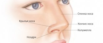
The nasal cavity consists of the nostrils and a hollow passage just behind the nose. The nasal cavity filters, warms and humidifies the air we breathe and provides a person's sense of smell. The paranasal sinuses are hollow chambers around the nose. These sinuses give shape to the face and eyes and help give the voice a unique sound.
Cells in the nasal cavity or sinuses sometimes change and no longer grow or behave normally. These changes can lead to malignant tumors or benign tumors such as nasal polyps or inverted papilloma.
Specialists who provide oncology treatment abroad note that in some cases, changes in the nasal cavity or in the cells of the nasal cavity can cause cancer. The nasal cavity and paranasal sinuses are made up of different types of cells that can become cancerous. Each type of cancer behaves or grows differently. Most often, nasal or paranasal cancers begin in thin, flat cells called squamous cells. These cells line the inside of the nasal cavity and paranasal sinuses. This type of cancer is called squamous cell carcinoma of the nasal cavity or paranasal sinuses. Sometimes cancer can start in the gland cells of the nose or sinuses. An example of this type of cancer is called adenocarcinoma of the nasal cavity or paranasal sinuses. Rare types of nasal cavity and paranasal sinus cancer may also develop. These include esthesioneroblastoma and sinus undifferentiated carcinoma.
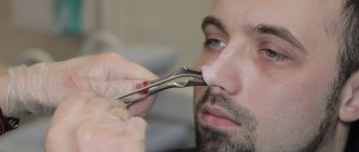
How is a nasal bulla removed?
In the classic version of the bulla resection operation, mechanical destruction of the walls of the vesicle and excision of the altered nasal concha using surgical instruments are performed. This uses a transnasal approach and requires general anesthesia.
After making an arcuate or contour incision along the edge of the modified concha and peeling off the mucous membrane along with the periosteum, the bulla is resection. The wound is covered with a flap of the mucous membrane, carefully removing excess tissue. Be sure to carry out a tight tamponade. This is necessary not only to prevent postoperative bleeding, but also to improve the survival of the mucous membrane. Relatively small nasal bullae are removed completely, while very large formations are partially resected. During the operation, they try to avoid damage to the mucous membrane of the upper part of the nasal cavity, which is necessary to preserve olfactory function.
This classic submucosal resection of the bullous turbinate is quite traumatic and the recovery period after it can take several weeks.
Types of nose cancer
First of all, in general, neoplasms in the nose can be divided into benign and malignant. Benign tumors:
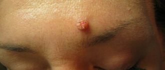
- Nasal polyps.
- Inverted papilloma: A unilateral, warty, slow-growing tumor that may resemble a nasal polyp with nasal congestion.
- Hemangioma is a collection of blood vessels that usually leads to nosebleeds and nasal congestion.
- Osteoma: bony, smooth tumor.
- Angiofibroma is a collection of fibrous tissue and blood vessels that appears on one side with nasal obstruction and occasional nosebleeds. It usually occurs in teenage boys.
Treatment options for nasal cancer will depend on the following factors:
- Where the tumor is located in the sinuses or nasal cavity and whether it spreads to other organs and tissues.
- Tumor size.
- Type of cancer.
- The patient's age, health status and medical history.
- Is it a primary tumor or a relapse of oncology?
After treatment, it is very important to be constantly and carefully monitored by qualified doctors, for example, in oncology centers in Israel, there are a lot of adequate specialists, because there is an increased risk of developing a secondary tumor in the head or neck.
There are two types of nasal cancer:
- Squamous cell carcinoma . Squamous cell carcinoma of the sinus and nasal cavity is usually treated with surgery. Surgery may be followed by radiation therapy and, in some cases, chemotherapy.
- Olfactory neuroblastoma . Treatment for olfactory neuroblastoma involves a combination of surgery and radiation therapy. Depending on certain characteristics, chemotherapy may also be indicated.
- Sinasal undifferentiated carcinoma . These are very aggressive tumors that require rapid intervention. Treatment includes surgical resection followed by chemotherapy and radiation therapy.
- Sinonal neuroendocrine carcinoma . It is a very aggressive type of oncology, and accordingly, aggressive and intensive treatment of nasal cancer abroad is also applied to them. Treatment involves aggressive, multidisciplinary therapy, including surgical resection followed by chemotherapy and radiation therapy.
- Melanoma of the mucous membrane . Treatment for mucosal melanoma is usually multidisciplinary and involves surgery followed by radiation and chemotherapy.
- Hemangiopericytoma . Previously, treatment of hemangiopericotomes was invasive, performed using large open surgeries with facial incisions. Nowadays, such tumors have already been treated endoscopically without facial incisions, which helps to significantly reduce the cost of cancer treatment abroad. Depending on the amount of blood supply, these tumors may also be embolized before surgery (a procedure performed by an interventional radiologist to reduce the amount of bleeding during surgery). If they extend into the intracranial cavity, a neurosurgeon is involved in the resection to ensure complete removal of the tumor. Surgery may be accompanied by radiation therapy to prevent recurrence, usually localized to the postoperative site, and especially in cases where the tumor has not been completely removed.
- Adenocarcinoma. Surgical resection is the usual treatment for adenocarcinoma, and advanced disease is treated with surgery combined with radiation therapy.
- Adenoid cystic carcinoma . Treatment is usually followed by radiation therapy. Chemotherapy is not usually used for adenoid cystic carcinoma.
- Chordomas . Treatment for chordoma depends on many factors, including your overall health and the size and location of the tumor. Whenever possible, surgery is used to remove the tumor. This can usually be performed endoscopically through the nasal cavity, avoiding any incision or craniotomy. If it is too difficult to completely remove the chordoma, radiation therapy and chemotherapy may be considered after surgery to destroy any remaining tumor cells. To date, chemotherapy has not been shown to be very effective in treating chordomas, but if surgery is not an option, it can be used in combination with radiation therapy.
- Tumors of the base of the skull . Many skull base tumors are extremely complex and require highly specialized treatment from multiple experts. Otolaryngologists work to create individualized treatment plans with the help of neurosurgeons, oncologists, endocrinologists, plastic and reconstructive surgeons, and many other specialists.
It is important to note that the effectiveness of cancer treatment in Israel, Turkey, Germany or any other country directly depends on the accurate determination of the type of tumor.
Removal of a bulla at Dr. Korenchenko’s Clinic
See also Treatment of ENT diseases Removal of a nasal turbinate bulla
In Dr. Korenchenko’s clinic, preference is given to a modern, minimally invasive method of bulla removal. In this case, the manipulation to remove a bulla in the nose is carried out on an outpatient basis, under local anesthesia and under the control of an endoscope. The doctor has the ability to accurately control the depth and size of the incisions made using a surgical laser.
A properly performed operation does not lead to the formation of synechiae and allows you to restore nasal breathing as much as possible, and the use of high-tech equipment can significantly shorten the recovery period and improve the patient’s quality of life.
Experienced, certified and highly qualified specialists possess all the necessary skills and carry out manipulations in the most gentle way possible. Specialists at Dr. Korenchenko’s Clinic accept appointments by appointment, which can be done using the feedback form or by calling the phone numbers listed on the website.
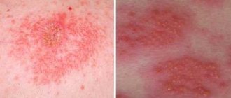
Symptoms of polyps in the nasopharynx
The polyp clinically manifests itself when the nasal passage is limited and the drainage of the nasal passages is impaired.
A polyp sometimes develops as a result of a sinus infection, and sometimes, on the contrary, a large polyp that causes airway obstruction itself contributes to the development of a sinus infection.
Clinically identified nasal polyps are characterized by wheezing, nasal congestion, loss of taste and smell, headache, toothache, pressure on the forehead and face, sneezing, and frequent nosebleeds. The patient constantly feels like he has a cold. Contact your doctor if the above symptoms persist for more than 7-10 days.
Nasal congestion
Call 911 immediately if you have severe respiratory distress, blurred or blurred vision, limited eye movement, severe swelling around the eyes, severe headaches, sudden worsening of symptoms: fever, neck stiffness.
Stages of development of neoplasms
The severity of signs and symptoms directly depends on the stage of the disease. When the tumor is small, a sign of its appearance is a feeling of congestion. This symptom looks like the beginning of a cold, so often at this stage a person does not diagnose and treat growths. Treatment boils down to the use of vasoconstrictor drops, which ultimately do not help. During this period, the body is more vulnerable to all kinds of infections, and otitis media, tonsillitis and other inflammatory diseases of the ENT organs can occur.
At the next stage, symptoms such as loss of smell and the appearance of a nasal voice appear. If the tumor blocks the opening of the auditory tube, signs of hearing loss appear.
In the third stage, the symptoms of the disease appear most intensely. Nasal discharge, headaches, constant congestion, breathing through the mouth - all these are signs that the polyp has reached an impressive size.
The symptoms of the disease are actually not as harmless as they look. To get rid of them, you must definitely contact an ENT doctor to prescribe competent treatment.
Unfortunately, few patients come to an otolaryngologist at the first signs of illness, when conservative treatment is still possible. Most of them turn to an ENT doctor when only surgical treatment can really help.
Symptoms of the disease
Often small polyps do not cause discomfort to patients. But, large or multiple, they can block the nasal passages, leading to breathing problems, decreased sense of smell, and recurring respiratory infections.
Most common symptoms:
- chronic runny nose;
- persistent deterioration in breathing through the nose;
- decreased or absent sense of smell;
- violation of taste sensitivity;
- facial pain, pain in other parts of the head;
- a feeling of heaviness, discomfort in the eyes, facial walls of the sinuses, head;
- snore.
Diagnostics
Due to similar symptoms, papilloma of the nasal vestibule must be distinguished from basal cell carcinoma (a type of skin cancer) and squamous cell carcinoma in the initial stage.
Friends! Timely and correct treatment will ensure you a speedy recovery!
Also, papillomas of the nasal cavity, if the disease constantly recurs, must be distinguished from the initial forms of cancer of the nasal cavity.
How can mycetoma be cured?
Is it possible to do without surgery? It is impossible to get rid of fungus in the maxillary sinus without surgical intervention, I will say this right away. No pills, no drops of “dancing with a tambourine” and everything else will give the desired therapeutic effect. First of all, it is necessary to surgically remove the fungal body, remove this mycelium, remove all the fungus from the maxillary sinus.
This can be done either through a nasal surgical approach or an intraoral approach.
How to remove fungus from the maxillary sinus
With intraoral access, a perforation is made in the vestibular wall of the maxillary sinus - access, and evacuation occurs through this hole, i.e. removal of the fungal body, mushroom mycelium. Then the maxillary sinus is well washed, treated with antifungal and antimicrobial drugs and sutured. Subsequently, the patient is prescribed antifungal therapy.
Surgical removal of mycetoma shows good results today, relapses are extremely rare.
Signs of a malignant process
The root cause of the development of nasal cancer has not been determined. There are risk factors that provoke the appearance of a tumor:
- Active (to a greater extent) and passive smoking.
- Work in hazardous industries: interaction with flour dust, wood, textiles, leather goods.
- Prolonged contact with radium, chrome and nickel dust, formaldehyde, solvents, and other chemical compounds.
- Chronic forms of various variants of sinusitis, rhinitis.
- Alcohol abuse.
- Long stay in a place with unfavorable ecology.
- Viral infection (for example, human papillomavirus).
When determining the stage of nasal cancer, experts pay attention to the following points:
- The spread of the process to other sinuses, soft tissues of the face, bone fragments.
- The presence or absence of metastases in regional lymph nodes - those closest to the diseased organ.
- The presence or absence of metastases distant from the lesion.
Possible complications
When we talk about cancer, complications almost always arise. But their character depends on the process itself, its distribution, and the anatomical structures that are nearby and involved in this process.
The most common complications of nasal cancer include the following:
- vision is impaired. When nasal cancer is in its final stage, the disease spreads to the eyes, brain, and skull. Due to this pathology, the functioning of organs, as well as vision, is disrupted;
- problems related to food arise. Since the tumor grows with nasal cancer, it becomes difficult for a person to open his mouth and chew food. These moments contribute to general exhaustion, weight loss;
- there is a risk that the disease will relapse;
- metastasis. If cancer of the paranasal sinuses is in the last stages, then metastases may occur, during which the lymph nodes will be affected.