Folliculitis is a common form of staph infection. A sign of pathology is pimples or bumps around the hair on the arms or chest.
Folliculitis is a superficial or deep infection by bacteria that enter from the outside through the sebaceous glands or the hair follicle itself. Particularly prone to this type of inflammation are areas of the body where the skin rubs against clothing, such as the neck or back.
Hair follicles - what are they?
Hair follicles are hollows
in the skin in the form of tubules in which the hair roots are located.
They also serve as an outlet for the sebaceous glands that produce secretions. Their function is to protect the epidermis. Irritation or damage
to the opening of the hair follicle causes germs to enter the hair follicle, leading to infection.
The infection causes inflammation of the hair follicle, which results in painful
and itchy patches in the growing hair area.
Abscessing undermining folliculitis and Hoffmann's perifolliculitis and follicular occlusion syndrome
Abscessing undermining folliculitis and Hoffman's perifolliculitis is an inflammatory recurrent disease of the scalp, characterized by the formation of abscesses, followed by cicatricial atrophy. The disease occurs predominantly in men and is characterized by a long course [1].
The cause of abscessing undermining folliculitis and Hoffmann's perifolliculitis is not fully known. Pathophysiological changes in the skin in this disease are associated with follicular occlusion, dilation and rupture of the follicular canal, which leads to an inflammatory reaction. Bacterial infection on the rash develops secondarily and is not the cause of this disease [2, 3].
The clinical picture of the disease is represented by multiple painful nodes with purulent discharge, which, merging, form interconnected abscesses and sinuses [1]. The process has typical localization sites with damage to the scalp (usually the parietal and occipital regions), and the back of the neck. The triggering mechanism in the formation of inflammatory elements is associated with occlusion of the sebaceous glands, which explains the characteristic location of localization [4–7]. According to topographic anatomy, the largest sebaceous glands are located on the skin of the scalp, with up to 5 lobules, deep-lying, with long convoluted ducts [8].
After resolution of the inflammatory process, hypertrophic or atrophic scars of the scalp remain, which leads to limited foci of cicatricial alopecia localized at the sites of inflammation [7].
Some authors indicate the possibility of developing complications, such as squamous cell carcinoma and marginal keratitis [2, 3, 7, 9].
Diagnosis of abscessing undermining folliculitis and Hoffman's perifolliculitis is based on the clinical picture, trichological examination, and, if necessary, histology.
The histological picture of the disease depends on the degree of inflammation. At an early stage, follicular hyperkeratosis is observed with blockage and dilatation of the follicle. As a result, apoptosis occurs and the follicular epithelium is destroyed. Subsequently, infiltrates of neutrophils, lymphocytes and histiocytes are formed. Later, an abscess forms, leading to the destruction of the hair follicle. Abscesses can penetrate the dermis and subcutaneous fat. As the process develops, granulation tissue is formed in the dermis and subcutaneous fat, containing lymphoplasmatic infiltrate and histiocytes, as well as multinucleated giant cells around keratin and hair shafts. The healing process ends with extensive fibrosis. In the later stages, scarring, fibrosis, subcutaneous sinuses, and chronic granulomas are noted, which are represented by lymphocytes, macrophages, giant cells of foreign bodies, and blood plasma cells [2, 4, 9, 10].
The trichoscopy method can detect characteristic signs: “3D” (“three-dimensional”) yellow dots in the area of dystrophic hair, yellow amorphous areas, white dots with halos, milky-red areas without follicular openings [4, 11].
The patients we observed with a diagnosis of “abscessing undermining folliculitis” and “Hoffmann’s perifolliculitis” are presented in the photographs (Fig. 1, 2). Clinical examples demonstrate abscesses and hypertrophic scars at the site of former rashes in the scalp in men 23 and 28 years old. In the presented patients, the process was localized only on the scalp, the skin in other areas was not affected, and there was no acne.
Abscessing undermining perifolliculitis may be one of the symptoms included in the follicular occlusion syndrome, which includes a combination of skin diseases similar in pathophysiological manifestations: conglobate acne, inverse acne, abscessing undermining perifolliculitis, pilonidal cyst. Currently, the literature describes double follicular occlusion syndrome, triad of follicular occlusion and tetralogy of follicular occlusion.
Double follicular occlusion syndrome combines abscessing undermining perifolliculitis and acne conglobata. A clinical example is presented in Fig. 3. A 25-year-old man has hypertrophic scars on the skin of the scalp at the site of former rashes in the scalp, post-acne scars on the skin in the face, grade 2-3 acne (Fig. 3).
The triad of follicular occlusion occurs in patients with conglobate acne, abscessing undermining perifolliculitis and inverse acne (or hidradenitis), and in the presence of a pilonidal cyst it is diagnosed as a tetrad of follicular occlusion [2, 9]. The exact pathogenesis of this group of diseases is unknown, but evidence suggests that they share the same pathological process initiated by follicular occlusion. Recent studies have shown that cytokeratin 17 (normally found in the sebaceous duct) is absent from the sebaceous duct epithelium of patients with acne inversis, contributing to epithelial fragility and leading to rupture of the follicular duct wall [12].
Follicular occlusion syndrome occurs more often in the third or fourth decade of life in males. However, this syndrome has been described in adolescence, as well as in women [2, 9, 13]. According to the literature, in rare cases, follicular occlusion syndrome can be combined with spondyloarthritis, osteomyelitis, Crohn's disease, sternoclavicular hyperostosis, Reid's syndrome (a rare combination of keratitis, ichthyosis and deafness) [2, 9, 14].
Inverse acne can be an independent disease, or it can be a symptom of follicular occlusion syndrome. According to the definition of European clinical guidelines, inverse acne is a chronic, inflammatory, recurrent dermatosis, with primary damage to the hair follicle, occurring after puberty and characterized by painful, deep rashes localized in parts of the body where apocrine glands are located (axillary, inguinal, anogenital areas) .
Inverse acne was first described in 1839 by Velpeau, who initially called the disease “hidradenitis suppurativa”, believing that inflammatory changes in this disease occur in the sweat glands [2, 9]. In 1939, Brunsting, based on a histological study, proved that the central link in the pathogenesis of hidradenitis, capitis folliculitis capitis and conglobate acne is hyperkeratosis of the follicular ducts of the pilosebaceous follicle [15]. In 1989, Plewigand and Steger introduced the term “inverse acne”, pointing out the incorrect term “hidradenitis”, since hyperkeratosis of the follicular duct, and not inflammation of the sweat glands, is pathogenetically significant. The difference between inverse acne and acne vulgaris, in addition to localization, is the absence of an increase in the secretion of the sebaceous glands [13, 16].
Currently, diagnostic criteria for inverse acne have been defined, in which basic and additional criteria are identified.
I. Basic:
- Recurrence of rashes two or more times within 6 months.
- The rashes are localized in the axillary, inguinal, anogenital and buttock areas, under the mammary glands (in women).
- The presence of nodes (inflammatory or non-inflammatory), sinuses (inflammatory or non-inflammatory), abscesses, scars (atrophic, hypertrophic or linear).
II. Additional:
- Having a family history.
2. Absence of pathogenic flora in a smear from the discharge (only the presence of normal microbiota) [7].
There are several stages of inverse acne:
- Stage I: abscesses, single or multiple, without sinuses or scars.
- Stage II: Recurrent abscesses with scarring, single or multiple, with areas of unaffected skin.
- Stage III: diffuse or almost diffuse lesions of the scalp, multiple interconnected sinuses and abscesses over the entire area (Hurley, 1989) [17].
Severe variants of follicular occlusion syndrome are associated with metabolic syndrome, obesity, eating large amounts of simple carbohydrates, smoking, and male gender [7].
We present clinical cases of the triad of follicular occlusion in Fig. 4, 5. Patients 43 and 52 years old complained of rashes on the skin of the scalp, face, back, chest, axillary area, and groin (Fig. 4, 5). The patients had similar medical histories and clinical presentations. They consider themselves sick since adolescence, when rashes first appeared on the skin of the face, chest and back. We were treated as an outpatient with a diagnosis of conglobate acne. At the age of about 30, painful rashes began to appear on the skin in the scalp and armpits. Patients were treated on an outpatient basis, doxycycline for 2–3 weeks, and excision of large nodes. The treatment brought temporary improvement, remission lasted less than six months.
Differential diagnosis of abscess folliculitis and Hoffmann's perifolliculitis
Differential diagnosis must be carried out, first of all, with diseases localized on the scalp. Acne-keloid (syn.: papillary dermatitis of the head, sclerosing folliculitis of the back of the head) is also observed in men in the back of the head and on the back of the neck. Initially, groups of small follicular pustules appear, which are located in the form of a cord. The skin around them is significantly thickened, the skin furrows are sharply expressed, which gives the impression of the existence of papillary tumors, and the hair grows in tufts. When the follicles resolve, keloid scars remain. The process proceeds slowly, without the formation of abscesses [1]. The absence of fluctuating nodules, large abscess formations, and fistula tracts is the main difference between acne keloids and abscessing undermining folliculitis and Hoffmann's perifolliculitis.
Often cases of cicatricial alopecia caused by previous folliculitis are usually diagnosed as Hoffmann's abscessive and undermining perifolliculitis. Decalvating (epilating) folliculitis was first described in 1888 by Quinqaud. Folliculitis decalvans also occurs in adults and is more common in men. Lesions in folliculitis decalvans are usually located on the scalp, but can sometimes be localized on other parts of the body covered with hair: beard, armpit area, pubis. The process begins with folliculitis surrounded by a zone of erythema. After inflammation resolves, small round or oval foci of scar atrophy form. Merging with each other, small lesions become larger, usually maintaining rounded outlines. Old areas where the inflammatory process has ended are represented by zones of scar atrophy. Thus, the clinical picture is represented by small foci of cicatricial alopecia with tufts of 5–10 hairs emerging from dilated follicular openings.
Mycotic lesions of the scalp are characterized by the absence of acute inflammatory phenomena and a positive fungal culture.
Other conditions that may mimic abscess folliculitis and Hoffmann's perifolliculitis include malignant proliferating pilar cysts, folliculotropic mycoses fungoides with gross cellular transformations, and erosive pustular dermatosis of the scalp [4, 18, 19].
Treatment of abscessing undermining folliculitis and Hoffmann's perifolliculitis and follicular occlusion syndrome
Therapy for follicular occlusion syndrome, as a rule, begins with treatment with antibacterial drugs prescribed orally (tetracycline, clindamycin, rifampicin) and externally (clindamycin). Some authors recommend ciprofloxacin 250–500 mg or clindamycin and rifampicin 300 mg [20].
However, antibacterial therapy has a short-term effect. According to some authors, it is more effective to combine antibiotic therapy with injections of corticosteroids into the lesions [21]. For the treatment of treatment-resistant cases, dapsone 50–150 mg daily is recommended [13].
However, according to the literature, surgical intervention is often necessary. Surgical treatment is carried out against the background of antibacterial therapy with complete resection of the affected skin of the scalp in step-by-step procedures, followed by skin reconstruction [22].
Currently, most publications devoted to the treatment of follicular occlusion claim the high effectiveness of isotretinoin in the treatment of this dermatosis. Isotretinoin is recommended to be prescribed in the same dosages as for acne vulgaris - 0.5–1 mg/kg for 3–12 months [20, 21].
Some authors recommend a combination of oral antibiotics and topical isotretinoin [20, 21].
In modern literature, there are isolated publications demonstrating the high effectiveness of genetically engineered biological drugs, such as adalimumab, infleximab, which are proposed as a third line of therapy [23, 24].
We observed a 52-year-old patient with a triad of follicular occlusion (acne conglobata, abscessing undermining perifolliculitis and inverse acne), who received treatment with isotretinoin at a dose of 30 mg per day for 6 months. As a result of therapy, long-term remission was observed for a year. The photographs show the results of therapy after 2 months of taking the drug (Fig. 6). The process has completely resolved and is represented by post-inflammatory spots and hypertrophic scars.
Thus, the presented clinical cases demonstrate various variants of follicular occlusion syndrome, as a result of which nodes of various types (inflammatory or non-inflammatory), abscesses and, as a consequence of the inflammatory process, the formation of various scars are formed on the skin. Currently, the most effective drug for the treatment of abscessing undermining folliculitis and Hoffmann's perifolliculitis and follicular occlusion syndrome is isotretinoin in the dosages recommended by the instructions.
Literature
- Berenbein B. A., Studnitsin A. A. Differential diagnosis of skin diseases. M.: Medicine, 1989. 672 p.
- Scheinfeld N. Diseases associated with hidranitis suppurativa: part 2 of aseriesonhidradenitis // Dermatol Online J. 2013, Jun 15; 19(6):18558.
- Badaoui A., Reygagne P., Cavelier-Balloy B., Pinquier L., Deschamps L., Crickx B. et al. Dissecting cellulitis of the scalp: a retrospective study of 51 patients and review of literature // Br J Dermatol. 2016, Feb. 174(2):421–423.
- Gaopande Vandana L., Maithili M. Kulkarni, Avinash R. Joshi, Ashish N. Dhande. Perifolliculitis Capitis Abscedens et Suffodiens in a 7 Years Male: A Case Report with Review of Literature Case Rep Surg. 2016: 21230 // Int J Trichology. 2015 Oct-Dec; 7 (4): 173–175.
- Mihic LL, Tomas D., Situm M., Krolo I., Sebetic K., Sjerobabski-Masnec I., Barišic F. Perifolliculitiscapitisabscedens et suffodiens in a caucasian: diagnostic and therapeutic challenge // Acta Dermatovenerol Croat. 2011; 19 (2): 98–102.
- Karpouzis A., Giatromanolaki A., Sivridis E., Kouskoukis C. Perifolliculitiscapitisabscedensetsuffodiens successfully controlled with topical isotretinoin // Eur J Dermatol. 2003, Mar-Apr; 13 (2): 192–195.
- Zouboulis CC, Desai N., Emtestam L., Hunger RE, Ioannides D., Juhász I. et al. European S1 guideline for the treatment of hidradenitis suppurativa/acne inverse // J Eur Acad Dermatol Venereol. 2015. Apr. 29 (4): 619–644.
- Sobolevskaya I. S. Some morphometric indicators of lipid-accumulating and lipid-synthesizing structures of human skin // Bulletin of VSMU. 2012, vol. 11, no. 2.
- Scheinfeld N. Review Dissecting cellulitis (Perifolliculitis Capitis Abscedense Suffodiens): a comprehensive review on new treatments and findings of the last decade with commentary comparing the therapies and causes of dissecting cellulitis to hidradenitis suppurativa // Dermatol Online J. 2014, May 16; 20(5):22692.
- Branisteanu DE, Molodoi A., Ciobanu D. The importance of histopathologic aspects in the diagnosis of dissecting cellulitis of the scalp // Rom J Morphol Embryol. 2009; 50 (4): 719–724.
- Rakowska A., Slowinska M., Kowalska-Oledzka E., Warszawik O., Czuwara J., Olszewska M., Rudnicka L. Trichoscopy of cicatricial alopecia // J Drugs Dermatol. 2012, Jun; 11(6):753–758.
- Kurokawa I., Nishijima S., Kusumoto K. et al. Immunohistochemical study of cytokeratins in hidradenitis suppurativa (acne inversa) // J Int Med Res. 2002; 30: 131–136.
- Vani Vasanth, Byalakere Shivanna Chandrashekar. Follicular occlusion tetrad // Indian Dermatol Online J. 2014, Oct-Dec; 5 (4): 491–493.
- Kohorst JJ, Kimball AB, Davis MD Systemic associations of hidradenitis suppurativa // J Am AcadDermatol. 2015, Nov; 73(5 Suppl 1):S27–35.
- Brunsting HA Hidradenitis suppurativa: abscess of the apocrine sweat glands // Arch fur Dermatol und Syph (Berlin). 1939. 39: 108–120.
- Plewig G., Steger M. Acne inversa (alias acne triad, acne tetrad, or hydradenitis suppurativa). Marks R., Plewig G., eds. Acne and Related Disorders. London: Martin Dunitz Ltd; 1989. 343–357.
- Hurley H. Axillary hyperhidrosis, apocrine bromhidrosis, hidradenitis suppurativa, and familial benign pemphigus: surgical approach. In: Roenigh RRH, ed. Dermatologic surgery. New York: Marcel Dekker, 1989: 729–739.
- Von Laffert M., Stadie V., Wohlrab J. et al. Hidradenitis suppurativa/acne inversa: bilocated epithelial hyperplasia with very different sequelae // Br J Dermatol. 2011; 164:367–371.
- Torok RD, Bellet JS Tinea capitis mimicking dissecting ce llulitis // Pediatr Dermatol. 2013, Nov-Dec; 30(6):753–754.
- Khaled A., Zeglaoui F., Zoghlami A., Fazaa B., Kamoun MR Dissecting cellulitis of the scalp: response to isotretinoin // J Eur Acad Dermatol Venereol. 2007 Nov; 21 (10): 1430–1431.
- Koudoukpo C., Abdennader S., Cavelier-Balloy B., Gasnier C., Yédomon H. Dissecting cellulitis of the scalp: a retrospective study of 7 cases confirming the efficacy of oral isotretinoin // Ann Dermatol Venereol. 2014 Aug-Sep; 141(8–9):500–506.
- Hintze JM, Howard BE, Donald CB, Hayden RE Surgical Management and Reconstruction of Hoffman's Disease (Dissecting Cellulitis of the Scalp) // Case Rep Surg. 2016; 2123037. DOI: 10.1155/2016/2123037. Epub 2016 Feb 7.
- Martin-García RF, Rullán JM Refractory dissecting Cellulitis of the Scalp Successfully controlled with Adalimumab // PR Health Sci J. 2015, Jun; 34 (2): 102–104.
- Sand FL, Thomsen SF Off-label use of TNF-alpha inhibitors in a dermatological university department: retrospective evaluation of 118 patients // Dermatol Ther. 2015, May-Jun; 28 (3): 158–165.
A. A. Kubanov1, Doctor of Medical Sciences, Professor, Corresponding Member of the Russian Academy of Sciences Yu. A. Gallyamova, Doctor of Medical Sciences, Professor T. A. Sysoeva, Candidate of Medical Sciences
FGOU DPO RMAPO, Moscow
1 Contact information
Causes of folliculitis
The cause of folliculitis is its superficial or deep infection
, causing inflammation.
Most often, bacteria are responsible for them, mainly Staphylococcus aureus
, which is physiologically found on the skin under normal conditions.
Folliculitis - risk factors
Factors that contribute to the appearance of folliculitis include: - wearing tight clothing; - frequent depilation;
- obesity;
- staying in one position for a long time;
- decreased immunity;
- diabetes;
- use of certain medications; - use of inappropriate cosmetics for skin care; - prolonged exposure of the body to the sun.
Men are at risk for chronic inflammation
hair follicles on the face in areas that are covered with hair. The disease has a long course. This is a special form of purulent inflammation of the hair follicle. It is known as sycamore.
Folliculitis in the groin in children
More often, children who often suffer from dermatitis are susceptible to folliculitis. Also children who have weak immunity.
The causes of folliculitis in children are:
- parents’ failure to comply with basic hygiene rules (when a child is bathed in dirty water)
- rarely examine the skin for microtraumas
- poor nutrition
- dressing a child in tight synthetic clothing
There are also ordinary reasons that do not depend on the parents. For example, climatic factors and profuse sweating of the child, resulting in skin irritation.
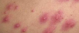
Symptoms of folliculitis
Symptoms of folliculitis - small yellow pustules
or papules, which are usually pierced by hair.
A red border around the lesion indicates inflammation. Over time, it may take the form of a purulent vesicle. Rashes occur singly or in groups
.
If the lesions extend into the deeper layers of the skin, soreness, pain and itching also develop. Facial folliculitis in men is usually very
severe, and the rash appears in the form of bumps.
Areas of the body where inflammation may occur
most often present as hairy skin that is removed by epilation or frequently rubbed against clothing.
This is why folliculitis occurs most often on the labia, buttocks, legs, face and armpits.
Inflammation of hair follicles on the head
Folliculitis of the scalp is usually
occurs in other dermatological diseases, mainly seborrheic dermatitis.
This also often applies to people who wear a hat for a long time during the day. Because of this, the scalp is constantly damp and warm, which promotes the growth of bacteria. Symptoms include pimples and redness around the hair shaft,
often filled with pus.
Proper care is important when treating an illness
behind the scalp.
It is recommended to wash your hair every day to prevent the accumulation of sebum, which is a breeding ground for bacteria. Shampoo for folliculitis on the head
should be gentle, natural, free of chemicals and have antibacterial or antifungal properties.
Features of folliculitis in the groin
The main causative agents of folliculitis in the groin are Staphylococcus aureus, herpes virus, Candida fungi, and herpes zoster.
Folliculitis in the groin mainly affects people who live in unsanitary conditions, in landfills or homeless people. But as described above, people who do not follow basic rules of personal hygiene are also susceptible to this.
The inflammation itself, as the name suggests, occurs in the pubic part of the abdomen. Reaching both the suprapubic part and below, it can even affect the external genitalia.
Initially, skin hyperemia occurs, after which a feeling of discomfort, pain, and swelling occurs. After which this swelling begins to gradually increase in size. Then it turns into a pustule (focus of inflammation), which contains pale yellowish and thick pus.
Depending on the microorganisms that actually “attacked” the follicle area, they are distinguished:
- staphylococcal
- gonorrheal
- herpetic
- syphilitic
- candidal folliculitis
If you open this pustule surgically, an ulcer appears at the site of the opening. Soon it dries out, becoming crusty. When the pustular component “resolves”, scars remain at the site of the ulcer.
With inadequate and untimely treatment, the stage of folliculitis can progress to the stage of a boil or carbuncle. In more severe cases, it even develops into an abscess. For minor manifestations of folliculitis or suspicious rashes, you should immediately consult a dermatologist. He will make an appropriate diagnosis and refer you for treatment.
How to cure folliculitis
Folliculitis should be treated
under the supervision of a dermatologist.
Therapy is based on the use of topical antibiotics in the form of ointments. In case of serious changes, general antibiotic therapy is recommended. Medicines for folliculitis are available with a doctor's prescription. You can also use urea creams for irritated skin, which will soften and moisturize the epidermis.
Herbs such as chamomile and sage are also effective against folliculitis.
What can you use at home for folliculitis?
Home treatment for folliculitis is primarily about hygiene and infection prevention measures.
To wash your body, use gentle lotions
that contain only natural ingredients.
Underwear should be airy and clothing should not be too tight
. To avoid inflammation of the hair follicles on the legs after depilation, it is necessary to disinfect shaving accessories or use disposable razors.
Local therapy for folliculitis in the groin
In addition to general measures to combat folliculitis, local treatment is used.
Various creams and ointments are applied to the inflammatory area of the skin. The most commonplace, but very common method in the fight against folliculitis is the application of tetracycline ointment. In addition to the ability to destroy folliculitis pathogens, tetracycline ointment has a good soothing effect on the skin.
In addition to tetracycline ointment, various disinfectant antimicrobial and antiseptic drugs are prescribed locally for folliculitis, for example:
- Zelenka or iodine
- Ichthyol ointment
- Erythromycin ointment
In addition to local measures, there are techniques for applying tea tree oil, calendula or chamomile decoction to inflammatory areas. Various compresses are also applied: from thistle, from various berries (viburnum, rose hips, green nut shells). Also a bread compress with regular homemade salt.
Folliculitis in the groin due to scabies
Folliculitis, which occurs in the form of scabies, is called ostiofolliculitis.
Manifests itself in the form of a purulent-inflammatory process of the skin. The main causative agent is Staphylococcus aureus.
After some time, a pustule appears at the site of inflammation, which has a cone shape with a diameter of 5 mm. It is painful when pressed.
On the top of the head, such a pustule has a yellow “cap” from which hair emerges. The rash with ostiofolliculitis occurs in the form of a scattered, ungrouped focus. Pustules are located in all parts of the body and are chaotic in nature, affecting unprotected areas of the body.
Diagnostic methods
First of all, the doctor collects the patient’s complaints and anamnestic data, specifying the time of onset of pathological symptoms, the presence of chronic diseases and similar problems in relatives. After this, a dermatoscopy (examination of the patient) is performed. Inflamed follicles are examined under multiple magnification, this allows you to determine the depth of the inflammatory process.
Also prescribed:
- Bacteriological culture of discharge from pustules. The study makes it possible to establish the type of pathogen, and in the future also determine its sensitivity to antibacterial drugs.
- PCR diagnostics. It is carried out to exclude the presence of diseases such as syphilis and gonorrhea.
- Test for the presence of fungal flora. To do this, scraping is carried out from the affected area and examined under a microscope.
- General blood analysis.
- General urine analysis.
It is imperative to carry out differential diagnosis; deep folliculitis must be differentiated:
- with a superficial form of the disease (ostiofolliculitis);
- with streptococcal impetigo;
- furunculosis;
- nodular cystic acne;
- drug toxicoderma.
If necessary, the patient is referred for examination to an immunologist and allergist.
The main differences between folliculitis and some other diseases
How to distinguish folliculitis from herpes?
What is the main distinguishing feature of folliculitis from herpes infection?
When the hair follicle becomes inflamed, it becomes infected and a pustule protrudes outward. Hair comes out of this very pustule.
There is no such feature with herpes. With a herpes infection, the virus infects the skin chaotically, without specifically affecting the hair follicle.
How to distinguish folliculitis from a boil?
At its core, a boil is a complication of folliculitis.
We can only say one thing: with a prolonged course and untimely treatment, folliculitis can progress to the stage of a boil. Then the process passes into an acute purulent necrotic stage, in which connective tissue is damaged.
Folliculitis in the groin with diabetes
Folliculitis has nonspecific features in various diseases. It can often be confused with a rash that occurs with diabetes. It must be remembered that the rash in diabetes mellitus is dry on palpation, its turgor is impaired and elasticity is reduced.
The rash in diabetes mellitus, as in folliculitis, is usually localized in the same areas. Both a diabetic rash and a rash characteristic of folliculitis can occur simultaneously. Those. a person is sick with both diseases, which is fraught with serious consequences.
Preventive actions
It is much easier to prevent any disease than to treat it. To avoid skin problems, it is recommended:
- Carry out personal hygiene manipulations in areas with active hair growth with caution.
- Regularly undergo medical examination if you have a history of diabetes.
- Men should shave, and women should depilate only with the use of special products with disinfectant properties.
- Treat damaged or abraded skin with antiseptic solutions.
- Conduct timely treatment of diseases that can provoke the development of Hoffmann folliculitis.
- Carefully ensure that chemical solutions and compounds do not come into contact with your skin.
- If even minor pathological symptoms appear that resemble disruptive folliculitis, seek help from a doctor.
- Maintain personal hygiene.
- Enrich the body with vitamins A and E.
- Avoid excessively greasy hair.
To summarize, I would like to focus on the fact that Hoffmann’s folliculitis is a rather dangerous disease that requires timely treatment. Remember this, be attentive to your body, and it will thank you with good health.
Treatment
First of all, it is necessary to direct all efforts to strengthen the body’s immune defense. You need to monitor your diet; your diet should include as many vitamins, vegetables and fruits as possible, as well as protein-rich foods. The consumption of sweets, baked goods, seasonings, spicy and salty foods should be kept to a minimum.
An important point is compliance with the drinking regime. During an exacerbation of the disease, it is recommended to drink at least 2 liters of water, because pathogenic microorganisms leave the body cavity with liquid.
Drug therapy
Of the medications, the following drugs are considered justified:
- Broad-spectrum antibiotics, preference is given to penicillins of semi-synthetic origin: Unazine, Augmentin, Clavulin, Amoxiclav, Claventin. Cephalesporin derivatives - Cephobid, Claforan, Kefzol, Cefuroxime - can also be used. If such antibiotic therapy is impossible for certain reasons, then sulfonamides are used: Septrin, Groseptol, Bactrim and Biseptol.
- Immunostimulants, such as Immunoglobulin, Bacteriophage, Antifagin.
- Vitamins and restoratives; vitamins E, A, C, and group B are considered the drugs of choice.
For topical use use:
- antibacterial ointments;
- Ichthyol;
- Fulevil;
- Levosin;
- Levomekol.
After opening abscesses and fistulas, clean them using antiseptic solutions, such as:
- Nitrofuran;
- Iodoform;
- Potassium permanganate;
- Hydrogen peroxide.
The course of treatment ranges from 7 to 12 days; to consolidate the therapeutic effect, it is recommended to repeat it after 10 days.
In advanced and complicated cases, surgical treatment may be necessary.
Hardware treatment methods
A high level of effectiveness is observed when combining drug treatment of acute folliculitis with physiotherapeutic methods. The use of ultraviolet rays is considered the most effective, because ultraviolet rays penetrate into the thickness of tissues, providing an anti-inflammatory and restorative effect. Good results are also observed when using:
- carbon dioxide laser;
- low-intensity UHF therapy;
- magnetic therapy;
- dry heat.
The main differences between folliculitis and eczema
Eczema is an inflammatory skin disease. Often of a chronic and allergic nature, in which the epidermis and dermis of the skin itself are affected.
If we talk about the differences between folliculitis, we can say that the hair follicles are not affected by eczema. In more severe cases, it is difficult to distinguish folliculitis from eczema.
When there are really a lot of papules and it is practically impossible to see whether hair comes out of them or not. Since with eczema there are also areas where it affects the scalp.