YOU MAY ALSO LIKE
Newborn care
Fever in babies and toddlers: recommendations and guidelines for action
The rosy cheeks of a baby usually only cause tenderness. True, sometimes pink cheeks are one of the symptoms of the disease. Fifth disease, or erythema infectiosum, is a viral disease, one of the manifestations of which is a rash on the face of a child. It can be mistaken for blush on the cheeks. As a rule, this disease is not dangerous, but it is worth learning more about it in case your baby becomes infected with it. In this article we will tell you about the fifth disease, its manifestations and symptoms, methods of prevention, and when you need to see a doctor.
What is fifth disease?
Fifth disease is an infectious disease caused by parvovirus B19. Most often it affects children aged 5 to 15 years. This disease is considered mild because children mostly feel well, even if they develop a rash. The medical name is erythema infectiosum, and the phrase “fifth disease” arose because erythema was fifth on the list of six common childhood diseases accompanied by a rash. Here is a list of these diseases:
- measles
- scarlet fever
- rubella
- Infectious mononucleosis
- erythema infectiosum
- roseola infantile (sudden exanthema)
Fortunately, having recovered from the fifth disease, your child will forever acquire immunity to it.
Causes
Erythema can be both physiological and non-physiological in nature.
In the first option, erythema may occur as a result of various psycho-emotional states or short-term abnormal phenomena. This kind of redness goes away very quickly on its own and is not the cause of certain disorders in the body. If erythema is of non-physiological origin, then it can be considered a separate disease, which is accompanied by prolonged redness of the skin and the presence of an inflammation process. Erythema can have many causes. These are viruses, infections, various diseases of the skin and connective tissues, as well as physiotherapeutic procedures that involve the use of sources of thermal or chemical action. Also, causes of erythema may include poor circulation, allergic reactions, regular rubbing of the skin, exposure to cold or chemicals.
Often reddened areas of the skin are a consequence of Crohn's disease, ulcerative colitis and pregnancy. Erythema can also be caused by medications such as penicillin, hormonal contraceptives (pills), non-steroidal anti-inflammatory drugs, anticonvulsants and sulfa drugs.
What are the symptoms and manifestations of fifth disease?
The main symptom of fifth disease is a rash. As a rule, it does not appear at the very beginning of the disease, but after a couple of days. First it appears on the face. After a few days, the rash spreads to the child's arms, torso, thighs, buttocks, and feet. In adults, the rash usually does not appear. The rash looks like pink spots that can merge with each other in the form of a kind of “lace”. Elements of the rash can be either true spots (that is, not felt by the fingers), or have edges slightly raised above the skin level. Often such satiety is accompanied by itching, especially severe in the area of the feet. The rash usually goes away within seven to ten days. It sometimes occurs again weeks or months later, often after exposure to sunlight, extreme heat, or extreme cold. At the very beginning of the disease, even before the rash appears, fifth disease can manifest itself with symptoms similar to those of the common cold:
- a sore throat
- headache
- stuffy or runny nose
- redness of the eyes
- weakness
- elevated temperature
- diarrhea
- indigestion
- itching
- enlarged lymph nodes
- joint pain or swelling (hands, wrists, knees, ankles), although this is more common in older children and adults
Erythema annular centrifugal Darier (ECDA) is a rarely diagnosed disease that occupies a special place in the structure of erythemas. Difficulties and diagnostic errors determine the relevance of the problem and indicate the need to improve differential diagnostic methods, and issues related to the etiopathogenesis of this disease require solutions. The basis for successful treatment of erythema annulare is the identification and elimination of the etiological or trigger factor.
Despite the fact that the role of infectious diseases in the pathogenesis of ECD is generally recognized, studies devoted to identifying the direct connection between the formation of this pathology and infectious agents are few [1–3]. The pathogenetic, immunological and clinical features of dermatosis have not been sufficiently studied; the data obtained are fragmentary and need to be systematized.
The term “erythema annular centrifugal” was first used by Daria in 1916. It characterizes clinically single or multiple annular erythematous rashes that spread, quickly increase in size, regress after 1-2 weeks, or exist for a long time in one place. The pathogenesis of the disease remains unclear [4]. For about 100 years, many authors have tried to analyze the possible causes of the disease. In most cases, trigger factors remained unidentified. In the literature, erythema annulare is described as a polyetiological allergic reaction. Erythema is often associated with infectious diseases, hormonal dysfunctions, autoimmune diseases, taking certain foods and medications (salicylates, antimalarials, cimetidine, amitriptyline), as well as cancer. Some authors consider ECCD as an allergic phenomenon within dermatomycosis along with such clinical forms as eczematous rashes, erythema multiforme exudative, psoriasiform dermatitis, erythema nodosum, urticaria [5, 6]. In 1986, K. Yoshikuni et al. [7] suggested an association of the superficial form of ECD with hereditary deficiency of lactate dehydrogenase. Detection of IgG deposits in the basement membrane zone of the epidermis suggests an autoimmune genesis of the disease. However, in most cases the diagnosis is “idiopathic erythema annular centrifugal”.
Increasingly, observations of the paraneoplastic form of ECD are found in the literature [8]. Isolated clinical cases of disseminated annular erythema in association with chronic lymphocytic leukemia and lymphoma have been described, in which chemotherapy contributed to the regression of skin rashes; with autoimmune hepatitis [9, 10]. Cases of ring-shaped erythema have been observed in relapsing polychondritis [11]. An association of erythema annulare with endocrine dysfunctions is often detected [12]. A number of works [2, 3] describe the occurrence of ECD due to viral infections caused by VZV. In 1974, foreign researchers [1] described a clinical case of ECD in a child due to EBV infection.
We present the results of our own observations and analysis of three clinical cases of ECD, in the etiopathogenesis of which the trigger factor, apparently, was the reactivation of chronic EBV infection.
Observation 1.
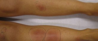
Patient L., 14 years old, presented with multiple rashes on the skin of the torso and limbs
(Fig. 1 a, b).
Figure 1. (a, b). Patient L., 14 years old. Erythema annular centrifugal Daria. The disease had a continuously relapsing course, the duration was 3 years. Relapses occurred, as a rule, against the background of or after suffering inflammatory diseases of the ENT organs, acute respiratory viral infection (ARVI). The duration of the elements' existence varied from 1 month to 1 year. There were no subjective sensations in the form of itching or burning. Previously, the patient was observed with diagnoses of granuloma annulare, eczema and repeatedly received courses of antihistamines and topical corticosteroids (without effect). An analysis of anamnestic data revealed a recurrent herpetic infection in the form of labial herpes with a relapse rate of 2-3 times a year and a relapse duration of 5-10 days. There is no allergic history. Infections suffered in childhood - rubella, chickenpox. Heredity is not burdened. Upon examination, the skin pathological process was generalized in the form of multiple infiltrated pink papules with a diameter of 0.2–4.0 cm and ring-shaped infiltrated lesions with a recess in the center (size 1–5 cm). Some rashes were arched in nature. The peripheral edge is slightly flattened, smooth and barely palpable. The rashes were localized on the torso, lower extremities in the area of the legs and thighs, and upper extremities in the area of the forearms. At the time of examination, a 0.2 cm focus of painful microvesicles with serous content was observed on the mucous membrane of the tongue in the area of the tip on the left. Palpation revealed a slight enlargement of the cervical lymph nodes. In a general clinical blood test, the number of monocytes (up to 9%) and segmented neutrophils (up to 62%) was increased. General clinical urine test, biochemical blood test - without pathological changes. As a result of the examination, no infections of bacterial etiology or parasitic infestations were identified. According to a virological study, a mixed herpesvirus infection was identified with the identification of EBV and human herpes simplex virus (HSV) type 1 in the secretions of the salivary glands using the DNA diagnostic method. When studying blood serum using enzyme-linked immunosorbent assay (ELISA), specific antibodies of the IgM class (2.5 OD) to the EBV capsid antigen (VCA), high titers of IgG antibodies to the capsid protein (25 OD), early EA antigen (2.2 OD) and nuclear antigen were detected EBNA (95.5 U/ml). An immunological study revealed a decrease in the relative and absolute content of activated NK cells with the CD16+ marker to 6.7% and 1.70·108 g/l, as well as a low level of activation molecules (CD25+), which are a receptor for interleukin-2 (IL-2). 2; 5.4%). Indicators of phagocytic activity of neutrophils are within normal limits. When studying the humoral component of immunity, a violation of the ratio of the main classes of immunoglobulins was noted, expressed in a decrease in the level of IgA to 1.2 g/l. When assessing interferon status, serum and spontaneous interferon (IFN) levels were within normal values, the level of induced IFN-α was 320 U/ml, induced IFN-γ was 80 U/ml, which is significantly lower than normal.
Histological examination of biopsy specimens from the lesion in the middle and deep layers of the dermis revealed a clearly defined perivascular infiltrate of mononuclear cells, predominantly lymphocytes (Fig. 2 a, b).
Figure 2. Microphotographs. Hematoxylin and eosin staining; a, b, d, d, f - vol. ×20; in about. ×10.
Based on the clinical and morphological picture, a diagnosis of ECD was made. Considering the identified signs of activation of chronic HSV-EBV infection, the patient was treated with the drug Valacyclovir at a dose of 500 mg 2 times a day for 30 days, for the purpose of immunocorrection - immunotherapy with the therapeutic multicomponent vaccine Immunovac-VP-4[] using the nasal-subcutaneous method according to the scheme in within 4 weeks. The rash began to regress on the 10th day of treatment and resolved completely after 25 days. Post-inflammatory hyperpigmentation was observed at the sites of the rash. Over the course of 6 months, no recurrence of erythema or signs of viral activity were observed.
Observation 2.
Patient S., 32 years old, complained of rashes on the skin of the forearms and abdomen for 6 months
(Fig. 3, a, b).
Figure 3. (a, b). Patient S., 32 years old. Erythema annular centrifugal Daria. The disease had a continuously relapsing course. There were no subjective sensations. Previously, the patient received therapy with topical corticosteroids (without effect). Allergological history and heredity are not burdened. Denies concomitant diseases. The general condition is satisfactory, the patient was worried about weakness, malaise, and sleep disturbances. The skin pathological process was widespread in the form of multiple erythematous ring-shaped rashes with clear boundaries in the form of an infiltrated ridge 1-7 cm in size. On the inner part of the peripheral border of individual lesions, thin peeling is noted. Regression of the rash was accompanied by hyperpigmentation of varying intensity. On palpation, a slight increase in the submandibular and cervical lymph nodes was noted. General clinical, bacteriological, virological, and immunological studies were carried out to identify concomitant pathologies of autoimmune and infectious origin. General clinical blood test without pathological changes. During a laboratory examination, HHV-6 and EBV DNA was identified in the secretions of the salivary glands. The ELISA method revealed IgM antibodies to the capsid antigen (1.8 OD), high titers of specific serum antibodies of the IgG class to the capsid antigen (32 OD), IgG to the early (1.8 OD) and nuclear antigen (83 U/ml) of the Epstein virus in the blood serum. —Barr and high titers of IgG antibodies to HHV-6 (96 U/ml).
When assessing the immune status, a decrease in the absolute and relative content of CD3+ T-lymphocyte subpopulations was revealed to 50% and 9.00·108 g/l and CD4+ helper T-lymphocytes to 30% and 4.50·108 g/l and T-lymphocytes with CD8+ marker up to 16% and 3.0·108 g/l, as well as a low level of activation molecules CD25+ (7.5%), which are a receptor for IL-2. The content of normal killer cells CD16+ and B-lymphocytes CD72+ was increased to 33% and 7.0·108 g/l, 48% and 6.0·108 g/l, respectively. Indicators of phagocytic activity of neutrophils are within normal limits. When assessing IFN status, an increase in the level of serum IFN to 4 U/ml was noted, while the level of spontaneous IFN was within normal limits, the level of induced IFN-α was 320 U/ml, induced IFN-γ was 64 U/ml, which was significantly below normal.
The histological picture of skin biopsies is represented by dense superficial perivascular lymphocytic infiltrates in the upper and middle parts of the dermis with mononuclear cells, spongiosis and focal parakeratosis in the peripheral zone (see Fig. 2, c, d)
. Based on clinical and histological signs, a diagnosis of “ECCD, superficial form” was made.
Based on a clinical and laboratory study, reactivation of chronic viral infection caused by EBV and HHV-6 was identified, associated with signs of immunological deficiency in the form of suppression of the cellular component of immunity. The course of treatment included vitamin therapy and immunocorrective therapy with the Immunovac-VP-4 vaccine. The rash completely resolved on the 14th day of treatment. There were no relapses of erythema within 6 months. There were also no signs of activation of persistent herpesvirus infection.
Observation 3.
Patient Ts., 32 years old, complained of slightly itchy rashes that appeared on the skin of the chest, neck and back
(Fig. 4, a, b).
Figure 4. (a, b). Patient Ts., 32 years old. Erythema annular centrifugal Daria. I first got sick at the age of 12. This relapse was preceded by a severe form of ARVI. The last relapse lasted 2 months against the background of a short-term increase in temperature to subfebrile levels. Analysis of anamnestic data revealed a previous toxoplasma infection diagnosed in 2004, for which in 2004-2005. 3 courses of therapy were carried out in the hospital. There is no allergic history. Heredity is not burdened. Denies concomitant diseases. No endocrine pathology was detected. General condition is satisfactory. The skin pathological process was widespread in the form of multiple erythematous ring-shaped rashes with clear boundaries in the form of an infiltrated ridge with a diameter of 3-8 cm, localized in the neck, chest, and upper back. Fine-plate peeling was noted on the inner part of the peripheral border of the lesions. On palpation, a slight enlargement of the cervical lymph nodes was observed.
A general clinical blood test revealed leukocytosis of 10.3·106 U/l, an increase in the erythrocyte sedimentation rate to 25 mm/h. General clinical urine test and biochemical blood test - without deviations from normal values. As a result of the examination, no infections of bacterial etiology or parasitic infestations were identified. The quantitative ELISA method detected IgG antibodies to Toxoplasma, which indicates a previous infection. A study of the endocrine status revealed no pathological changes. Virological research methods revealed a persistent herpesvirus mixed infection with the identification of EBV and HHV-6 in the secretions of the salivary glands using DNA diagnostics. An enzyme immunoassay of blood serum revealed IgM to the capsid antigen (1.7 OD), high titers of IgG antibodies to the capsid protein (69.3 OD), “early” IgG antibodies (0.319 OD) and IgG to the nuclear antigen (61.4 g/l ) Epstein-Barr virus, which may indicate the activity of chronic persistent EBV infection. When assessing the immune status, a decrease in the relative content of CD3+ T-lymphocyte subpopulations was revealed to 58%, CD4+ helper T-lymphocytes to 34% and 5.30·108 g/l and T-lymphocytes with the CD8+ marker to 27%, as well as a low level of activation CD25+ molecules (5.9%). The content of normal killer cells CD16+ and B-lymphocytes CD72+ was increased (33% and 7.0·108 g/l, 48% and 6.0·108 g/l, respectively). Indicators of phagocytic activity of neutrophils are within normal values. When assessing IFN status, serum and spontaneous IFN levels were within normal limits, the level of induced IFN-α was 320 U/ml, induced IFN-γ was 80 U/ml, which was significantly lower than normal.
A histological examination of a skin biopsy in the superficial dermis revealed dense perivascular lymphocytic infiltrates. Covering epithelium with signs of spongiosis, focal hyper- and parakeratosis (see Fig. 2, e, f)
.
Based on the clinical and histological picture, a diagnosis of “ECCD, superficial form” was made.
Based on a clinical laboratory study, reactivation of a chronic viral infection caused by EBV and HHV-6 was established.
The patient received a course of antiviral therapy with Valaciclovir
1000 mg/day for 30 days against the background of immunotherapy with IFN
Viferon
in the form of rectal suppositories at a dose of 1 million IU/day for 10 days, then 3 times a week at the same dose for 1 month in combination with an IFN inducer – Kagocel (7-day cycles for 1 month). The rash resolved completely on the 13th day of treatment.
In all the described clinical cases, the presence of signs of reactivation of a chronic viral infection of the Herpesviridae
during the period of relapse of the skin disease. All examined patients had insufficiency of the IFN system of 2-3rd degree in the production of IFN-α and 2-3rd degree in the production of IFN-γ by blood leukocytes, expressed in a decrease in the stimulated production of IFN-α and the production of IFN-γ stimulated by phytohemagglutinin, which may indicate long-term persistence of viruses. In one patient, a more pronounced decrease in these parameters was observed against the background of an increase in serum IFN levels, which likely reflects the degree of viral replication during activation of EBV-HHV-6 and infection. In 2 patients, there was a deficiency in the cellular component of immunity, depletion of the T-lymphocyte population, manifested in a decrease in total T-lymphocytes CD3+, CD4+, a decrease in the level of cytotoxic T-lymphocytes CD8+ against the background of an increase in the content of natural killer cells CD16+ and B-lymphocytes CD72+. All patients had a decrease in the content of lymphocytes with the CD25+ marker, which is a receptor for IL-2. The described changes in immune status indicators probably reflect the activity of the viral process, which acts as a trigger factor in the etiopathogenesis of ECD. After a course of antiviral and immunomodulatory therapy, when examining patients using DNA and culture diagnostics, as well as determining titers of specific antibodies, no signs of viral replication and, accordingly, activity of the infectious process were detected, skin manifestations regressed. When studying the dynamics of immune status indicators 1.5 months after treatment, there was a significant increase in lymphocyte subpopulations (CD3+, CD4+, CD8+, CD72+) and the production of IFN-α and IFN-γ.
In the presented clinical cases, ECCD is a peculiar reaction of skin hypersensitivity to infectious agents of viral etiology, which are the Epstein-Barr virus and HHV-6. Timely identification of the trigger factor and the prescription of combination therapy using immunomodulatory drugs contributed to the correction of immunological disorders, a decrease in the activity of the viral process, regression of skin rashes and long-term clinical remission.
Histological examination of skin biopsies was carried out in the Department of Pathomorphology, State Budgetary Healthcare Institution Consultative and Diagnostic Center No. 6 of the Northern Administrative District, Moscow, Head of the Department of Pathomorphology, Doctor of Medical Sciences, Prof. P.G. Malkov.
[]The vaccine was developed at the Research Institute of Veterinary Medicine named after. I.I. Mechnikov RAMS (Moscow), approved for use in healthcare practice (Order of the Ministry of Health of the Russian Federation No. 270 of November 17, 1993, registration certificate No. 93/270/20).
How long can fifth disease be contagious?
The fifth disease is transmitted to other people within a week from the onset of the disease (that is, the appearance of the first symptoms similar to the manifestations of ARVI) until the appearance of a rash in the child. Once the rash appears, the child is no longer contagious. Just in case, do not allow your baby to come into contact with other people, even when he has a rash, especially if he has a fever. This way the virus will not spread further. If your baby has erythema infectiosum, try to keep him away from pregnant women. If an expectant mother becomes infected with this virus during pregnancy, it can lead to serious problems in the development of the fetus.
Why does toxic erythema occur?
“The causes of toxic erythema in infants remain unknown ,” says Olesya Anatolyevna Kluban, neonatologist at the ISIDA clinic. – The most common hypotheses for the occurrence of toxic erythema are as follows :
- activation of the newborn’s immune system in response to stress (childbirth, hypoxia, cooling);
- inflammatory response to skin colonization by normal bacterial flora at birth;
- acute hypergic skin reaction caused by maternal lymphocytes transferred shortly before or during childbirth;
- allergic reaction of the body to external and internal irritants;
- reaction of adaptation mechanisms of newborn skin in relation to mechanical or thermal stimulation.”
How can a child become infected with fifth disease?
Erythema infectiosum, like many other viral diseases, is transmitted by airborne droplets, that is, through the ingestion of respiratory secretion products (saliva, sputum, mucus) by virus carriers when sneezing or coughing. Infected people are most contagious when the symptoms of the disease are similar to those of a typical cold. Fifth disease is also transmitted through blood, so pregnant women are advised to avoid sick people, as the pathogen can be transmitted through the blood to the fetus.
What are the means to prevent fifth disease?
There is no vaccine for fifth disease. However, there are several recommendations that you, your family members and your children can follow to reduce the likelihood of infection in the family:
- Wash your hands frequently with soap and water for at least 20 seconds.
- Cover your nose and mouth when sneezing or coughing.
- Avoid touching your face, especially your nose, eyes and mouth.
- Try not to contact potentially infected people.
- If you have fifth disease, stay home and avoid contact with other people to avoid infecting them.
Etiology
Herpes viruses contain double-stranded DNA and have a glyco-lipoprotein envelope. The sizes of viral particles are from 120 to 220 nm.
Today, 8 types of herpes viruses that have been identified in humans have been described:
- two types of herpes simplex virus (HSV-1, HSV-2),
- varicella zoster virus (VZV or HHV-3),
- Epstein-Barr virus (EBV or HHV-4),
- cytomegalovirus (CMV or HHV-5), HHV-6, HHV-7, HHV-8.
Based on the biological properties of viruses, 3 subfamilies of herpes viruses have been formed: (alpha herpes viruses, beta herpes viruses and gamma herpes viruses). A-herpesviruses include HSV-1, HSV-2, VZV.
Betaherpesviruses include CMV, HHV-6, HHV-7. They, as a rule, multiply slowly in cells, cause an increase in the affected cells (cytomegaly), are capable of persistence, mainly in the salivary glands and kidneys, and can cause congenital infections. Gammaherpesviruses include EBV and HHV-8.
When should you contact a pediatrician?
If your child's condition worsens (for example, a sudden increase in temperature) or if his symptoms do not go away, contact your doctor as soon as possible. In children with weakened immune systems, erythema infectiosum is more severe. If your child has a weakened immune system, it is recommended to consult a pediatrician at the first symptoms of the disease.
With fifth disease, the baby may feel weak. To help him feel better in a few days or weeks, surround your child with care and love. But there is good news - the fifth disease goes away quite quickly, so everything will soon return to normal.
How this article was written The information presented in this article is based on expert advice published by trusted (medical and government) sources such as the American Pediatric Association and the American College of Obstetricians and Gynecologists. A complete list of links to sources used to write this article can be found at the end of the article. The information on this page is not a substitute for professional medical advice. Always consult your doctor for diagnosis and treatment.
Treatment
Regardless of whether viral erythema is detected in children or infectious, it is still worth following the doctor’s recommendations. Note that therapy, depending on the type of disease, is prescribed individually. The characteristics of the child’s body are also taken into account.
More often, treatment of erythema in children includes:
- taking antihistamines;
- taking vitamin complexes;
- treatment of local lesions with ointments, creams, aerosols;
- taking antibiotics (in certain cases).
Erythema nodosum, infectious and viral erythema in children is treated comprehensively. The child must be under the supervision of the attending physician. You can make an appointment with a highly qualified specialist in the center of Moscow by contacting JSC "Medicine" (clinic of Academician Roitberg), which is located at 2nd Tverskoy-Yamskaya lane 10 (not far from the Mayakovskaya, Belorusskaya, Novoslobodskaya, Tverskaya, Chekhovskaya metro stations) .