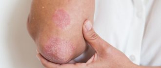
Treatment of psoriasis with hydrogen peroxide is a method of alternative medicine. Some patients note its effectiveness.
Despite the fact that peroxide has no contraindications, consult your doctor before taking the described course. Each organism is individual and sometimes it is unpredictable how someone will react to a treatment method.
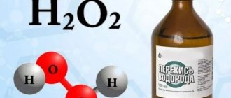
Hydrogen peroxide has a disinfecting effect: it kills fungi, viruses, bacteria. That's why we use this remedy if we cut a finger or break a knee. This drug is in everyone's medicine cabinet.
Treatment of psoriasis with hydrogen peroxide: recipe
Professor Neumyvakin
Professor I.P. Neumyvakin is studying hydrogen peroxide and its effect on the body. For more than 30 years he was associated with space medicine, then he opened his own medical and health center (which is closed at the time of writing).
The professor spoke about his experience in the book: “Hydrogen peroxide. Myths and reality".
Recipe for treating psoriasis with hydrogen peroxide according to Neumyvakin:
- Take peroxide daily orally , 3 times a day. You need to start with a drop of peroxide (3%), which is diluted in a tablespoon of water. Every day, increase the amount of water and peroxide by two. That is, on the second day - two drops of peroxide per two tablespoons of water, on the third - three, and so on. You can increase up to 10 parts of peroxide and water. After this, take a break for a week, then repeat the course.
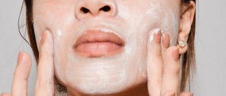
- topically to psoriasis spots twice a day.
Make compresses : mix 2 tablespoons of peroxide (3%) with 50 ml of water, soak a cotton pad and apply to the stain.
The author says that treatment takes 3-4 weeks. During treatment with peroxide, it is recommended to refrain from other methods in order to objectively evaluate the results.
Side effects
Before treating psoriasis, possible side effects such as severe burning, pain and general discomfort should be taken into account. In addition, fainting is possible, which is associated with oxygen oversaturation of tissues and muscles.
Before neutralizing the disease with peroxide, it is necessary to make sure that there is no allergic reaction to this drug, and if manifestations are minimal, it is recommended to reduce the dose of peroxide by 2 times, followed by a gradual increase in the dosage.
Treatment of psoriasis with hydrogen peroxide: reviews
Reviews about this treatment method are conflicting. Some patients claim that psoriasis on their scalp went away thanks to paint with peroxide. Others say that psoriasis remains despite everything. If this is your first time visiting the official 1xBet website, the functionality seems unclear and questions arise, then you don’t even have to call and contact technical support yourself. The bookmaker company, making the comfort and convenience of customers a priority, takes another step towards ideal service by offering the functionality of the so-called call back. You just need to leave your number on the 1x Bet website or mirror so that a support representative will call and explain in detail what needs to be done, how to bet on sports, work with coupons and other nuances. Technical support usually responds within a few minutes.
Review of the use of hydrogen peroxide by a Creole user
Feedback on the use of hydrogen peroxide from user Nadezhda39
Feedback on the use of hydrogen peroxide by Stator user
Review of the use of hydrogen peroxide by user Renina
What is seborrheic dermatitis?
The best environment for seborrhea is sebum, or rather the presence in its composition of certain fatty acids, where a number of microbes prefer to be active. Seborrhea can develop due to comedones, which appear when sebum is not completely and not promptly removed from the surface of the skin, resulting in clogged pores.
There are two types of seborrhea:
- oily (more often occurs in adolescents during puberty, as well as with gastrointestinal disorders or chronic infections and even caries): this type is characterized by acne and oily hair;
- dry skin is characterized by a decrease in sebum secretion, which causes complications such as comedones, acne and dandruff.
Sometimes mixed seborrheic dermatitis is diagnosed, which combines the signs of both forms of the disease.
Seborrheic dermatitis in infants and children of other ages
In infants, the disease most often manifests itself in the first weeks of life and is characterized by round, scaly spots of varying sizes. Treatment at this age is usually not required, since the dermatitis goes away on its own and leaves no traces.
Seborrheic dermatitis can appear in children aged 2–4 years (most often diagnosed at this age), but this does not mean that other age groups are not at risk, since the main factors that provoke the occurrence and spread of the disease are weak immunity , chronic or recent infections and genetic predisposition.
If in newborn children seborrheic dermatitis often goes away without treatment, then in an older child the disease cannot be left without control and a visit to a pediatric dermatologist. Treatment under the supervision of a doctor is necessary to get rid of the disease as quickly as possible and prevent the occurrence of exacerbations and undesirable consequences.
Conclusion
Hydrogen peroxide for treating psoriasis is an alternative method that can affect the body both positively and negatively.
It is safer to treat the disease with proven and effective methods.
- To exfoliate scales and crusts, use: non-hormonal ointments against psoriasis, salicylic ointment, grease-based ointments.
- To influence psoriasis at the cellular level : ultraviolet 311 nm and PUVA therapy. Also, read about other phototherapy treatments for psoriasis. They are not so common, but perhaps they will be effective for you.
- For general strengthening and healing of the body : diet for psoriasis. Eliminate fatty, floury, and too salty foods. Dr. Pegano also recommends eliminating nightshades (tomatoes, potatoes) and red meat from your diet.
- For an optimistic mood (which is especially important for recovery): listen to your favorite music, walk in the fresh air, communicate with pleasant people, watch good films, avoid stress.
Stages of gouty arthritis
Gouty arthritis occurs in 4 stages:
- Asymptomatic
- Increased levels of MUN in the blood without the presence of crystals and gout attacks. - Asymptomatic
- increased levels of MUN in the blood with the presence of crystals in the synovium and joint fluid, but without signs of gouty arthritis and the presence of tophi. - Intermittent
- deposition of MUN crystals in tissues in combination with attacks of acute gouty arthritis. - Chronic tophi
- the presence of tophi in articular and periarticular tissues in combination with chronic arthritis, destruction of cartilage tissue, impaired joint function and kidney damage.
Any form of arthritis has serious complications, so you should not delay treatment.
See how easily the disease can be cured in 10-12 sessions.
If gouty arthritis is not treated
If you suspect gouty arthritis, you should immediately contact a rheumatologist. The disease requires treatment, both during an attack and in the interictal period. The main reason why the development of gout attacks cannot be brought under control is the refusal of patients to undergo treatment in the inter-attack period, which inevitably leads to:
- hyperuricemia – increased urate levels in the blood;
- resumption of attacks of gouty arthritis;
- transition of acute gouty arthritis to chronic;
- joint destruction and disability;
- severe complications from the kidneys and cardiovascular system.
What to do during exacerbations
If severe joint pain occurs in combination with severe swelling and redness of tissues, increased body temperature, and malaise, you should:
- take any sedative + medicine from the group of non-steroidal anti-inflammatory drugs (NSAIDs) - Diclofenac (oral tablet or rectal suppository), Ibuklin, Nise, etc. Apply ointment or gel from the same group (Voltaren, Pentalgin, etc.) to the skin over the sore joint. ;
- call a doctor at home;
- lie down and take a position that minimizes joint pain.
Drugs for the treatment of exacerbation of gouty arthritis
The best remedies for Psoriasis
Add to cart
Antipsoriasis cream 990 rub.
Add to cart
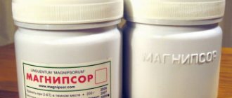
Magnipsor ointment RUB 1,490
Add to cart
Ultraviolet lamp Dermalight ® RUB 14,900.
Localizations
With gout, gouty arthritis of the joints of the lower extremities most often develops. There may be other localizations, including damage to the joints of the upper extremities. Gout is also characterized by asymmetrical joint lesions.
Gouty arthritis of the lower extremities
During a primary gouty attack, the pathological process in half of the cases involves the 1st metatarsophalangeal joint of the foot. And even if this joint is not the first to be affected, gouty arthritis will still develop in it later. The periarticular tissues swell, the skin turns red. Subsequently, small and large tophi appear on the dorsum of the foot.
Gouty arthritis of the ankle is less common and most cases occur with repeated attacks. The ankle becomes inflamed, swollen and red, and the inflammation spreads to the heel. There is severe pain and the inability to step on the foot.
The knee is often affected, the lesions are asymmetrical, often combined with lesions of the 1st metatarsophalangeal and elbow joints. Severe pain, swelling and redness are initially combined with impaired limb function due to pain, but with prolonged gout, joint deformation and ankylosis (immobility) occur.
Hip gouty arthritis is rare and the redness and swelling are not so noticeable under the thick layer of muscles and ligaments. But the pain can be severe.
Chondroprotectors: what are they, how to choose, how effective are they?
Joint pain at rest
Gouty arthritis of the upper extremities
The small joints of the hand and fingers often become inflamed, and the fingers become like sausages. The pain, inflammation and swelling are very severe. Large tofuses appear on the back of the hand.
The elbow is no less often affected. The lesions are asymmetrical and are often combined with the involvement of small joints of the hand and foot. Small and large tophi appear on the extensor surface of the shoulder and forearm.
Brachial gouty arthritis develops much less frequently, but is painful. Swelling and redness are not expressed, tophi appear on the flexor surface of the shoulder.
Lesions in gouty arthritis of the upper extremities are usually asymmetrical
Tofus lesion of the spine
In the mid-50s of the last century, spinal damage due to gout was first identified. In this case, tophi grow in the soft tissues and joints of the spine with the destruction of their structures.
The lumbar region is most often affected, followed by the cervical region. Pain appears in the back, which is often mistaken for symptoms of osteochondrosis. When the vertebrae are destroyed and the spinal nerves and spinal cord are compressed, neurological symptoms appear. When the cervical spine is affected, this results in paresis and paralysis of the upper limbs, and radicular pain.
When the lumbosacral region is affected, it can be complicated by compression of the final part of the spinal cord - the cauda equina. In this case, the function of the pelvic organs is disrupted - involuntary urination, defecation, and potency disorders occur.
Diagnostics
Despite the fact that gouty arthritis has pronounced symptoms, only 10% of patients can be correctly diagnosed during the first attack. In other cases, a diagnosis of other types of arthritis is made. Diagnostic criteria for gout are:
- acute arthritis of the 1st toe;
- the presence of large and small tophi;
- increased levels of uric acid in the blood;
- detection of EOR crystals in joint fluid and tissues.
If at least two criteria are identified, the diagnosis of gouty arthritis is considered reliable.
Laboratory research:
- general blood test
- signs of inflammation; - biochemical blood test
- uric acid content more than 0.32 mmol/l; increased levels of C-reactive protein (a sign of an inflammatory reaction); - general urine analysis
; - examination of synovial fluid using polarization microscopy
- identification of MUN crystals and a large number of leukocytes.
Instrumental studies:
- Ultrasound of joints
- detection of EOR crystals on the surface of cartilage and tophi; - radiography of the joints
- in the early stages there are no changes; later, bone changes are revealed; - computed tomography (CT)
- reveals the presence of changes in the spine.
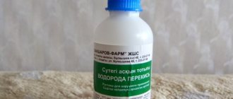
Release form
Solution for local and external use 3%.
40, 100 ml in glass bottles or glass dropper bottles with a screw neck, sealed with PE stoppers and screw caps or caps with gaskets.
40, 100 ml in drug bottles made of high or low pressure PE, sealed with screw-on plastic caps with gaskets or with a special nozzle.
Each bottle and dropper bottle is placed in a cardboard pack.
Glass bottles of 40 and 100 ml are allowed to be packaged in cardboard boxes with partitions or grids made of corrugated cardboard.
It is allowed to pack PE bottles of 40 and 100 ml in PE shrink film or in cardboard boxes.
500, 1000 ml in bottles, sealed with screw-on plastic caps with gaskets (for hospital use).
12 fl. 500 ml, 8 fl. 1000 ml are packed in PE shrink film or in corrugated cardboard boxes (for hospitals).