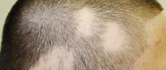
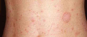
For a long time, doctors have been arguing about the reasons why a patient is affected by asbestos-like lichen. The pathological formation is also called follicular keratosis or absestoid pityriasis. The disease violates the integrity of the skin and spreads to the scalp. The patient experiences yellowish discharge on the skin in the form of scales; they cling to the roots of the hair, which provokes itching, redness and pain.
Causes of the disease
All the reasons for the appearance of lichen are completely unknown; sometimes patients are affected by pityriasis for no apparent reason. Doctors have found that asbestos disease occurs due to the following known root causes:
- decrease in the body's immune forces;
- stressful situations;
- instability of emotional calm;
- skin reaction to trauma;
- consequences of streptococcal infection;
- advanced seborrheic dermatitis;
- allergic reaction;
- psoriasis;
- eczema.
Symptoms
Itching of the scalp is characteristic of this disease.
Asbestos lichen affects the frontal and parietal lobes of the head, sometimes occurring near the neck at the back of the head. The disease causes the following symptoms:
- Gray lumps appear on the scalp; they stick to the skin and hair roots. It is difficult to separate these scales from the hairs; they often come out with the bulbs.
- The skin is inflamed, reddened, and the patient also suffers from constant itching.
- The skin becomes covered with a white crust, which is difficult to separate.
- Blood circulation worsens. Hair follicles do not receive the required amount of oxygen and vitamins due to asbestos disease, which results in excessive hair loss.
- Asbestos-like skin lesions have the ability to spread to nearby areas, for example, behind the ears.
Lichen asbestos
Gadzhimuradov M.N., Israfilova G.O., Asadulaeva Z.G.
Republican Dermatovenerological Dispensary of the Ministry of Health of the Republic of Dagestan, Makhachkala
Lichen asbestos (tinea amiantacea; syn: pityriasis asbestiform, keratosis follicularis asbestos, seborrhea psoriasiformis) is a disease of unknown etiopathogenesis. Currently, several possible causes of lichen asbestos are indicated. It is believed that the basis of this disease is a severe form of seborrhea. According to other opinions, the disease is considered one of the clinical forms of seborrheic eczema of the scalp or lichen simplex caused by pyococcal microflora. Sometimes it develops against the background of psoriasis, eczema, keratosis pilaris and other dermatoses. Lichen asbestos is even considered as an eczematous reaction of the scalp to the influence of infection or exposure to trauma. The disease can occur for no apparent reason.
It is observed more often in children and young people in the crown area, less often in the back of the head and temples. The scales resemble asbestos fibers, which are difficult to separate. Lichen asbestos is accompanied by itching of varying intensity. The hair is adjacent to the skin, dry, shrouded in small whitish scales.
The histological picture of asbestos-like lichen is characterized by: hyperkeratosis, parakeratosis and slight lymphocytic infiltration around the hair follicles, as well as dystrophic changes in the sebaceous glands. Diagnosis is based on clinical data. The course of the disease is chronic, long-term. Differential diagnosis is carried out with mycoses of the scalp, seborrhea, psoriasis, eczema.
In therapy, general strengthening agents are indicated, locally 3-5% salicylic ointment (on OL. Vaselini), sulfur-tar, sulfur-salicylic ointment. Long-term ingestion of purified sulfur, aevita and B vitamins is beneficial. Local applications of antifungal ointment (clotrimazole) to the scalp after preliminary cleansing of scales are effective.
A 6-year-old boy with massive asbestos-like scales on the scalp was hospitalized at the RKVD RD hospital. She has been sick for about a month, when whitish scales appeared on the scalp. Intense itching occurs in the affected areas. Previous diseases: suffers from intracranial hypertension.
The child's condition is satisfactory. The boy is of average height, regular build. The tongue is moist, the mucous membranes are pink.
Skin lesions are widespread: the parietal, temporal and occipital areas of the head are covered with difficult-to-remove massive gray scales riddled with hair. When the layer is removed, pink-red skin is exposed. Hair is dry, dull.
Laboratory tests: no pathogenic fungus was found in scrapings from the lesions. General analysis of urine and blood without pathological changes.
Diagnosis. Lichen asbestos.
Treatment.
Pikovit 1 tablet. x 2 times a day, vitamins B2 and B6 alternated intramuscularly, Aevit 1 drop. x 2 times a day; locally – 2% salicylic ointment. As a result of treatment, the scales were rejected, and mild itching and flaking of the scalp persisted. ‹ Anti-aging therapy with hyaluronic acid and DNA-RNA complexes Up Association of autoimmune bullous dermatoses with herpes simplex virus ›
Treatment of the disease
Treatment of asbestos-like lichen includes a complex of methods, they consist of drug therapy, external effects on lichen, traditional medicine and proper nutrition.
In addition to drug treatment, it is necessary to comb out the scales of lichen from the hair.
The course of treatment is long and takes several months. In addition to using medications and folk remedies, the patient needs to get rid of silvery lumps; to do this, they are softened and then combed out of the hair with a thin comb. It is impossible to comb out dried scales without harming the skin; they stick to the skin and hair like a crust. To soothe irritated skin, herbs and ointments are used, patients also refuse soaps and shampoos with a chemical composition, and use baby shampoo.
Medication
To eliminate painful symptoms, the patient is prescribed the following medications:
| Group of drugs | Titles | Description |
| Anti-allergenic | "Suprastin" | Eliminate allergic reactions, which may be the root cause of the development of lichen |
| "Claritin" | ||
| Local treatment | "Clotrimazole" | Combat painful symptoms, eliminate itching and help soften the resulting crust of lichen asbestos. |
| "Lamisil" | ||
| Salicylic ointment | ||
| Tar soap | ||
| Vitamins | "B-complex" | Raises the body's immune strength during asbestos-like skin lesions |
| "Retinol" | ||
| "Tocopherol" |
Traditional methods
A decoction of flax seeds is taken for two weeks.
To overcome asbestos-like lichen on the head, the following folk methods are used:
- Grape seed oil. The product is rubbed into the affected part of the body several times a day for 30 days.
- Tar and sulfur. Mixed products eliminate itching and redness. These components are mixed in equal proportions and applied to the lichen.
- Apple vinegar. Pure, undiluted apple cider vinegar is used to treat lichen once a week, for 5 weeks in a row.
- Fish oil and kerosene. These products are used to make medicinal lotions. The components are taken in unequal proportions; 2 tablespoons of fat are used per spoon of kerosene. Lotions soften dried lumps and help remove them from the scalp.
- Calendula and castor oil. Infused calendula is mixed with castor oil in equal proportions, the mixture is applied to the head, and after 1 hour it is washed off with warm water.
- Sea buckthorn oil. Patients are recommended to consume a tablespoon of sea buckthorn oil every morning.
- Flax seeds. Pour a tablespoon of flaxseeds into a glass of water and cook until boiling, drink the broth 3 times a day for 2 weeks.
Website of dermatovenerologist Betekhtin M.S.
Continuing the topics about non-contagious lichens and figurative names in dermatology, this time we will talk about lichen asbestos.
A woman came to the appointment with complaints about long-existing seals, which seemed to have glued her hair into buns and which were very difficult to remove even after washing her hair. During the examination, lichen asbestos was identified - one of the skin conditions that in the vast majority of cases is diagnosed by appearance and rarely requires additional diagnostic methods.
Lichen asbestos
Lichen asbestos (AL) is a peculiar inflammatory reaction of the scalp, which is characterized by the formation of dense, thick areas of flaking of a silvery or yellow color, which in turn surround and adhere tightly to the hair tufts. When these dense areas of scaling are removed, temporary or scarring alopecia may occur. The disease acquired its name in 1832, when the French dermatologist Jean Alibert described AL as “la porrigine amiantacee” (asbestos-like porrigo (porrigo is a Latin disease of the scalp). Since then, AL has acquired several more names, including asbestos-like pityriasis, psoriasiform seborrhea, follicular asbestos keratosis, tinea amiantacea (the latter is used in English-language literature).
AL can occur at any age, but is more common in adolescents. It occurs in both sexes with a predisposition among women (60-70%) (1,2). Clinically, all cases of AL present with dry, scaly eruptions that may be localized or diffuse and may be accompanied by pruritus, erythema, and non-scarring alopecia (3,4).
The pathogenesis of AL remains largely unexplored; currently available evidence indicates that AL is a pronounced inflammatory response with the possible involvement of genetic and environmental factors. Researchers suggest that AL is a skin response to various inflammatory diseases. AL is most often described in association with seborrheic dermatitis (one third of all cases), psoriasis (another one third), as well as lichen planus, lichen simplex chronicus, atopic dermatitis, Darier's disease, ringworm capitis and bacterial infections (1,3 ,5-7). Despite the connection with dermatoses and diseases, the mechanism of the appearance of peeling remains unclear.
The role of microorganisms in the development of AL is currently being actively discussed. Staphylococci were detected in 97% of patients, most often it was Staphylococcus aureus (S. aureus), coagulase-negative staphylococci and micrococci were also often detected (1). In addition, according to some researchers, various types of fungi have been identified, incl. Microsporum canis, Trichophyton violaceum, Trichophyton rubrum, Trichophyton schoenleinii and Trichophyton verrucosum(8). But the question of the primary or secondary nature of these microorganisms in pathological areas of the skin remains open. In any case, these microorganisms may participate in the maintenance of the disease by secreting inhibitors of epidermal cell differentiation, leading to the persistence of the disease. Additional evidence of this is the acceleration of resolution of rashes when antibiotics and antifungals are added to treatment regimens. In some cases, precipitating factors have been drugs (tumor necrosis factor alpha, interferon alpha, vilproic acid, vemurafenib), as well as anxiety and sudden changes in environmental conditions (9-12).
Histological analysis reveals the following changes: pronounced spongiosis, acanthosis, hyperkeratosis, parakeratosis, follicular keratosis and mixed inflammatory infiltrate (13).
AL can be a problem because It can be quite resistant to treatment, and no uniform approaches to its treatment have been created. However, researchers agree that treatment should be aimed at the underlying inflammatory disease (eg, seborrheic dermatitis, psoriasis, ringworm, etc.). The first published clinical cases demonstrated the effectiveness of the use of immunosuppressants (primarily local and systemic steroids) and keratolytics. In recent publications, treatment has been successful when combining the above approaches with systemic antibiotics against S. aureus and antifungals. For severe and persistent disease, topical and systemic retinoids are used (1,4).
1. Abdel-Hamid IA, Agha SA, Moustafa YM, El-Labban AM. Pityriasis amiantacea: A clinical and etiopathologic study of 85 patients. Int J Dermatol 2003;42:260-4.
2. Amorim GM, Fernandes NC. Pityriasis amiantacea: A study of seven cases. An Bras Dermatol 2016;91:694-6.
3. Ring DS, Kaplan DL. Pityriasis amiantacea: A report of 10 cases. Arch Dermatol 1993;129:913-4.
4. Gupta LK, Khare AK, Masatkar V, Mittal A. Pityriasis amiantacea. Indian Dermatol Online J 2014;5:S63-4.
5. Verardino GC, Azulay-Abulafia L, Macedo PM, Jeunon T. Pityriasis amiantacea: Clinical-dermatoscopic features and microscopy of hair tufts. An Bras Dermatol 2012;87:142-5.
6. Hansted B, Lindskov R. Pityriasis amiantacea and psoriasis. A follow-up study. Dermatologica 1983;166:314-5
7. Udayashankar C, Nath AK, Anuradha P. Extensive Darier's disease with pityriasis amiantacea, alopecia and congenital facial nerve palsy. Dermatol Online J 2013;19:18574
8. Ginarte M, Pereiro M Jr., Fernández-Redondo V, Toribio J. Case reports. Pityriasis amiantacea as manifestation of tinea capitis due to microsporum canis. Mycoses 2000;43:93-6.
9. Shiiya C, Nomura Y, Fujita Y, Nakayama C, Shimizu H. Psoriasis vulgaris with fibrokeratoma from pityriasis amiantacea. JAAD Case Rep 2017;3:243-5
10. Hussain W, Coulson IH, Salman WD. Pityriasis amiantacea as the sole manifestation of Darier's disease. Clin Exp Dermatol 2009;34:554-6
11. Ettler J, Wetter DA, Pittelkow MR. Pityriasis amiantacea: A distinctive presentation of psoriasis associated with tumor necrosis factor-α inhibitor therapy. Clin Exp Dermatol 2012;37:639-41.
12. Bilgiç Ö. Vemurafenib-induced pityriasis amiantacea: A case report. Cutan Ocul Toxicol 2016;35:329-31.
13. Knight AG. Pityriasis amiantacea: A clinical and histopathological investigation. Clin Exp Dermatol 1977;2:137-43.
Consequences of the disease
Absestosis lichen - a chronic disease, weakened immunity and other reasons can provoke its secondary appearance. Therefore, you need to monitor your health, not succumb to stressful situations and adhere to the rules of hygiene. A completed course of treatment will help you get rid of painful, discomforting symptoms. After an illness, the patient may be left with bald patches or abrasions; various methods are used to restore hair; they can be prescribed by a trichologist.
Advanced asbestos-like disease will subsequently lead to the development of seborrheic dermatitis.
Causes
There is currently no consensus on the causes of seborrheic dermatitis.
However, the development of the disease is usually associated with the functioning of the sebaceous glands. This disease is characterized by changes in the qualitative composition and quantity of sebum, and disruption of the epidermal barrier. Also, with seborrhea, there is a high degree of colonization of fungi of the genus Malassezia - yeast-like lipophilic fungi. Fungi of this genus require a fatty nutrient medium. That is why these microorganisms parasitize in the upper layers of human skin, where the sebaceous glands are most represented. As a result of excessive colonization with Malassezia fungi, the stratum corneum of the skin is damaged, peeling appears, and a local allergic reaction and inflammation develop. Interestingly, an increase in the number of fungi of this genus is not the root cause of the development of seborrheic dermatitis, but a consequence of excessive activity of the sebaceous glands. Also, patients with seborrheic dermatitis often experience an increased level of colonization of fungi of the genus Candida (especially in children of the first year of life). Typically, among the most common causes that provoke the development of seborrheic dermatitis are the following:
- Diseases of the gastrointestinal tract.
- Immune and endocrine disorders. Seborrheic dermatitis is often one of the first symptoms of HIV infection. So seborrheic dermatitis may indicate the development of immunosuppression (immune suppression).
- Psycho-emotional stress.
- Obesity.
Prevention methods and forecasting
If the root cause of the development of asbestos disease is established, prevention will be aimed at curing the cause. It is important to maintain good personal hygiene and use natural soaps and sulfate-free shampoos. Diet is an important part of the course of treatment and prevention of lichen. Refusal of harmful foods will increase the body's immune strength, improve the condition of the skin, nails and hair. It is possible to cure asbestos-type lichen so that it does not return again; to do this, you should adhere to the course of treatment, avoid stressful situations, eat right and not drink alcohol. Cured lichen will restore self-confidence to the victim and get rid of unpleasant scales, which are the ones that cause the main discomfort.

