Causes
In most cases, athlete's foot is diagnosed in men. Cases among children and adolescents are extremely rare.
The causative agent of the disease (fungus) is transmitted through contact and household contact. You can become infected with athlete's foot, for example, in a bathhouse or after using someone else's personal hygiene products.
Factors that provoke the development of the disease include:
- high temperature and humidity of the environment;
- excess weight;
- skin microtraumas;
- increased sweating;
- sedentary lifestyle.
results
ECS was recorded significantly more often (1.8 times) in men compared to women (63.8 and 36.2%, respectively; p
<0.05).
The largest number of patients belonged to the age group 35-55 years (48.1%; p
<0.05); aged 18-35 years - 25.1%; over 55 years old - 22.5%; rarely the disease was registered before 18 years of age (4.3%).
The distribution of ECS patients, taking into account the duration of the disease, is presented in Fig. 1. More than 1/3 (37.6%) of patients had a disease duration of up to 1 month, up to 2 months - 21.6%, up to 6 months - 20%, over 6 months - 20.8%.
Rice. 1. Distribution of ECS patients taking into account the duration of the disease, % (n=235).
In 146 (62.1%) patients, ECS occurred against the background of concomitant somatic pathology. The spectrum and incidence of somatic pathology are presented in Fig. 2. In the structure of concomitant ECS somatic diseases, diabetes mellitus was in the lead (40.6%), gastrointestinal pathology accounted for 20.6%, vegetative-vascular dystonia 15.1%, bronchial asthma 11.6%, 6.3% - chronic venous insufficiency, 7.1% - other diseases. Against the background of dermatological pathology, ECS was registered in 41 (17.4%) patients. Its structure was dominated by acne (19.5%), microbial eczema (17.1%), psoriasis (14.5%), rosacea (12.2%). The proportion of other dermatoses was minimal: seborrhea (9.8%), basal cell carcinoma (4.9%), true eczema (4.9%). It is important that almost half of the patients (45.1%) with ECS received treatment with TA of various groups before the start of the survey. The choice of drug depended on the qualifications, experience and personal priorities of the doctor. Preference was given to azoles (64.2%), less often allylamines were prescribed (18.8%), as well as drugs of other groups (17%). Repeated visits to the doctor during the multicenter study clearly indicate a relapse of the disease, the cause of which could be non-compliance with treatment regimens, self-medication, re-infection, lack of disinfection measures, as well as the development of fungal resistance to TA.
Rice. 2. Spectrum and incidence of concomitant somatic pathology in patients with ECS, % (n=235).
Features of the course of ECS at the present stage have been identified. The main subjective symptom was itching, which was reported in almost all patients ( n
=231;
98.3%), including weak ( n
=113; 48.9%), moderate (
n
=80; 34.6%) and strong (
n
=38; 16.5%).
Only a few people reported pain, mainly during ECS with complications ( n
= 21; 8.9%).
The frequency of lesions of various large folds (by anatomical region) was determined (Fig. 3). Almost all patients ( n
=224;
95.3%) localization of rashes was identified in the area of the inguinal folds (Fig. 4, a, b). Less frequently infected were the intergluteal fold ( n
=37, 15.7%) (see Fig. 4, c), folds under the mammary glands (
n
=23; 9.8%) and axillary folds (
n
=11; 4.7% ) (see Fig. 4, d).
One atomic region of the folds was affected in ¾ ( n
= 180; 76.6%) of patients, two - in 40 (20%), three - in 8 (3.4%) patients (Fig. 5). The average number of regions per ECS patient was 1.26±0.25. In most cases, the damage to large folds with epidermophytosis was symmetrical (61.3%). With a unilateral process, an experienced clinician, with a certain degree of probability, assumes the possibility of mycocarriage and/or a hidden course of the process on the opposite side. Therefore, in order to prevent relapse of the disease, prophylactic treatment of a symmetrical, externally unchanged large fold is carried out.
Rice. 3. Frequency of damage to various large folds (by anatomical region) with epidermophytosis, % (n=235).
Rice. 4. Localization of the process during ECS. a - inguinal folds with the process spreading to the thighs and scrotum; b - inguinal folds with the process spreading to the thighs and labia majora; c — epidermophytosis in the area of the intergluteal fold; d — epidermophytosis in the axillary region.
Rice.
Fig. 5. Distribution of ECS patients, taking into account the number of large folds involved in the process, % (n=235). An assessment was made of the characteristics and occurrence of rashes during ECS (Fig. 6). The main clinical manifestations of ECS were erythema ( n
=235, 100%), scales (
n
=208; 88.5%), infiltration in the area of the lesion (
n
=159; 67.7%), papules (
n
=134; 57%), maceration (
n
=75 ; 31.9%), cracks (
n
= 73; 31.1%).
Exudative morphological elements were recorded less frequently - pustules ( n
= 51; 21.7%) and vesicles (
n
= 39; 16.6%), accompanied by oozing (
n
= 69; 29.4%). The clinical characteristics of the rashes indicate that in 70.6% of patients with ECS, TA in the form of a cream was initially indicated.
Rice.
6. Characteristics and incidence of rashes in patients with ECS, % (n=235). The main clinical characteristics of ECS are the presence of pink or red erythema, with a bluish tint in the chronic form of the disease. The spots have peripheral growth and merge with each other. Along the periphery of the lesion there is a pronounced continuous edematous ridge, on the surface of which papules and vesicles are located. It’s not for nothing that ECS is called “fringed eczema” ( exezema
marginatum
).
In the center of the lesion, resolution of the process is observed with pronounced fine-plate peeling and weak erythema (Fig. 7, a). This classic course of ECS was recorded in 1/3 of patients (32.7%). It should be noted that in 2/3 (67.3%) of cases, erythema was combined with skin infiltration. The spread of the process along the upper third of the thighs occurred in more than half of the patients ( n
= 135; 57.7%) (see Fig. 7, b). Clinically verifiable damage to the scrotum in the form of erythema of varying intensity was detected in 101 (67.3%) men (Fig. 8).
Rice. 7. Inguinal athlete's foot. a - the classic version of the course, designated as “fringed eczema” (eczema marginatum); b - peripheral growth of the focus of inguinal epidermophytosis with the spread of the process to the thighs.
Rice.
8. Inguinal epidermophytosis in men with lesions of the scrotum of varying degrees of intensity. a - light; b - moderate; c - strong. Concomitant ECS mycosis of the feet was present in 1/3 of the patients ( n
=77; 32.7%) (Fig. 9). Among the clinical forms of mycosis of the feet, the leading form was the intertriginous form (66.2%) (Fig. 10, a, b). Dyshidrotic (19.5%) and squamous (14.3%) forms were equally common. In men, mycosis of the feet was recorded 1.7 times more often than in women (63.6% versus 36.4%).
Rice. 9. Incidence of concomitant mycosis of the feet in patients with ECS, % (n=235).
Rice.
10. Mycoses of the feet identified in patients with athlete's foot. a - intertriginous athlete's foot with severe maceration of the epidermis; b - intertriginous athlete's foot with peeling and single blisters. Onychomycosis was diagnosed in ¼ of patients ( n
=58;
24.7%), as well as its combination with mycosis of the feet - in 55 (71.4%) patients. Onychomycosis was recorded significantly 1.9 times more often in men (65.5% versus 34.5%). The nail plates of the fifth finger were most often affected ( n
= 31; 53.4%), less often - II (
n
= 18; 31%), and in some patients - III (15.5%). The normotrophic type of onychomycosis prevailed 2.2 times over the hypertrophic type (69% versus 31%).
The data obtained indicate that ECS often occurs as a multifocal process, when not only the inguinal folds are affected, but also the folds of other localizations, the scrotum, thighs, feet, and nail plates (Fig. 11).
Rice. 11. Patient V., 47 years old. Multiple foci of epidermophytosis. a - in the intergluteal region; b - in the area of the inguinal folds; c — intertriginous and dyshidrotic athlete’s foot; d — vesiculobullous epidermophytids on the hands.
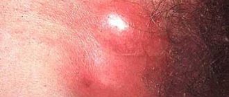
Complications of pacemaker were registered in 1/3 ( n
=78;
34.6%) patients. In the structure of complications, secondary pyoderma predominated ( n
= 38; 48.7%) (Fig. 12, a), mycotic eczema (
n
= 27; 34.6%) took second place (see Fig. 12, b, c) , allergic dermatitis was recorded less frequently (
n
= 13; 16.7%). Epidermophytids were detected in 20.4% of patients: they predominated on the hands in 54.2% of patients (see Fig. 12, d), less often recorded on the hips - in 29.2% (Fig. 13, a, b) and on buttocks - in 14.7%. Mycotic sensitization in men was recorded significantly more often (1.5 times) than in women (60.4% versus 39.6%).
Rice. 12. Complications of epidermophytosis of large folds. a - inguinal epidermophytosis, complicated by secondary pyoderma, epidermophytida on the thigh; b — epidermophytosis of the intergluteal fold, complicated by mycotic eczema (left buttock) after local therapy with zinc paste, epidermophytida on the right buttock; c — epidermophytosis of the intergluteal fold, complicated by mycotic eczema on both buttocks after local therapy using furatsilin solution.
Rice.
13. Epidermophytids. a - erythematous-urticarial rashes on the thighs with damage to the inguinal folds; b - erythematous-squamous rashes on the thighs with damage to the intergluteal and inguinal folds. The effectiveness of sertaconazole has been assessed ( Zalaina
) in the treatment of ECS, taking into account the clinical features of the disease. Treatment regimens for patients with ECS occurring alone or in combination with epidermophytosis of other localizations have been unified.
1. For uncomplicated ECS, sertaconazole cream ( Zalain
) rubbed into lesions on the skin (folds, thighs, scrotum, feet) and nails 2 times a day (morning and evening) until clinical manifestations are completely resolved. To enhance the penetration of the cream into the nail plates, patients were asked to file off the shiny layer after a soda-soap bath.
2. For ECS complicated by mycotic eczema or allergic dermatitis, and in the presence of epidermophytis, suprastinex or parlazin was prescribed orally, 1 tablet at night. The course of treatment was 10 days.
3. In the presence of pustules (athlete's foot, complicated by secondary pyoderma or mycotic eczema), betadine solution (area of folds) or betadine ointment (outside the folds) was used externally.
Recovery was considered to be the resolution of all clinical manifestations of dermatophytosis in the patient; significant improvement - reduction in the area of lesions, predominance of erythema in the clinical picture of the disease, disappearance of papules, vesicles, epithelization of erosions; improvement - preservation of the area of lesions with positive dynamics of resolution of rashes.
After 1 week, doctors noted improvement in 80.9% of cases (Fig. 14). In 15.7% of patients, no signs of recovery or positive dynamics were recorded. After 2 weeks, recovery occurred in 11.1% of patients, significant improvement in 73.2%. The overall therapeutic effect (recovery and significant improvement) was 84.3%.
Rice. 14. The effectiveness of treatment with sertaconazole in patients with epidermophytosis of large folds in the entire sample (n=235, %).
After 3 weeks, ECS was completely resolved in 45.5% of patients, and significant improvement was noted in 54.5% of patients. The overall therapeutic effect was 100%. After 1 month, almost all patients recovered (98.7%).
The use of Spearman's correlation coefficient (SCC) made it possible to identify the dependence of the timing of resolution of clinical manifestations of ECS and mycosis of other localizations on a number of factors.
— The recovery time correlated with the age of the patients (CCR = 0.276) (Fig. 15). After 2 weeks, recovery occurred 2.4 times faster in the age group under 35 years than in older patients (18.8% versus 7.8%); after 3 weeks - 1.8 times faster (66.7% versus 36.7%).
Rice. 15. Recovery time for patients with ECS, taking into account the age of the patients (CCR = 0.276).
— The recovery time was associated with the duration of the pathological process on the skin (up to 2 months or more) (KCR = 0.204) (Fig. 16). In patients with a disease duration of up to 2 months, after 2 weeks the process resolved 1.6 times faster (13.2% versus 8.1%), and after 3 weeks - already 3.8 times faster (50% versus 13.1 %).
Rice. 16. Recovery time for patients with ECS, taking into account the duration of the disease (CCR = 0.204).
— The presence of concomitant somatic pathology seriously influenced the recovery time of patients (CMR = 0.411) (Fig. 17). After 2 weeks, recovery was recorded mainly in patients with no somatic pathology (23%), and after 3 weeks this figure increased to 79.8%. In patients with somatic pathology, recovery after 3 weeks was recorded in 27% of cases, but only in isolated cases in the presence of diabetes mellitus (6.8%).
Rice. 17. Recovery time for ECS patients, taking into account concomitant somatic pathology (CCR = 0.411).
— The recovery time of patients was significantly influenced by previous TA therapy (CCR = 0.207) (Fig. 18). If patients had not previously received local TA therapy, then after 2 weeks the process resolved 6.4 times faster (17.8% versus 2.8%), and after 3 weeks - 3.6 times faster (67.4% versus 18.9%).
Rice. 18. Recovery time for patients with ECS, taking into account previous therapy with topical antimycotics of other groups (CCR = 0.207).
— The recovery time of patients depended on the number of large folds (anatomical areas) involved in the process (KCR = 0.411) (Fig. 19). After 2 weeks and after 3 weeks, the process resolved mainly when the rash was localized in the folds of one anatomical area (13.9 and 57.2%, respectively). When two or more folds were involved in the process (according to anatomical areas), recovery occurred mainly after 1 month.
Rice. 19. Recovery time for ECS patients, taking into account the number of anatomical areas involved in the process (KCR = 0.411).
— A correlation was established between the timing of resolution of ECS occurring with and without mycosis of the feet (CMR = 0.334) (Fig. 20). After 2 weeks, recovery was achieved in 27.3% of patients without concomitant mycosis of the feet and in a few patients (3.2%) with it. After 3 weeks, these differences were 8.9%. The presence of onychomycosis also influenced the recovery time of patients (CCR = 0.234). After 2 weeks, in the absence of it, 13.5% of patients recovered, and in its presence, only 3.4%. After 3 weeks, these indicators differed by 1.8 times (52% versus 28.9%).
Rice. 20. Recovery time for patients with ECS, taking into account the presence of mycosis of the feet (CCR = 0.334).
— Complications accompanying ECS also influenced the recovery time of patients (CCR = 0.418) (Fig. 21). After 2 weeks, recovery was observed mainly in patients with no complications (13.6%). After 3 weeks, in this group, complete resolution of clinical manifestations was registered 4.3 times more often than in the presence of complications (58% versus 13.6%). The presence of mycogenic sensitization lengthened the recovery time of patients (CMR = 0.334). Recovery of patients after 2 weeks in the absence of epidermophytis was observed in 13.9% of cases, and in their presence such cases were not registered. After 3 weeks, in the absence of epidermophytids, 3.5 times more patients recovered (45% versus 12.5%).
Rice. 21. Recovery time for patients with ECS, taking into account complications (CCR = 0.418).
Prevention
To avoid the possibility of inguinal athlete's foot, you need to follow the rules of personal hygiene, especially in saunas and swimming pools. Use a personal towel, washcloth, and underwear.
If a person has suffered this disease, his household items, linen and clothing must be thoroughly disinfected.
If symptoms of inguinal athlete's foot appear, SM-Clinic specialists recommend not delaying going to the doctor and not delaying treatment. You can make an appointment in St. Petersburg by calling the phone number listed on the website.
Symptoms
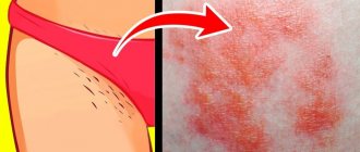
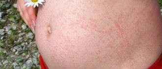
Inguinal dermatophytosis is characterized by an acute onset of the disease. The lesions are pink-red in color and are sharply limited from healthy skin. The surface is peeling. The lesions reach 15 cm in diameter and can unite. Swelling occurs, papules, pustules, vesicles and scales appear.
Since fungi affect keratin-containing tissues (hair, skin, nails), one of the first signs may be the appearance of a hairless area with scales and a rash. There may be lesions with the formation of crusts. Pain and itching appear. Discomfort increases while moving.
Inflammation of the skin most often affects the inguinal folds, the surface of the thighs, and the scrotum, without affecting the genitals.
At-risk groups
Men are more susceptible to dermatophytosis inguinalis than women. The following also increase the risk of infection:
- history of fungal infections;
- weakened immunity;
- disruption of the endocrine and digestive systems;
- stressful conditions;
- climatic conditions (high humidity and air temperature);
- close-fitting clothing;
- presence of chronic diseases;
- increased sweating (hyperhidrosis);
- frequent visits to public baths and saunas;
- obesity;
- genetic predisposition.
What fungus causes athlete's foot?
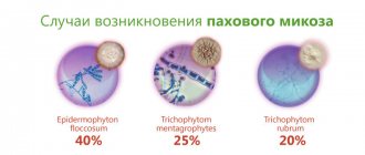
The causative agent of the disease is the fungus Epidermophyton floccosum, which forms its micromycelium in skin scales and nails. It is also found in liquid cellular aggregates formed when tissue is destroyed by mycosis. In terms of its prevalence among the causes of fungal diseases, this pathogen is in 4th place.
Most often, the disease is recorded in regions with tropical and subtropical climates. The fungus is predominantly found in the human population, so laboratory experiments on animals cannot be used to study the pathogen. Fungal spores are resistant to drying and heat.
Diagnostics
For diagnosis, the patient’s medical history and symptoms are studied, and a number of studies are carried out:
- CON test. Belongs to the group of microscopic studies. Before microscopy, the affected area is treated with a 10% potassium solution or 25% sodium hydroxide solution to clear the preparation. The test identifies the spores or mycelium of the fungus. The method accurately confirms the diagnosis, but has low sensitivity and may show a false negative result.
- Culture method. The material is planted on a nutrient medium. Based on the results of the analysis, the type of pathogen and its susceptibility to drugs are determined. Microscopy of the culture is performed. They make a diagnosis. The disadvantage of the method is the long duration of the study (up to 7 weeks).
- Direct DNA diagnosis of dermatophytosis. This is a new method using polymerase chain reaction. The analysis requires specialized laboratory equipment.
- Histological examination of the skin by taking a biopsy. Used in difficult to diagnose cases.
- Wood's lamp examination. Used when it is necessary to distinguish dermatophytosis from erythrasma. When examining erythrasma, the lesions have a pinkish glow.
Traditional medicine recipes
In folk medicine, many recipes are used to treat fungus. Traditionally, herbs with antiseptic properties, essential oils and tannins are used for this.
Phytoncides, which are found in plants such as onions, garlic (ramson), horseradish, and eucalyptus, are highly active against fungus. However, it must be borne in mind that the skin in the groin is softer and more sensitive than in other places. Therefore, such folk recipes must be used with caution.
Herbal infusion
For patients with chronic mycosis, a herbal infusion is recommended, which is taken orally for 20 days, 1/2 tbsp. 3 times a day. Its composition includes the components listed in the table below.
| Medicinal plant | Active substances | Therapeutic effect | Quantity, tsp. |
| Violet tricolor, flowers | Flavonoids, salicylic acid, anthocyanins | Antioxidant, antimicrobial, anti-inflammatory | 1 |
| Chamomile, flowers | Essential oils, quercetin, bitters | Antiseptic, anti-inflammatory | 3 |
| Yarrow, herb | Ascorbic and aconitic acids, tannins, coumarin | Antiseptic, antioxidant | 2 |
| Lingonberry, leaves | Phenologlycosides | Antiseptic | 3 |
| St. John's wort, herb | Flavone glycosides, essential oil | Antibacterial, antiseptic, regenerating | 4 |
| Eucalyptus, leaves | Essential oil | 2 |
All components are mixed, take 4 tbsp. l. mixture, pour 2 tbsp. boiling water and leave for half an hour.
Blend of herbs and flax seeds
The ingredients for this recipe are shown in the table below.
| Medicinal plant | Main active components | Therapeutic effect | Quantity, tsp. |
| Line, grass | Flavonoids, ascorbic acid, tannins | Antiseptic, bactericidal, anti-inflammatory | 2 |
| Yarrow, herb | Coumarin, ascorbic and aconitic acid, tannins | Antiseptic, antioxidant | 3 |
| Oak, bark | Tannins | Antiseptic, anti-inflammatory | 2 |
| Flax, seeds | Fatty oil, organic acids | Emollient | 2 |
The above components are mixed, 2 tbsp. l. mixture is poured into 1 tbsp. boiling water and leave for half an hour. The resulting product is used as a lotion on the affected areas. They need to be done 3-4 times daily until the weeping and inflammation stop.
Radish seeds
Radish seeds contain a large amount of essential oils that have antiseptic properties. Fatty oils help soften the skin. Allyl mustard oil, which has a toxic effect on fungi, is contained in smaller quantities than in root vegetables. Thanks to this, compositions based on it have a milder effect.
To prepare the medicine, the seeds of the plant are ground in a mortar, poured with warm boiled water, and mixed. This paste is applied to the affected areas of the skin. It must be taken into account that the plant can cause irritation, so the procedure should be carried out no more than 5-10 minutes.
Garlic paste
Ramson, or wild garlic, contains alliin, which when the clove or green tissue of the plant is destroyed turns into allicin. This substance has a strong bactericidal and antifungal effect.
To prepare a compress, garlic is crushed and mixed with a small amount of olive oil. The paste is applied to the groin area and left for 5-10 minutes. If there are deep cracks or other violations of the integrity of the skin, then this recipe is not recommended.
Birch and poplar buds
Birch and poplar buds are rich in flavonoids, saponins, tannins, phytoncides, and essential oils. They have antimicrobial, anti-inflammatory, wound-healing and general strengthening effects.
To prepare a medicinal product, the buds of these plants are mixed in equal proportions, filled with alcohol (vodka), and kept in a cool, dark place for 1 week. Then filter through cheesecloth or a sieve. Alcohol infusion is used to wipe the affected areas.
Fig oil
Fig (or fig) seed oil is a rich source of vitamins and bioactive molecules for the skin. It has anti-inflammatory and antioxidant properties, promotes faster recovery of damaged areas.
In the treatment of athlete's foot, it is used as an emollient that has a protective effect. The oil is applied several times during the day, and the excess is blotted with a paper towel.
Celandine
Celandine is a poisonous plant that has a bactericidal and antifungal effect. It retains its properties even in a dried state, so you can use pharmaceutical preparations (but not concentrated extracts, which are used to remove warts and burn the skin).
To prepare the product, dry leaves need to be poured with a small amount of boiling water, wait until the infusion cools to room temperature. Place gauze or a bandage on the affected area of skin, and then a paste of herbs. The bandage is secured with a bandage and kept for 10 minutes.
Saline solution
Table and sea salt have antiseptic properties. Solutions with a concentration of 10-15% significantly reduce the development of putrefactive bacteria and fungi. This property can also be used in the treatment of athlete's foot.
To prepare the solution you need to take 9 parts boiled water and 1 part salt. The product is used as a lotion on the affected areas. The duration of the procedure is 10-15 minutes. Saline solution should not be applied to open wounds on the skin.
Baking soda
Baking soda in folk medicine is better known as a means for disinfecting teeth and gums, for sore throats and for reducing heartburn. It can also be used for fungal diseases as an antiseptic.
For this, 1 tbsp. l. powder is stirred in 1 tbsp. water. The resulting solution is used to wipe the areas affected by the fungus and leave for 15-20 minutes. After completing the procedure, the soda solution must be washed off. For greater effectiveness, you can add ½ tsp to the solution. table vinegar 9%
Herbs and white wine
There is also a recipe for making lotions from herbs and white wine:
- Burdock roots (fresh or dried) are mixed with sage herb in equal proportions (3-4 tbsp each). Burdock has an anti-inflammatory and wound-healing effect, and sage has an antimicrobial effect.
- The mixture of herbs is poured into 2 tbsp. white wine.
- Boil the solution for 20 minutes.
- Cool and filter.
This solution is used to wipe the skin affected by the fungus several times a day.