How to treat Epstein-Barr virus.
August 25, 2022
7487
4.6
2
Content
- Why do diseases occur due to the Epstein-Barr virus?
- How are EBV classified?
- Symptoms of Epstein-Barr virus
- How to diagnose Epstein-Barr virus
- How to treat Epstein-Barr virus
- Complications of Epstein-Barr virus
- Prevention of Epstein-Barr virus
- How to treat Epstein-Barr virus
- Groprinosin
- Proteflazid
- Novirin
- Laferobion
- Isoprinosine
For most people, Epstein-Barr virus (EBV) remains in the body for life after exposure to infection. EBV often triggers the development of various diseases, and the first contact with the virus usually occurs in the first 10 years of a child’s life, causing a sluggish infectious disease. At an early age, symptoms of EBV are practically absent or blurred. When exposed to the virus before the age of 10, a child develops respiratory diseases in 40% of cases, and infectious mononucleosis in 10-25%.
Epstein-Barr virus is a representative of herpes viruses; it is capable of infecting various cells, including B cells of the immune system (these are white blood cells) and epithelial cells of the mucous membranes.
Relevance
One of the pressing problems of modern medicine is the high incidence of herpesvirus infections, which are quite widespread in the human population.
The complex structure of the genome of viruses of the herpes family, compared to other DNA-containing viruses, determines the main differences in their replication cycle. Genes encoding structural proteins make up only 15% of the DNA in herpes viruses, while the majority of the genome consists of areas responsible for the synthesis of regulatory proteins and enzymes, and it is this feature that allows them to implement a completely unique program, including the possibility of a latent, persistent and reactivated state in infected organism [1]. A special place among herpes viruses is occupied by Epstein-Barr virus (EBV), which infects 95% of the population and, like all herpes viruses, it is capable of infecting almost all organs and systems of the body, causing latent, acute and chronic forms of infection, prone to reactivation in conditions of immunosuppression . Active proliferation of the virus in all organs and systems that have lymphoid tissue leads to structural changes that have an adverse effect on the body as a whole. The key role of EBV in the development of diseases such as acute and chronic mononucleosis, interstitial pneumonitis, myocarditis, hepatitis, tumors of lymphoid and epithelial tissues, hemophagocytic lymphohistiocytosis, leukoplakia of the tongue and post-transplantation lymphoproliferative complications has been proven.
Why do diseases occur due to the Epstein-Barr virus?
EBV is transmitted by airborne droplets. That is why infectious mononucleosis, which develops against the background of a virus, is called the “kissing disease.”
Another way to become infected with the Epstein-Barr virus is through household contact (dishes, toothbrush, towel, etc.). EBV is also transmitted through semen and blood.
The Epstein-Barr virus multiplies in epithelial cells and in B-lymphocytes, so the manifestations of this infection are very diverse. The peculiarity of EBV is that it attacks the cells of the immune system - they begin to clone the DNA of the virus. An inflammatory process develops in the body, causing various symptoms.
Main symptoms of chronic EBV infection
After the primary infection is complete, the virus remains in the human body for life. It is found in absolutely healthy people in saliva, urine and even blood, which is why taking smears for EBV infection is not advisable. In most cases, the virus is in an inactive form and this condition is called asymptomatic carriage.
Chronic active EBV infection is detected only in people with impaired immunity.
It manifests itself with the following symptoms
- prolonged enlargement of several groups of lymph nodes;
- an unreasonable increase in temperature within 37.5 degrees;
- unmotivated fatigue;
- muscle and joint pain.
The role of the virus in the development of malignant tumors of the nasopharynx and Burkitt's lymphoma has been established.
How are EBV classified?
Since there is no official classification of the Epstein-Barr virus, in practice its classification looks like this:
- by time of infection - acquired and congenital;
- according to the form of the disease - typical (infectious mononucleosis) and atypical (asymptomatic, erased, affecting internal organs);
- according to the severity of the course - mild, moderate and severe;
- according to the duration of the disease - acute infection, protracted and chronic;
- by activity - active virus and inactive;
- mixed type infection - this type is usually diagnosed if cytomegalovirus is associated with EBV.
Epstein-Barr virus
Photos from open sources
Symptoms of Epstein-Barr virus
Epstein-Barr virus causes the following diseases:
- Infectious mononucleosis (Filatov's disease, glandular fever).
This is a common infectious disease in which there is a strong increase in temperature, enlarged lymph nodes, inflamed pharyngeal mucosa, and enlarged liver and spleen. In mononucleosis, the virus enters the body through the upper respiratory tract. - Lymphogranulomatosis (Hodgkin's disease) and some types of non-Hodgkin's lymphomas.
In these diseases, malignant cells appear in the gastrointestinal tract, spleen, liver, lymph nodes and bone marrow. - Chronic fatigue syndrome. In this condition, a person feels tired for a long time, even if he gets enough rest.
- Alice in Wonderland syndrome.
In this condition, a person’s sense of his own body is disturbed: the whole body or its parts seem to him very small or, conversely, too large. - Hepatitis due to EBV.
Hepatitis often develops with mononucleosis. Symptoms of hepatitis include a yellowish tint to the skin and mucous membranes, nausea, general weakness, and enlarged liver. - Herpes on the lips or genitals.
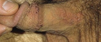
In addition, the Epstein-Barr virus can cause stomatitis. Symptoms are a burning sensation, pain, and then small blisters. - Multiple sclerosis.
When infected with EBV, an autoimmune disease such as multiple sclerosis can develop, which affects nerve fibers in the brain and spinal cord. Symptoms include decreased muscle strength, loss of reflexes, and paralysis. - Hairy leukoplakia.
Rough, painless areas appear on the tongue. They do not cause discomfort, but can become cancerous. - Nasopharyngeal carcinoma.
This is a malignant lesion of the pharynx, the symptoms of which are hearing loss, frequent ear infections, nasal congestion, blood in the saliva and from the nose, headaches and enlarged lymph nodes. - Autoimmune thyroiditis.
With this chronic disease, antibodies are formed to the thyroid tissue. The gland enlarges, swelling, fatigue, drowsiness, hair loss, dry skin, and frequent constipation appear.
Manifestations of EBVI
The most well-known and studied manifestation of Epstein-Barr virus infection (EBVI) is infectious mononucleosis (IM). At present, the immunopathological basis for individual differences in the course of MI and its outcomes is unclear. Clinical manifestations, diagnosis and treatment of MI are presented in Table 1.
A meta-analysis of 5 randomized placebo-controlled trials involving 339 patients showed no effect of acyclovir in patients with MI of EBV etiology [5].
EBV has multiple mechanisms of immunosuppression and evasion of the immune response in the human body, which can lead to the formation of chronic EBVI (CEBVI) [6], during which immunological disorders are aggravated, the production of interferons is suppressed, apoptosis mechanisms are blocked, which forms a secondary immunodeficiency that contributes to the development of autoimmune and tumor processes in genetically predisposed individuals.
Clinical forms of CHEBVI are presented in Table 2.
A lot of data has been accumulated on the etiological role of EBV in the formation of chronic fatigue syndrome, the development of systemic vasculitis, specific colitis, and there is evidence of the trigger role of EBV in the development of multiple sclerosis and systemic lupus erythematosus [15, 16].
Skin manifestations of CHEBVI have been described, such as hypersensitivity to mosquito bites and light pox (Hydroa vacciniforme, HV) (see Table 2). Lightpox is a disease characterized by the development of a staged polymorphic necrotic rash on areas of the skin exposed to solar radiation. The disease is often familial, but in a number of patients, histochemical analysis of skin lesions shows infiltration of T cells expressing small RNA encoded by EBV. The coincidence of the presence of CEBVI and lightpox (HV) can develop into the development of malignant lymphoma, which was included in the 2008 WHO classification as HV-like lymphoma [14].
Active EBVI (see Table 2) can serve as a trigger for the development of idiopathic thrombocytopenic purpura [17], an autoimmune hematological disease based on thrombocytopenia with the development of hemorrhagic syndrome. Characteristic signs of this disease are multiple, polymorphic hemorrhages in the skin and mucous membranes, as well as bleeding of various locations (nose, gums, etc.).
Hemophagocytic lymphohistiocytosis (HLH) is one of the most dangerous, life-threatening complications of EBVI, the main symptoms of which are: fever, refractory to antimicrobial therapy, hemorrhagic and edematous syndromes, jaundice, exanthema, hepatosplenomegaly, symptoms of damage to the central nervous system (excitability, depression of consciousness, convulsions , meningeal signs) [18]. The development of HLH is associated with a disorder of immune regulation as a result of uncontrolled activation and proliferation of macrophages and T cells, which is manifested by excessive production of cytokines, inflammation and tissue damage. There are primary HLH, characterized by a family history and a certain genetic defect, and secondary HLH, associated with infection, autoimmune and oncological diseases, as well as an immunodeficiency state [19].
The key role of EBV in the development of lymphoproliferative diseases has been proven. One of the key lymphomas associated with EBV is Burkitt's lymphoma [20]. Clinical manifestations of lymphomas include enlarged lymph nodes, splenomegaly, cytopenia, and fever. It is believed that the presence of latent EBVI in the nasopharyngeal epithelium is an early stage of the pathogenesis of undifferentiated nasopharyngeal carcinoma [21].
How to diagnose Epstein-Barr virus
To identify EBV in the body, the doctor collects an epidemiological history, takes into account the clinical picture of the disease and refers the patient to certain tests. Typically, laboratory diagnosis of EBV includes the following markers:
- Epstein-Barr virus, determination of DNA in blood (Epstein Barr virus, DNA);
- Epstein-Barr virus, DNA determination in scrapings of epithelial cells of the nasal mucosa (Epstein Barr virus, DNA);
- Epstein-Barr virus, DNA determination in scrapings of epithelial cells of the oropharynx (Epstein Barr virus, DNA);
- Epstein-Barr virus, determination of DNA in blood serum (Epstein Barr virus, DNA);
- Epstein-Barr virus, determination of DNA in saliva (Epstein Barr virus, DNA);
- Epstein-Barr virus, determination of DNA in saliva (Epstein Barr virus, DNA);
- Epstein-Barr virus, determination of DNA in prostate secretion, ejaculate (Epstein Barr virus, DNA);
- IgG class antibodies to the nuclear antigen of the Epstein-Barr virus (EBV NA IgG, Epstein-Barr Virus Nuclear Antigen IgG, EBNA IgG);
- IgM class antibodies to the capsid antigen of the Epstein–Barr virus (EBV VCA-IgM, Epstein-Barr Virus Capcid Antigen IgM, EBV VCA-IgM);
- IgG class antibodies to the capsid antigen of the Epstein-Barr virus (anti-Epstein-Barr viral capsid antigens IgG, EBV VCA IgG);
- IgG class antibodies to the early antigen of the Epstein-Barr virus (anti-EBV EA-D IgG; Epstein-Barr Virus Antibody to Early D Antigen (EA-D), IgG; anti-EBV EA-D IgG).
Only a doctor can tell which of the above tests should be taken for a patient with suspected Epstein-Barr virus. Diagnosis and treatment should be carried out by a therapist, pediatrician or infectious disease specialist. If a patient has one or more lymph nodes enlarged, he constantly feels tired, coughs for a long time, the abdomen is enlarged, there is pain in the abdomen or bones, a consultation with a hematologist, oncologist and other specialists is needed.
Symptoms of viral mononucleosis
Photos from open sources
Clinical observations
Case 1.
Patient Yu., 26 years old, was taken to Infectious Diseases Clinical Hospital No. 2 (ICH No. 2) with complaints of increased body temperature, weakness, shortness of breath, and a feeling of palpitations during physical activity.
From the anamnesis it is known that she fell ill 15 days ago, when an increase in body temperature to 39.0 °C, sore throat, weakness, and shortness of breath during physical activity appeared. In the following days, the elevated body temperature persisted, heaviness in the right hypochondrium and darkening of the urine appeared. She was examined on an outpatient basis: a clinical blood test revealed lymphomonocytosis, atypical mononuclear cells, and a decrease in hemoglobin level to 73 g/l. She was hospitalized at ICH No. 2 with a diagnosis of “Infectious mononucleosis. Severe anemia."
Upon admission, the patient's condition was moderate. Body temperature 38.0 °C. Marked weakness. The skin is pale, there are no rashes, hemorrhages, or bleeding. The sclera is injected, and marginal icterus is noted. The mucous membrane of the oropharynx is slightly hyperemic, the tonsils are enlarged to degree II, there are purulent deposits in the lacunae. The submandibular and cervical lymph nodes up to 1–1.5 cm are palpable, dense, painless. Breathing through the nose is free. There is no nasal discharge. Breathing in the lungs is weakened. No wheezing can be heard. NPV 20 per minute. Heart sounds are muffled and rhythmic. A systolic murmur is heard at the apex of the heart. Heart rate 80 per minute. Blood pressure 110/70 mm Hg. Art. The tongue is moist, covered with a white coating. The abdomen is soft on palpation, sensitive in the right hypochondrium. Peristalsis is heard. There are no peritoneal signs. The liver protrudes from under the edge of the costal arch by 4 cm, is dense, sensitive to palpation. An enlarged spleen is palpable, dense, painless. The symptom of tapping in the lumbar region is negative on both sides. Physiological functions are not impaired. There are no focal or meningeal signs. The patient was prescribed treatment with ceftriaxone 2.0 g × 2 times/day intravenously, rinsing the oropharynx with furacillin solution, intravenous detoxification therapy with a 5% glucose solution with ascorbic acid, and diclofenac injections for fever.
During the examination: a general blood test shows a decrease in the number of erythrocytes to 1.96 × 10¹²/l, hemoglobin to 64 g/l, leukocytosis 17.5 × 10⁹/l, lymphocytosis 82%, 18 atypical mononuclear cells were found among the lymphocytes. A biochemical blood test shows a moderate increase in liver transaminases (ALT 106.3 U/L; AST 165.4 U/L). Serum iron content is normal. A PCR test of the blood revealed EBV DNA; the ELISA test for antibodies to VCA showed positive IgM and negative IgG.
An ultrasound of the abdominal organs was performed: diffuse changes were detected in the liver parenchyma and pancreatic parenchyma, a significant increase and diffuse changes in the spleen parenchyma.
The patient was consulted by a hematologist, the conclusion: the blood picture corresponds to a leukemoid reaction of the lymphocytic type against the background of infectious mononucleosis of EBV etiology. Therapy with epoetin beta, folic acid, and vitamin B12 is recommended. The patient received 1 injection of epoetin beta (2 thousand units subcutaneously); then, due to the patient’s severe pain syndrome and the patient’s categorical refusal, the injections were stopped.
On the 11th day of the patient’s stay in the hospital, there is no positive dynamics from the treatment: in the general blood test, a decrease in red blood cells remains to 1.84 × 10¹²/l, hemoglobin to 62 g/l, a biochemical blood test shows an increased level of liver transaminases (ALT 110.4 U/l; AST 160.1 U/l). The patient was prescribed therapy with ganciclovir 250 mg 2 times/day intravenously.
On the 17th day of hospital stay, the patient complained of abdominal pain, and therefore a repeat ultrasound of the abdominal organs was performed, revealing a small splenic infarct area in the lower pole. The patient was consulted by a surgeon, and dynamic monitoring was recommended.
Subsequently, against the background of the therapy, positive dynamics are noted in the form of normalization of body temperature and regression of tonsillitis. In the general blood test, attention is drawn to an increase in hemoglobin to 96 g/l, red blood cells to 3.01 × 10¹²/l, and normalization of leukocyte levels.
On the 21st day of hospitalization, the patient was discharged under the supervision of an infectious disease specialist and hematologist at her place of residence.
Case 2
. Patient Zh., 25 years old, was admitted to ICH No. 2 with complaints of an increase in body temperature to 37.7 °C and a dry cough.
From the anamnesis it is known that he fell ill on March 10, 2019: weakness, dizziness, increased body temperature to febrile levels. 03/11/2019: increase in body temperature to 38.5 °C. He was treated independently: he took tilorone and azithromycin without effect. 03/14/2019: examined by a therapist at home, diagnosed with ARVI, oseltamivir was prescribed. 03/15/2019: the symptoms persisted, a single loose stool appeared without pathological impurities, a runny nose, and a dry cough. On March 19, 2019, he was examined on an outpatient basis: a plain chest x-ray was performed - no pathological changes, a general blood test showed slight leukocytosis (11×109/l), other indicators were normal. Levofloxacin was prescribed without effect. On March 21, 2019, he was hospitalized at ICH No. 2 by the ambulance service with a diagnosis of fever of unknown etiology.
Upon examination, the patient's condition is moderate. Body temperature 37.7 °C. The skin has a physiological color, moderate humidity, and no rash. The mucous membrane of the oropharynx is hyperemic, the follicles on the back wall of the pharynx, the tonsils are not enlarged, free from overlap. On palpation: peripheral lymph nodes are not enlarged. There is no peripheral edema. Breathing through the nose is free. There is no nasal discharge. In the lungs, breathing is harsh, carried out in all parts, wheezing is not heard, respiratory rate is 18 per minute. Heart sounds are clear, rhythmic, heart rate 78 per minute, blood pressure 100/60 mm Hg. Art. The abdomen is soft and painless on palpation in all parts. Peristalsis is active. There are no peritoneal signs. The liver protrudes from under the edge of the costal arch by 2 cm, the spleen is not enlarged, painless. The symptom of tapping in the lumbar region is questionable on both sides. According to the patient, the stool is shaped and discolored. Urination is not impaired. Urine is dark. There are no focal or meningeal signs.
During the examination: a general blood test revealed leukocytosis of 10×109/l, lymphocytosis of 74%, 17 atypical mononuclear cells were found among the lymphocytes, the rest of the indicators were normal. A biochemical blood test shows a moderate increase in liver transaminases (ALT 61 U/L; AST 60.4 U/L). PCR testing of blood and oropharyngeal smear did not detect EBV DNA; ELISA for antibodies to VCA showed a positive reaction to IgM and IgG.
According to ultrasound of the abdominal cavity and kidneys, there is an increase and diffuse changes in the liver parenchyma, an increase and diffuse changes in the spleen parenchyma, and moderate diffuse changes in the kidney parenchyma.
Cefotaxime 2 g 2 times/day intramuscularly, rinsing the oropharynx with a solution of chlorhexidine, cetirizine, a complex of lactobacilli acidophilus and kefir fungi were prescribed. On the 3rd day of hospital stay, due to persistent fever, antimicrobial therapy was replaced with ciprofloxacin 400 mg 2 times/day intravenously.
On the 5th day of the patient’s stay in the hospital, positive dynamics were noted in the form of disappearance of fever, and on the 9th day the patient was discharged from the hospital under the supervision of an infectious disease specialist at the place of residence.
How to treat Epstein-Barr virus
There is no specific treatment for EBV. Typically, treatment for Epstein-Barr virus involves eliminating symptoms. The patient needs to drink enough fluids, take antipyretic and painkillers, interferons, antiviral drugs, antibiotics (only if there is a viral sore throat or bacterial complications), corticosteroids. You need to get plenty of rest and reduce physical activity. If a person has a severe form of EBV, doctors prescribe medications against herpes infection.
Treatment of herpes type 4
Timely therapy can prevent the development of the virus, which is achieved by blocking its activity. To prevent the development of dangerous pathological conditions, a diagnosis is carried out, and based on the test results, the doctor prescribes a treatment regimen.
Like any virus, herpes infection is blocked by drugs of 2 groups: immunostimulating, antiviral.
A standard treatment regimen for this pathological condition of the body has not yet been developed. Complex therapy is carried out: antioxidants are prescribed to help eliminate toxins; alpha interferons.
The treatment regimen can be adjusted depending on the degree of damage to internal organs, the condition of the body and immunity. In case of a generalized form of the disease, treatment is carried out inpatiently. The pathological condition in this case may be accompanied by damage to the nervous system, which means that observation by a neurologist is necessary.
Read also: How herpes is transmitted About the psychosomatics of herpes Read more
about the herpes simplex virus here .
Therapy for latent and mild forms of the disease is carried out on an outpatient basis. If a secondary bacterial infection has developed, antibiotics are prescribed. For pain and redness of the mucous membranes in the throat: gargle and take medications that will help eliminate these symptoms. If the temperature rises, it is necessary to take antipyretic drugs.
During treatment, when infected with herpes type 4, it is recommended to reduce physical activity. It is important to adjust your diet, which will reduce the load on the liver. Remove fatty, highly salted, spicy foods. It is important to drink a lot of water - more than 2 liters per day (more details here).
Diagnostics
The presence of herpes type 4 in the body is determined in several ways. Tests for the virus:
- General blood analysis. Allows you to determine the increased content of leukocytes, band neutrophils, mononuclear cells.
- Blood chemistry. Makes it possible to detect changes in several indicators: increased ALT, AST (in case of liver damage).
- PCR. The DNA of the herpes virus and its quantity are detected, which makes it possible to assess the extent of the infection.
- Immunological research. Makes it possible to determine the immunoregulatory index.
- An enzyme-linked immunosorbent assay that detects antibody titers to the virus. If IgG EA is elevated, it means the infection has occurred recently.
An immunological study allows you to determine the immunoregulatory index.
To determine the extent of damage to the spleen and liver, an ultrasound is performed.
Drug treatment
When infected with herpes, different types of drugs are prescribed:
- immunostimulants: Alfaglobin, Viferon, Cycloferon (more details here);
- antiviral: Acyclovir, Valacyclovir, etc. (more details here);
- NSAID medicine: Paracetamol, Tylenol;
- antibiotics: from the group of cephalosporins, lincosamides, macrolides (more details here);
- glucocorticosteroids (in case of complications, for example, laryngeal edema): Hydrocortisone, Prednisolone, Dexamethasone.
Cycloferon is used to treat type 4 herpes.
Prevention
Considering that herpes type 4 can be transmitted in several ways, it is difficult to protect against it. Prevention measures:
- the likelihood of contact with infected people is eliminated: even after recovery, a person can pose a danger, since the virus is released for several months after symptoms have disappeared;
- If blood transfusions are given, donors should be carefully screened for the presence of the herpes virus and other infections.
Groprinosin
An antiviral drug with immunomodulatory properties, “Groprinosin” is prescribed to patients with viral infections: herpes types 1 and 2, cytomegalovirus, chickenpox, Epstein-Barr virus, mumps and measles virus, etc. The drug stimulates the functioning of the immune system, if it is reduced, it reduces symptoms of viral diseases, speeds up recovery and helps reduce the number of relapses of viral infections. The dosage of the drug is determined by the doctor based on the weight and age of the patient. Side effects include increased levels of uric acid in the blood plasma and urine, and general malaise. You can buy Groprinosin in tablets and syrup.
Groprinosin
Gedeon Richter, Hungary
The active substance of the drug Groprinosin, inosine pranobex, has a direct antiviral and immunomodulatory effect.
The direct antiviral effect is due to binding to the ribosomes of virus-affected cells, which slows down the synthesis of viral messenger RNA (mRNK) and leads to inhibition of the replication of RNA and DNA genomic viruses; the indirect effect is explained by the powerful induction of interferon formation. from 1200
5.0 1 review
1104
- Like
- Write a review
Epstein-Barr viral infection in children: modern approaches to diagnosis and treatment
Epstein-Barr virus infection (EBVI) is one of the most common human infectious diseases. Antibodies (Abs) to the Epstein-Barr virus (EBV) are detected in 60% of children in the first two years of life and in 80–100% of adults [3, 13]. The incidence of acute form of EBVI (EBVI) in different regions of the world ranges from 40 to 80 cases per 100 thousand population [2]. The chronic form of EBVI (CEBVI) develops in 15–25% of individuals after EBVI [1, 5, 15]. The role of EBV in the development of malignant neoplasms, autoimmune diseases and chronic fatigue syndrome has been established [3, 5, 14, 15]. All this indicates the relevance of the problem of EBVI.
EBV, discovered in 1964 by M. Epstein and Y. Barr, belongs to the γ-herpes viruses [3]. EBV has 3 antigens: capsid (VCA), early (EA) and nuclear (EBNA). The uniqueness of the pathological process in EBVI is determined by the ability of EBV to transform B-lymphocytes, lifelong persistence in the human body, induction of a secondary immunodeficiency state (IDS), autoimmune reactions, and malignant tumors [1, 3, 5, 12].
The source of EBV infection is patients with manifest and asymptomatic forms. 70–90% of people who have had EEBVI shed the virus in the next 1–18 months. Routes of transmission of EBV: airborne, household contact, parenteral, sexual, vertical. OEBVI is characterized by epidemic rises once every 6–7 years, and is more often registered at the age of 1 to 5 years, in organized groups [4, 7, 9].
The entrance gate for EBV is the mucous membrane of the upper respiratory tract: the virus penetrates the lymphoid tissue, infects B-lymphocytes, polyclonal activation of B-lymphocytes develops, dissemination of the pathogen within B-lymphocytes, the synthesis of antibodies (Ab) in response to antigenic stimulation is reduced. EBV primarily affects lymphoid organs (tonsils, liver, spleen).
The next stage is the formation of a clone of sensitized cytotoxic CD8 cells, the sequential synthesis of Abs to the VCA, EA and EBNA antigens of the virus. Due to disruption of the immune response and functional activity of innate resistance factors (neutrophils, macrophages, NK cells, interferon system), secondary IDS is formed [2–4, 12].
The immune status of 109 patients with OEBVI aged 5 to 14 years in our work revealed signs of activation of the T-cell component of the immune system - an increase in the number of T-lymphocytes (CD3), cytotoxic T-lymphocytes (CD8), cells with markers of late activation (HLA- DR); polyclonal activation of B lymphocytes - an increase in the number of CD20 cells, immunoglobulins (Ig) IgA, IgM, IgG, circulating immune complexes (CIC). Signs of suppression of the immune system were found: normal levels of T-helper cells (CD4), a decrease in the immunoregulatory index CD4/CD8, the number of natural killer NK cells (CD16), and an increase in the readiness of immunocompetent cells for apoptosis (CD95). Activation of oxygen-dependent metabolism of neutrophils and a reduction in its adaptive capabilities were observed.
In a third of the examined children (33.9%), EEBVI occurred in the form of a mixed infection with cytomegalovirus (CMV), herpes simplex viruses types 1 and 2 (HSV-1, HSV-2). During bacteriological examination of oropharyngeal smears, Streptococcus (S.) viridans was isolated in 41.3% of patients, Candida albicans in 11.9%, Staphilococcus (Staph.) epidermidis in 8.2%, S. pyogenes, in 2.7% - Klebsiella (Kl.) pneumoniae, in 41.3% - an association of bacteria. 43.1% of patients had serological markers of an active form of chlamydial infection, and 30.3% had mycoplasmosis.
The following outcomes of EBVI are possible: latent infection, EBVI, IDS, cancer, autoimmune diseases, chronic fatigue syndrome [5, 8, 10, 11]. The transition to CHEBVI is associated with a complex of unfavorable factors in the ante-, intra- and postnatal periods, disruption of neuroimmune-endocrine regulation, and genetic predisposition.
Our examination of 60 children aged 5 to 14 years with CEBVI showed that in this group 86.7% of mothers had a burdened obstetric history; In 83.3% of children, perinatal and postnatal pathologies of the central nervous system, ENT organs, etc. were detected.
The immune status of patients with CHEBVI revealed an increase in the content of interleukin-1 antagonist (IL-1RA), insufficient activation of immunocompetent cells (decrease in HLA-DR) and an increase in their readiness for apoptosis (increase in CD95). There was a disturbance in the functional activity of type 1 T helper cells (Th1) (decreased levels of interferon γ (IFN γ)); decrease in the total pool of T cells (CD3), the number of lymphocytes with receptors for IL-2 (CD25) and NK cells (CD16); the content of cytotoxic CD8 lymphocytes was increased. The persistence of EBV replication markers for a long time in this group indicated a violation of virus elimination; At the same time, an increase in the functional activity of Th2, polyclonal activation of B-lymphocytes (CD20), an increase in the content of IgA, IgM, IgG, CEC, a decrease in the level of neutrophil chemotactic factor (IL-8), and a change in their metabolism were noted.
Immune status disorders led to the activation of opportunistic microflora, viral and fungal infections. In the microbial spectrum of the oropharyngeal mucosa of patients with CHEBVI, S. Viridans (30%), Candida albicans (28.3%), Staph. Epidermidis (25%), S. Pyogenes (20%), Kl. Pneumoniae (8.4%), bacterial association (41.7%); 28.3% had serological markers of the active form of chlamydia, 26.7% had mycoplasmosis. In 90% of patients, the disease occurred in the form of a mixed infection involving herpes viruses: EBV + CMV, EBV + HSV-1, HSV-2.
Classification . There is no generally accepted classification of the disease; We recommend using the working classification of EBVI that we have developed.
- By period of occurrence: congenital, acquired.
- Form: typical (infectious mononucleosis), atypical: erased, asymptomatic, visceral.
- By severity: light, medium, heavy.
- According to the course: acute, protracted, chronic.
- By phase: active, inactive.
- Complications: hepatitis, splenic rupture, meningoencephalitis, polyradiculoneuropathy, myocarditis, sinusitis, otitis, hemolytic anemia, thrombocytopenia, neutropenia, pancreatitis, etc.
- Mixed infection.
Examples of diagnosis:
- Main: Acquired EBVI, typical severe form (infectious mononucleosis), acute course, active phase. Osl.: Acute hepatitis.
- Main: Acquired EBVI, visceral form (meningoencephalitis, hepatitis, nephritis), severe chronic course, active phase. Osl.: acute hepatic-renal failure. Associated with: Respiratory chlamydia (rhinopharyngitis, bronchitis, pneumonia).
The clinical picture of acute EBVI was first described by N. F. Filatov (1885) and E. Pfeiffer (1889). The incubation period lasts from 4 days to 7 weeks. A complete symptom complex is formed by 4–10 days of illness [4, 7].
We examined 109 children with OEBVI. In most patients, the disease begins acutely, with an increase in body temperature and the appearance of symptoms of intoxication; less often, a gradual onset is observed: for several days there is malaise, weakness, lethargy, and loss of appetite. Body temperature is subfebrile or normal. By days 2–4 of illness, the temperature reaches 39–40 °C; fever and symptoms of intoxication may persist for 2–3 weeks or more.
Generalized lymphadenopathy is a pathognomonic symptom of EBVI and from the first days of the disease manifests itself in the form of systemic damage to 5–6 groups of lymph nodes (LNs), with a predominant increase of up to 1–3 cm in diameter of the anterior and posterior cervical, submandibular LNs. LNs are slightly painful on palpation, are not fused to each other and the surrounding tissues, and are arranged in the form of a “chain” or “package”; visible when turning the head, giving the neck a “scalloped” outline. Sometimes there is a pastiness of the soft tissues over the enlarged lymph nodes.
Tonsillitis is the most common and early symptom of OEBVI, accompanied by enlargement of the tonsils to degree II-III. The lacunar pattern is emphasized due to infiltration of tonsil tissue or smoothed due to lymphostasis. On the tonsils there are plaques of yellowish-white or dirty gray color in the form of islands and stripes. They come from gaps, have a rough surface (reminiscent of lace), are easily removed without bleeding, rubbed, and do not sink in water. There is a discrepancy between the size of plaque and the degree of enlargement of regional lymph nodes. With the fibrinous-necrotic nature of the plaques, if they spread beyond the tonsils, a differential diagnosis with diphtheria is necessary. Plaques on the tonsils usually disappear after 5–10 days.
Signs of adenoiditis are found in the vast majority of patients. There is nasal congestion, difficulty in nasal breathing, snoring with an open mouth, especially during sleep. The patient’s face takes on an “adenoid” appearance: puffiness, pastiness of the eyelids, bridge of the nose, breathing through an open mouth, dry lips.
Hepatomegaly can be detected from the first days of the disease, but is more often detected in the second week. Normalization of liver size occurs within six months. In 15–20% of patients, hepatitis develops as a complication.
Splenomegaly is a late symptom and occurs in most patients. Normalization of the size of the spleen occurs within 1–3 weeks.
Exanthema with OEBVI appears on the 3rd–14th days of the disease, has a polymorphic character - spotted, papular, maculopapular, roseolous, punctate, hemorrhagic. There is no specific localization. The rash lasts for 4–10 days, sometimes leaving pigmentation. In children treated with ampicillin or amoxicillin, the rash appears more often (90–100%).
Hematological changes include leukocytosis (10–30 x 109/l), neutropenia with a band shift to the left, an increase in the number of lymphocytes, monocytes, and atypical mononuclear cells up to 50–80%, an increase in ESR up to 20–30 mm/hour. A characteristic hematological sign is atypical mononuclear cells in an amount of 10–50%: they appear by the end of the first week of the disease and persist for 1–3 weeks.
Chronic EBVI is the outcome of EBVI or develops as a primary chronic form [2, 5, 8, 10, 11, 15]. We examined 60 children with CHEBVI, the clinical picture of which included chronic mononucleosis-like syndrome and multiple organ pathology. All patients were found to have lymphoproliferative syndrome (generalized lymphadenopathy, hypertrophy of the palatine and pharyngeal tonsils, enlarged liver and spleen) and signs of chronic intoxication (prolonged low-grade fever, weakness, loss of appetite, etc.). Due to the development of IDS, acute infections of the respiratory tract and ENT organs were observed with exacerbations up to 6–11 times a year: nasopharyngitis (28.3%), pharyngotonsillitis (91.7%), adenoiditis (56.7%), otitis (11. 7%), sinusitis (20%), laryngotracheitis (18.3%), bronchitis (38.3%), pneumonia (25%). Noteworthy was the high frequency of multiple organ pathologies caused by long-term replication of EBV, secondary IDS, and autoimmune reactions (CNS pathology; chronic gastritis, biliary dyskinesia; cardiac syndrome, arthralgia).
In recent years, congenital EBVI has been described. It has been established that its risk with primary EBVI during pregnancy is 67%, with reactivation - 22%. The clinical picture of congenital EBVI is similar to that of CMVI.
The role of EBV in the development of cancer and paraneoplastic processes has been established - Burkett's lymphoma, nasopharyngeal carcinoma, lymphogranulomatosis, tumors of the stomach, intestines, salivary glands, uterus, leukoplakia of the tongue and oral mucosa, as well as a number of autoimmune diseases - systemic lupus erythematosus, rheumatoid arthritis, syndrome Sjögren, lymphoid interstitial pneumonitis, chronic hepatitis, uveitis, etc. [3, 5, 14, 15]. EBV, along with human herpes viruses types 6 and 7, is the etiological factor of chronic fatigue syndrome and the most common cause (15%) of the development of prolonged fever of unknown origin.
Diagnosis of EBVI is based on taking into account risk groups, leading clinical syndromes and laboratory data [8–11]. Risk groups in the mother include a burdened medical history, markers of herpes viral infections, etc., in the child - perinatal damage to the central nervous system, allergic phenotype, IDS, markers of herpes viral infections, etc. The leading clinical syndromes of EBVI are mononucleosis-like, general infectious syndromes, exanthema, syndrome of multiple organ pathology.
The mandatory standard for diagnosing EBVI includes a clinical blood test, a general urine test, a biochemical blood test, a bacteriological examination of the mucus of the oropharynx and nose, serological markers of EBV, other herpes viruses, chlamydia, mycoplasmas, ultrasound of the abdominal organs, consultation with an ENT doctor, if indicated. - radiography of the paranasal sinuses, chest organs, ECG. Additional diagnostic standard (in a specialized treatment and prophylactic institution): markers of EBV, other herpes viruses, chlamydia, mycoplasmas using polymerase chain reaction (PCR), second-level immunogram, consultation with an immunologist, if indicated - coagulogram, morphological picture of sternal puncture, consultation with a hematologist , oncologist.
The method of enzyme-linked immunosorbent assay (ELISA) is used to determine Abs to EBV antigens, which allows for laboratory diagnosis of EBV and determining the period of the infectious process.
IgM class antibodies to VCA appear simultaneously with the clinical manifestations of EBV, persist for 2–3 months, and are re-synthesized during EBV reactivation. Long-term persistence of high titers of these Abs is characteristic of CHEBVI, EBV-induced tumors, autoimmune diseases, and IDS.
IgG class antibodies to EA reach a high titer at 3–4 weeks of OEBVI and disappear after 2–6 months. They appear during reactivation and are absent in the atypical form of EBVI. High titers of Abs of this class are detected in cases of CHEBVI, EBV-induced oncological and autoimmune diseases, and IDS.
IgG antibodies to EBNA appear 1–6 months after the primary infection. Then their titer decreases and persists throughout life. When EBVI is reactivated, their titer increases again.
The study of IgG class Ab avidity (the strength of antigen binding to Ab) is of great importance. During primary infection, Abs with low avidity (avidity index (AI) less than 30%) are first synthesized. The late stage of primary infection is characterized by Abs with medium avidity (IA - 30–49%). High-avidity Abs (IA - more than 50%) are formed 1–7 months after EBV infection.
Serological markers of the active phase of EBVI are IgM Abs to VCA and IgG Abs to EA, low and medium avidity IgG Abs to markers of the inactive phase, IgG Abs to EBNA.
The material for PCR is blood, cerebrospinal fluid, saliva, smears from the oropharyngeal mucosa, organ biopsies, etc. The sensitivity of PCR for EBVI (70–75%) is lower than for other herpesvirus infections (95–100%). This is due to the appearance of EBV in biological fluids only during immune-mediated lysis of infected B lymphocytes.
Treatment. The principles of treatment for EBVI are complex, the use of etiotropic drugs, continuity, duration and continuity of treatment measures at the stages “hospital → clinic → rehabilitation center”, monitoring of clinical and laboratory parameters.
Based on the experience of treating 169 children with EBVI, we have developed a standard of treatment for this disease.
Basic therapy: protective regime; therapeutic nutrition; antiviral drugs: virocidal drugs - inosine pranobex (Isoprinosine), abnormal nucleosides (Valtrex, Acyclovir), Arbidol; IFN preparations - recombinant IFN α-2β (Viferon), Kipferon, Reaferon-ES-Lipint, interferons for intramuscular administration (Reaferon-EC, Realdiron, Intron A, Roferon A, etc.); IFN inducers - Amiksin, ultra-low doses of antibodies to γ-IFN (Anaferon), Cycloferon, Neovir. According to indications: local antibacterial drugs (Bioparox, Lizobakt, Stopangin, etc.); systemic antibacterial drugs (cephalosporins, macrolides, carbapenems); immunoglobulins for intravenous administration (Immunovenin, Gabriglobin, Intraglobin, Pentaglobin, etc.); vitamin and mineral complexes - Multi-tabs, Vibovit, Sanasol, Kinder Biovital gel, etc.
Intensification of basic therapy according to indications:
Immunocorrective therapy under the control of an immunogram - immunomodulators (Polyoxidonium, Likopid, Ribomunil, IRS-19, Imudon, Derinat, etc.), cytokines (Roncoleukin, Leukinferon); probiotics (Bifiform, Acipol, etc.); metabolic rehabilitation drugs (Actovegin, Solcoseryl, Elcar, etc.); enterosorbents (Smecta, Filtrum, Enterosgel, Polyphepan, etc.); second generation antihistamines (Claritin, Zyrtec, Fenistil, etc.); hepatoprotectors (Hofitol, Galstena, etc.); glucocorticosteroids (prednisolone, dexamethasone); protease inhibitors (Kontrikal, Gordox); neuro- and angioprotectors (Encephabol, Gliatilin, Instenon, etc.); “cardiotropic” drugs (Riboxin, Cocarboxylase, Cytochrome C, etc.); homeopathic and antihomotoxic remedies (Ocillococcinum, Aflubin, Lymphomyosot, Tonzilla compositum, etc.); non-drug methods (laser therapy, magnetic therapy, acupuncture, massage, physical therapy, etc.)
Symptomatic therapy.
For fever - antipyretic drugs (paracetamol, ibuprofen, etc.); if there is difficulty in nasal breathing - nasal medications (Isofra, Polydexa, Nazivin, Vibrocil, Adrianol, etc.); for a dry cough - antitussive drugs (Glauvent, Libexin), for a wet cough - expectorants and mucolytic drugs (AmbroHEXAL, bromhexine, acetylcysteine, etc.).
| Rice. 1. Scheme of complex therapy for Epstein-Barr viral infection in children |
For several years, for the treatment of EBVI, we have successfully used a combination stepwise etiotropic therapy regimen, which includes inosine pranobex (Isoprinosine) and recombinant interferon α-2β (Viferon) (Fig. 1, 2). Inosine pranobex (Isoprinosine) suppresses the synthesis of viral proteins and inhibits the replication of a wide range of DNA and RNA viruses, including EBV [3]. The drug has immunocorrective activity - modulates the immune response according to the cellular type, stimulates the production of Abs, cytokines, IFN, increases the functional activity of macrophages, neutrophils and NK cells; protects affected cells from post-viral decrease in protein synthesis. Inosine pranobex (Isoprinosine) was prescribed at 50–100 mg/kg/day orally in 3–4 divided doses. Three courses of treatment were carried out for 10 days with an interval of 10 days. Recombinant IFN α-2β (Viferon) inhibits viral replication by activating endonuclease and destroying viral messenger RNA [6]. The drug modulates the immune response, promotes the differentiation of B-lymphocytes, stimulates the production of cytokines, and increases the functional activity of macrophages, neutrophils and NK cells. The natural antioxidants it contains (vitamins E and C) stabilize cell membranes. The drug was prescribed according to a prolonged regimen (V.V. Malinovskoy et al., 2006) [6].
The effectiveness of etiotropic therapy for OEBVI was assessed in two groups of patients. Patients of the 1st group (52 people) received inosine pranobex (Isoprinosine) in combination with recombinant IFN α-2β (Viferon), patients of the 2nd group (57 children) received monotherapy with recombinant IFN α-2β (Viferon). Clinical and serological parameters before the start of treatment and after 3 months of therapy are presented in Table. 1. Over time, patients in both groups showed a significant decrease in symptoms such as generalized lymphadenopathy, tonsillitis, adenoiditis, hepatomegaly and splenomegaly. However, against the background of combination therapy, the positive dynamics of clinical indicators were more significant; acute respiratory infections (ARI) only in 19.2% of patients in group 1 and in 40.3% of patients in group 2 (p < 0.05). During combination therapy, a more rapid disappearance of serological replication markers was observed.
| Rice. 2. Mechanisms of etiopathogenetic action of the combination of inosine pranobex (Isoprinosine) and recombinant interferon α-βb (Viferon) in Epstein-Barr viral infection in children |
Combination therapy for OEBVI contributed to the modulation of the immune response by cell type (increase in CD3-, CD4-, CD8-, CD16- and HLA-DRT lymphocytes). The readiness of immunocompetent cells for apoptosis (CD95) decreased. There was stimulation of IgA production, switching of Ab synthesis from IgM to IgG, a decrease in CEC content, and improved neutrophil metabolic rates.
The effectiveness of etiotropic therapy was studied in 60 patients with CHEBVI. Patients of group 1 (30 children) received inosine pranobex (Isoprinosine) and recombinant IFN α-2β (Viferon), group 2 (30 people) received monotherapy with recombinant IFN α-2β (Viferon). Regardless of the treatment regimen, 3 months after the start of therapy, there was a significant decrease in the frequency of generalized lymphadenopathy, hypertrophy of the palatine and pharyngeal tonsils, splenomegaly, intoxication, infectious and vegetative-visceral syndromes (Table 2). The combination of inosine pranobex (Isoprinosine) with recombinant IFN α-2β (Viferon) contributed to more significant dynamics of clinical parameters. The number of ARI episodes decreased from 6–11 (7.9 ± 1.1) to 4–6 (5.2 ± 1.2) per year during monotherapy with recombinant IFN α-2β (Viferon), and to 2–4 ( 2.5 ± 1.4) per year during combination therapy (p < 0.05). In both groups, the frequency of EBV replication decreased, but with the combined use of antiviral drugs, this effect was more pronounced.
The use of a combination of inosine pranobex (Isoprinosine) and recombinant IFN α-2β (Viferon) in patients with CEBVI led to more pronounced positive dynamics of immune status indicators (decrease in IL-1RA content, normalization of the expression of activation markers of immunocompetent cells (HLA-DR) and apoptosis receptors ( CD95); increased functional activity of Th1 (increased IFN-γ), restoration of the number of T-lymphocytes and NK-cells, higher content of CD8-lymphocytes than with monotherapy. There was no complete normalization of the expression of the receptor for IL-2 (CD25). during combination antiviral therapy, the functional activity of Th2 decreased (normalization of IL-4 levels). The number of B cells at the end of treatment was normal. An increase in the level of IgA and a switch in Ab synthesis from the IgM class to IgG were recorded; the content of CEC decreased. Indicators of neutrophil metabolism improved The content of neutrophil chemotactic factor (IL-8) did not reach the norm, but was higher than with Viferon monotherapy.
There were no side effects when using inosine pranobex (Isoprinosine) and recombinant IFN α-2β (Viferon).
The results of the study indicate potentiation of the effects of the combination of inosine pranobex (Isoprinosine) with recombinant IFN a-2b (Viferon) in patients with EBVI.
Potentiation of the antiviral, immunomodulatory and cytoprotective effects of these drugs leads to more significant positive dynamics in the manifestations of clinical symptoms of EBVI than with monotherapy, and to the disappearance of serological markers of the activity of the infectious process. It should be noted the high efficiency and safety of combination therapy using inosine pranobex (Isoprinosine) and recombinant IFN α-2β (Viferon).
Rehabilitation. The child is observed by a local doctor and an infectious disease specialist, and is removed from the register 6–12 months after the disappearance of clinical and laboratory indicators of the activity of the infectious process. The frequency of inspections is 1 time per month. According to indications, a consultation with an ENT doctor, immunologist, hematologist, oncologist, etc. is recommended. Laboratory and instrumental studies of patients include: clinical blood test once a month for 3 months, then once every 3 months, more often if indicated; serological markers of EBV using ELISA once every three months, more often if indicated; PCR of blood, oropharyngeal smears once every 3 months, more often if indicated; immunogram - once every 3–6 months; biochemical and instrumental studies - according to indications.
Rehabilitation therapy includes: protective regime, nutritional therapy, antiviral drugs according to prolonged regimens. Under the control of the immunogram, immunocorrection is carried out. According to indications, local antibacterial drugs, courses of vitamin-mineral complexes, pro- and prebiotics, metabolic rehabilitation drugs, enterosorbents, antihistamines, hepato-, neuro- and angioprotectors, cardiotropic drugs, enzymes, homeopathic remedies, and non-drug treatment methods are prescribed.
Thus, EBVI is characterized by a wide distribution, a long course with periodic reactivation of the infectious process in some patients, the possibility of developing complications and adverse outcomes (oncological diseases, autoimmune pathology). The formation of secondary IDS plays an important role in EBVI. The leading clinical syndromes of EBVI are acute and chronic mononucleosis-like syndromes, intoxication, infectious, cerebral, gastrointestinal, cardiac and arthralgic syndromes. Diagnosis of EBVI is based on analysis of risk groups, identification of leading clinical syndromes and laboratory testing. Treatment of EBVI is complex and includes etiotropic drugs (virostatic drugs, interferon and its inducers), drugs for pathogenetic, immunomodulatory, and symptomatic therapy. The combined prolonged use of inosine pranobex (Isoprinosine) and recombinant IFN α-2β (Viferon), potentiating their immunocorrective and cytoprotective effects, significantly increases the effectiveness of treatment. Patients with EBVI need long-term rehabilitation with mandatory monitoring of clinical and laboratory indicators of the activity of the infectious process.
For questions regarding literature, please contact the editor.
E. N. Simovanyan , Doctor of Medical Sciences, Professor V. B. Denisenko , Candidate of Medical Sciences L. F. Bovtalo , Candidate of Medical Sciences A. V. Grigoryan Rostov State Medical University, Rostov-on-Don
Proteflazid
This antiviral agent is effective against herpes virus types 1 and 2, hepatitis, papillomavirus, HIV infection, influenza and ARVI. "Proteflazid" stimulates local immunity, protecting the mucous membranes. The drug is prescribed for Epstein-Barr virus, for the prevention and treatment of herpes (including shingles), for chickenpox, and cytomegalovirus. Side effects include abdominal pain, headache, general weakness, and a slight increase in temperature. Contraindications: stomach and duodenal ulcers, hypersensitivity to the components of the drug. You can buy "Proteflazid" in the form of drops and suppositories.
Proteflazid
PrJSC Fitofarm, Ukraine; Fitofarm NC, Russia; JSC "EcoPharmPlus", Russia
The drug Proteflazid contains flavonoids from wild cereals Calamagrostis epigeios L. and Deschampsia caespitosa L., capable of inhibiting DNA polymerase and thymidine kinase (specific enzymes of viruses) in cells that are infected with the virus.
Inhibition of these enzymes leads to the cessation of viral DNA replication, making it impossible for the virus to reproduce. In addition, Proteflazid improves nonspecific immunity by increasing the level of endogenous interferon, which improves the body's resistance not only to viruses, but also to bacteria. The drug has antioxidant properties, preventing the accumulation of lipid peroxidation products. from 2700
5.0 1 review
818
- Like
- Write a review
Principles of treatment for EBVI
Treatment of any form of EBVI, and especially CHEBVI, is a big problem for modern medicine. To date, there are no clear criteria to predict the outcome of primary EBV infection, there is also no pathogenetically based treatment regimen for patients with EBV, and the data of domestic researchers on the effectiveness of therapy do not have a sufficient evidence base and all recommendations are advisory in nature [1, 24–26].
The leading place among etiotropic drugs in the treatment of herpesvirus infections is occupied by acyclic analogs of guanosine (evidence level A), interferon and immunoglobulin preparations, which are an important additional component of treatment (evidence level B). The sensitivity of various herpes viruses to acyclovir can be arranged in descending order as follows: HSV-1, HSV-2, VH-3 > EBV, CMV-5 > HHV-6, HHV-7, HHV-8.
The evidence base shows that acyclovir is most effective against infections caused by alphaherpesviruses (HSV-1, 2, VH-3), has limited effectiveness against EBVI and is prescribed only in cases of severe MI with a high viral load. However, it is practically ineffective against infections caused by HHV-6, 7, 8. Different sensitivity to acyclovir is due to different levels of viral thymidine kinase in herpes viruses. Unlike acyclovir, all herpes viruses are sensitive to valacyclovir, and the highest sensitivity is in the alpha subfamily [26].
Treatment of patients with MI is carried out on an outpatient basis; persons with prolonged fever, severe intoxication syndrome, severe tonsillitis, hepatitis, jaundice, anemia, airway obstruction and the development of complications are hospitalized [24, 26]. Basic therapy for MI (see Table 1) includes a protective regimen, symptomatic therapy: adequate rehydration (drinking plenty of fluids), gargling with antiseptics and, if necessary, antipyretics. The use of antibiotics is necessary only in cases of bacterial infection with symptoms of lacunar or necrotizing tonsillitis, and the drugs of choice will be 2-3rd generation cephalosporins, macrolides, and carbapenems [1, 26].
Antiviral drugs are ineffective against latent CHEBVI [25, 26], because they inhibit DNA polymerase and replication of the lytic phase of the virus in infected cells that express viral DNA polymerase. In case of CHEBVI, the virus is in the latent phase in infected cells, expressing latent proteins, the synthesis of which does not require DNA polymerase [2, 23, 27].
Opinions also differ regarding the prescription of glucocorticosteroids (GCS). They are recommended for patients with severe MI, airway obstruction, and neurological hematological complications (severe thrombocytopenia, hemolytic anemia) [27, 28]. In latent EBV, GCS lead to reactivation of the infection with lytic replication of EBV through the induction of the early gene BZLF1 and the lytic transactivating protein ZEBRA. Of interest are studies on the use of monoclonal antibodies to CD20 (rituximab) in patients with immune thrombocytopenic purpura [26]. Immunosuppressive drugs provide a short-term positive effect in the form of a reduction in the symptoms of CHEBVI without long-term remission and can be prescribed in the initial phase of hemophagocytic lymphohistiocytosis, which often complicates CHEBVI [18, 19]. Positive results in the treatment of CHEBVI have been achieved using hematopoietic stem cell transplantation. It is recommended to start this therapy at the onset of the disease due to its severe tolerability [27, 28].
Based on the study of pharmacodynamics and their own extensive clinical experience, many researchers consider α-interferon drugs (level of evidence D) to be the drug of choice in the treatment of severe forms of EBVI and neuroinfections as an addition to specific drugs, especially indicated for children in the first 3 years of life and adolescents. These drugs inhibit viral replication by destroying viral mRNA, modulate the immune response, stimulate the production of cytokines, increase the functional activity of macrophages and neutrophils, and stabilize cell membranes. Thus, numerous studies [28–33] have shown the effectiveness of α-interferon in infectious mononucleosis in children compared to the control group, with the activation of chronic mononucleosis with intermittent fever, cytopenia, liver dysfunction, hepatosplenomegaly, abnormal titers of specific antibodies and a positive viral genome in a patient with CHEBVI, with post-transplant lymphoproliferative syndrome associated with EBV, as well as in patients with CHEBVI with common variable immunodeficiency.
β-Interferon drugs reduce the permeability of the blood-brain barrier and may have some benefits in relapsing-remitting EBV-associated multiple sclerosis, but the evidence base for such drugs is significantly less than for α-interferons [32, 33].
There are several studies indicating the effectiveness of recombinant interferon-γ preparations in EBVI, which have limited direct antiviral effects. Their clinical effect is mediated by enhancing cellular immunity that controls the endogenous virus. The therapeutic effect of this group of drugs was demonstrated in severe active EBV [34–38], recurrent nasopharyngeal carcinoma, and severe lymphoproliferative syndrome caused by EBV. Given their synergistic effect, they can be used as an addition to α-interferon therapy [35, 37]. It has been shown that the antiviral and immunomodulatory effects of α-interferons are partially mediated by the secondary induction of γ-interferon synthesis by T lymphocytes [23, 36, 37]. Interferon inducers have a low evidence base for EBVI, despite their widespread use in clinical practice in post-Soviet countries.
A number of authors recommend complex therapy for EBVI with the administration of class G immunoglobulin preparations. Unlike interferons, immunoglobulin preparations act primarily on the extracellular virus in the form of virions. The etiotropic effect of immunoglobulins is expressed in the virucidal and virusstatic effects of the drug and the development of antibody-dependent complement-mediated cytotoxicity. Their effectiveness has been convincingly proven in primary or secondary hypoimmunoglobulinemia, as well as as basic therapy for autoimmune complications (Guillain-Barre syndrome, thrombocytopenic purpura, Kawasaki syndrome, chronic demyelinating polyneuropathy and systemic lupus erythematosus) [36, 37]. A controlled, non-randomized study demonstrated the effectiveness of combination immunotherapy for EBVI, manifested by hypertrophy of the lymphoid organs of the lymphopharyngeal ring, for reactivated EBVI with various organ lesions and in difficult-to-treat forms of the disease [37, 39], as well as in the prevention of viremia in EBVI. Many researchers [26, 38, 39] consider it appropriate to use specific immunoglobulin in complex therapy for the prevention of EBV-associated lymphoproliferative syndrome in solid organ recipients with hemophagocytic syndrome associated with EBV, indicating the need for further research. Some patients require surgical treatment: removal of the spleen is sometimes performed in case of MI of EBV etiology when there is a threat of rupture or internal bleeding has begun; tracheostomy is possible in patients with severe MI in the presence of pharyngeal obstruction and asphyxia.
In clinical practice, the diagnosis and management of patients with EBVI are always complex and ambiguous. We present two clinical cases.
Novirin
Novirin has an increased interferon content, so it has a direct antiviral effect. You can buy Novirin in syrup and tablets. Novirin is prescribed for influenza, adenoviruses, mumps, measles, viral bronchitis, herpes, Epstein-Barr virus, hepatitis B, papillomavirus and cytomegalovirus. "Novirin" is also indicated for urinary tract infections. Contraindications include renal failure, urolithiasis, gout. "Novirin" is not suitable for treating children under one year old, pregnant and breastfeeding women.
Novirin
PJSC “Kyiv Vitamin Plant”, Ukraine
Novirin is a direct-acting antiviral drug.
Novirin contains inosine pranobex (a complex molecular complex of inosine; N,N-dimethylamino-2-propanol; p-acetamidobenzoic acid in a ratio of 1:3:3), which has a pronounced immunomodulatory and antiviral effect. Novirin has a direct antiviral effect, which is caused by the binding of the active substance to the ribosomes of infected cells, as a result of which the processes of synthesis of viral mRNA and the replication of RNA and DNA genomic viruses slow down. from 1100
5.0 1 review
610
- Like
- Write a review
EBV-associated malignancies.
The strongest associations between EBV and cancer tumors were found in Burkitt's lymphoma and nasopharyngeal carcinoma. Burkitt's lymphoma belongs to a group of non-Hodgkin's lymphomas that are most common in equatorial Africa and coexist with malaria. Malaria infection causes a weakening of immunological surveillance in cells containing EBV, which subsequently leads to their proliferation. This proliferation increases the likelihood of mutations leading to the appearance of lymphoma. Burkitt's lymphoma most often affects the jaw bones, which leads to the formation of massive tumor formations. These cells are easily treated with cytostatics (cyclophosphamide), but very often recur.
In some other cases, B cell lymphomas grow in people who have had a bone marrow transplant.
Associated with EBV is nasopharyngeal carcinoma, a form of cancer of the upper respiratory system (usually the nasopharynx). Nasopharyngeal carcinoma is found predominantly in southern China and Africa due to genetic and environmental factors. People of Chinese descent are genetically predisposed to this form of cancer. The prevalence of this tumor variant may also be explained by the characteristics of the traditional Chinese diet, rich in smoked fish. Smoked fish contains known carcinogens - nitrosamines
Laferobion
The antiviral and immunomodulatory drug "Laferobion" is prescribed to adults and children for acute respiratory viral infections, hepatitis B (acute and chronic), chronic hepatitis C, viral, bacterial infections, septic lesions, herpes, laryngeal papillomatosis, multiple sclerosis, Epstein-Barr virus, renal -cellular carcinoma, mycosis fungoides, etc. Contraindications: serious problems with the cardiovascular system, kidneys and liver, epilepsy and other diseases of the central nervous system, cirrhosis of the liver, etc. When taking Laferobion, chronic fatigue and muscle pain are possible , temperature increase. You can buy Laferobion in the form of suppositories, powder, or nasal spray.
Laferobion
PJSC "Biopharma", Ukraine
The drug Laferobion is an antiviral agent, increases immunity against viral diseases, has antitumor and immunomodulatory effects.
Laferobion increases immunomodulatory activity, suppresses the proliferation and growth of viruses in infected cells. from 700
824
- Like
- Write a review
Diagnostics
The diagnosis of acute and chronic Epstein-Barr infection is made on the basis of complaints, clinical manifestations and laboratory data:
1. General blood test
Increased leukocytes, ESR, increased lymphocytes and monocytes, detection of atypical mononuclear cells. There may be a decrease or increase in platelets and hemoglobin (hemolytic or autoimmune anemia).
2. Biochemical blood test
Increased AST, ALT, LDH and other enzymes, detection of acute phase proteins (CRP, fibrinogen), increased bilirubin and alkaline phosphatase.
3. Immunological study
The state of the interferon system, immunoglobulins, etc. is assessed.
4. Serological reactions
The method of enzyme immunoassay is used, through which the quantity and class of immunoglobulins (antibodies to the Epstein-Barr virus) are assessed. In the acute stage or during exacerbation, IgM predominates, and later, after 2-4 months, IgG predominates.
Reference values:
IgM antibodies
- <20 U/ml – negative;
- > 40 U/ml - positive;
- 20 – 40 U/ml – doubtful*.
IgG antibodies:
- <20 U/ml – negative;
- > 20 U/ml – positive*.
according to independent laboratory Invitro
5. DNA diagnostics
Using the polymerase chain reaction (PCR) method, the presence of Epstein-Barr virus DNA is determined in various biological materials (saliva, cerebrospinal fluid, smears from the mucous membrane of the upper respiratory tract, biopsies of internal organs).
6. If indicated, other studies and consultations
Consultation with an ENT doctor and immunologist, X-ray of the chest and paranasal sinuses, ultrasound of the abdominal cavity, assessment of the blood coagulation system, consultation with an oncologist and hematologist.
Isoprinosine
Isoprinosine is an immunostimulant with an antiviral effect. Isoprinosine is prescribed for herpes types 1 and 2, chickenpox, cytomegalovirus, Epstein-Barr virus, ARVI, papillomavirus (skin, mucous membranes, vulva, vagina, cervix), acute viral encephalitis, viral hepatitis, etc. Contraindications include exacerbation of gout and intolerance to the components of the drug. Side effects: temporary increase in the level of uric acid in the blood plasma and urine, headache, dizziness, general fatigue, drowsiness, nausea.


