Testicular cyst (spermatocele) is a fairly common disease of the scrotum. It is found in every third man during an ultrasound.
A testicular cyst in men is a cystic formation in which fluid accumulates. Usually there is a smooth, soft, well-defined cyst that is located in the area of the epididymis. Cyst formation occurs in the excretory ducts through which sperm move.
Epididymal cysts are benign. Sometimes a testicular cyst is confused with hydrocele, varicocele, hernia or other tumors.
According to scientists, if a patient develops a cyst on the left testicle, the volume of fluid in it will be much greater than when a testicular cyst forms on the right side. Most often, there is very little or no fluid in the right testicular cyst.
Classification
The most common of all testicular tumors are germ cell tumors; they develop from germ cells. With the right tactics, these tumors can be completely cured, but an important condition is their timely detection and treatment in a specialized center.
Germ cell tumors (account for 95% of testicular tumors)
- seminoma,
- nonseminoma (embryonic carcinoma, yolk sac tumor, postpubertal teratoma).
Sex cord and stroma tumors (<5% of all testicular cancers)
- Leydig cell tumor
- Sertoli cell tumor
- granulosa cell tumor.
Other tumors (the proportion of all cases of testicular tumors is not precisely determined)
- hemangioma,
- adenocarcinoma.
Complications
Removal of spermatocele, although a radical method, is very effective. If this pathology is not treated, it will develop and can lead to impaired reproductive function and infertility. With timely treatment, a man's reproductive health is completely restored.
Complications are also possible after surgery. Among the most common are the following:
- hydrocele;
- bleeding on the scrotum;
- pronounced scarring process;
- obstruction of the seminal tract;
- damage to testicular vessels.
To prevent complications and make treatment effective, contact specialists. The vast experience of the doctors at the Dr.AkNer clinic guarantees excellent results and qualified medical care.
Risk factors for developing testicular tumors
There are no clear risk factors for developing testicular tumors. One of the most likely causes is cryptorchidism - undescended testicle. With normal development, the testicles are located in the abdominal cavity before birth and by the time the child is born, they descend independently into the scrotum. In about 3% of children, one or both testicles may not descend into the scrotum. In such cases, surgery is performed. Surgical treatment of cryptorchidism before puberty reduces the risk of developing testicular tumors.
Cryptorchidism
Occupational activity may increase the likelihood of developing a testicular tumor; it is believed that workers in the gas and oil industries, miners and firefighters have an increased risk of developing a testicular tumor. White men are 5 to 10 times more likely to develop a testicular tumor than African American men. Asian and African men have a very low risk of developing a testicular tumor. Trauma and surgery on the genitourinary organs do not significantly increase the risk of developing testicular tumors.
Testicular torsion
When your boy suddenly starts crying, do not rush to attribute the pain to ordinary colic. Check the reaction to touch in the groin area. If the source of pain is there, doctors strongly advise promptly going to the hospital. There may be a rather dangerous complication of cryptorchidism called testicular torsion, volvulus, or entrapment. With torsion, blue skin of the scrotum is often observed.
The bloat causes the boy very severe pain and can lead to very unpleasant consequences. Only a qualified doctor can help in this situation. If help is not provided in a timely manner, then after a few hours the testicle can be damaged, and after about a day the changes will become irreversible. Typically, testicular impingement occurs in infancy or between 10 and 14 years of age. Unfortunately, surgery cannot be avoided for such a problem.
Do not try to treat this disease yourself. Ointments and compresses will not help here. You will only miss time that can cause serious complications, for example, necrosis (death) of the testicle.
Diagnostics
A testicular tumor may appear as a painless mass on the scrotum or testicle. The table below lists possible symptoms.
With a widespread process, the patient may be bothered by various symptoms: cough, shortness of breath, back pain, headaches, nausea, vomiting. If you experience these symptoms, you should urgently consult an oncologist or urologist.
Our doctors
Khromov Danil Vladimirovich
Urologist, Candidate of Medical Sciences, doctor of the highest category
Experience 36 years
Make an appointment
Perepechay Dmitry Leonidovich
Urologist, Candidate of Medical Sciences, doctor of the highest category
41 years of experience
Make an appointment
Kochetov Sergey Anatolievich
Urologist, Candidate of Medical Sciences, doctor of the highest category
35 years of experience
Make an appointment
Testicular tumor symptoms
A testicular tumor can be detected by the patient or his sexual partner. If any formation is detected in the testicle, you should immediately consult a doctor.
To detect a testicular tumor in a timely manner, we recommend performing a self-examination.
Testicular self-examination
Most often, testicular tumors are confused with inflammatory diseases. Epididymitis is an inflammation of the epididymis, in most cases of an infectious nature, requiring anti-inflammatory therapy. If tenderness and swelling persist after a course of treatment, including antibiotics, further diagnosis is necessary.
Testicular tumor
Examination of a patient at a doctor's appointment begins with an examination. The doctor will perform an examination, palpation of the chest for gynecomastia (enlarged mammary glands), abdominal cavity, testicles, and assess the condition of the inguinal and peripheral lymph nodes.
2. Reasons
The main routes of infection into the testicle are hematogenous (through the bloodstream) and lymphogenous (through the lymphatic ducts); The pathogenic agent almost always enters the appendage with blood. It is generally accepted that up to half of all cases of orchiepididymitis are caused by chlamydia, but the causative agent of the inflammatory process in the testicle can be almost any pathogenic or opportunistic microorganisms - viruses, bacteria, intracellular parasites, fungal cultures - delivered from the primary foci of acute or chronic infectious diseases. inflammatory process (lungs, intestines, urethra, skin, oral cavity, nasopharynx, etc.). Trauma, partial torsion, seminal granulomatosis, treated or untreated STDs and other factors that disrupt the integrity, anatomy or natural protection of the testicle can play a provoking role.
Visit our Urology page
Instrumental methods
Ultrasound examination (US) of the scrotum is the preferred initial imaging study for assessing testicular mass. Ultrasound can confirm the presence of a neoplasm, determine its location and assess the condition of the opposite testicle (Fig. 4). The sensitivity of the method is quite high - from 92% to 98%.
Computed tomography of the chest, abdominal cavity, and pelvis is used to assess the spread of the tumor process (Fig. 5).
As an additional diagnostic procedure, magnetic resonance imaging of the brain can be used.
A prerequisite for diagnosing testicular tumors is a blood test for biological tumor markers. These include AFP (alpha fetoprotein), β-hCG (β unit of human chorionic gonadotropin) and LDH (lactate dehydrogenase).
The presence of a tumor formation in the testicle, detected by ultrasound, and an increased level of tumor markers (all or one of them) require immediate referral of the patient for surgical treatment to a specialized oncology center.
If a primary tumor is detected in the retroperitoneal space, it is necessary to undergo a full examination provided for a testicular tumor.
Ultrasound image of the testicle
Stages
When a malignant tumor is detected, it is necessary to find out how far the tumor process has spread, that is, to determine the stage of the disease. Further treatment depends on this.
Benign testicular tumors are not staged.
Stages of malignant disease range from I to III. The earliest stage of testicular cancer is stage 0 (or germ cell neoplasia in situ). Stage III of testicular malignancy is distinguished. Depending on the stage of the disease, prognostic signs (level of tumor markers, the presence of secondary tumor changes in organs), according to developed standards, a decision on treatment tactics is made.
Etiology of the disease
The diagnosis of spermatocele can only be made by a doctor. The disease can be congenital or acquired. The first group includes pathologies of intrauterine development. The second is a consequence of the following reasons:
- genital trauma;
- scrotal disease;
- hormonal changes in the body (puberty or androgen pause);
- inflammatory processes of the testicle, vas deferens, seminal vesicles or appendages.
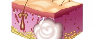
The cause of spermatocele is obstructed fluid outflow and its accumulation in the epididymis. This provokes the formation of a cystic cavity where secretions accumulate. The latter may contain epithelium, seminal plasma, leukocytes, and sperm.
Due to damage to the ducts, the timely evacuation of secretions is impaired, which leads to the accumulation of fluid and the growth of the cyst. In some cases, the formation may have several chambers.
Surgery
The surgical method is the primary step in the treatment of all types of testicular tumors. At the first stage, when a tumor is detected, surgery is performed to remove the testicle (orchidectomy). As a rule, a radical inguinal orchidectomy (surgical removal of the testicle) is performed. The operation is of a therapeutic and diagnostic nature, since in addition to removing the tumor, it allows you to establish a morphological diagnosis (determine the type of tumor), which is necessary to determine further tactics.
Therapy
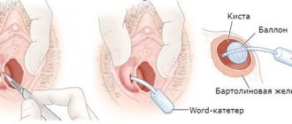
Treatment of small spermatoceles involves a wait-and-see approach. If the tumor does not bother you and does not increase, you should regularly see a urologist. If the cyst progresses, there is a risk of rupture of the cystic formation or suppuration, the doctor will recommend surgical intervention.
Don't be scared: spermatoclecectomy is a minor operation performed under local anesthesia. Using optical technology, the doctor makes a minimal incision in the scrotum and removes the tumor to the surface. With this intervention, neither the testicular tissue nor the epididymis is damaged.
After removal of an epididymal cyst, it must be submitted for histological examination in order to study its structure and exclude an oncological nature.
It is important to understand that although the intervention is minor, the success of the treatment and the absence of complications depend on the quality of the operation. The Dr.AkNer Urology Center offers qualified assistance in the treatment of spermatocele. We employ experienced specialists and have the most up-to-date medical equipment.
Radical inguinal orchidectomy
Surgery to remove a testicle for cancer is called a radical inguinal orchidectomy. The tumor is removed along with the testicle and spermatic cord.
Orchidectomy may be postponed if the patient is in extremely critical condition (in this case, treatment begins with chemotherapy for life-saving reasons).
Tumor in the testicle
After receiving a histological report on the nature of the tumor, examination data: CT of the chest, abdominal cavity, CT/MRI of the pelvis, the level of tumor markers, staging is carried out, the prognosis is determined, and a team of specialists makes a decision on treatment tactics.
When planning chemotherapy, you should discuss with your doctor the risk of fertility problems and the need for sperm cryopreservation. The procedure must be performed before starting chemotherapy.
Chemotherapy
Chemotherapy is one of the main methods of treatment for malignant testicular tumors. These tumors are highly sensitive to chemotherapy, and complete cure is possible even with a large spread of the tumor process. Chemotherapy is not used to treat benign testicular tumors.
Chemotherapy is usually given in cycles of 3 weeks. For malignant testicular tumors, it is very important to strictly observe the intervals between chemotherapy cycles.
The regimen and duration of treatment depend on the stage of the disease, prognosis group, treatment received previously, and the individual characteristics of the patient.
Let's sum it up
The testicles of a newborn boy should be in the constant attention of his parents. Not only the health and proper development of the baby, but also its ability to lead a normal sex life and have children in the future depends on proper child care, hygiene and timely treatment of diseases of the genitourinary system.
If you punctually and regularly examine your baby, take into account the advice of doctors, and do not neglect hygiene, then the risk of male problems and inflammatory diseases will be minimal.
Possible side effects of chemotherapy
Chemotherapy drugs act on rapidly growing cells and dividing tumor cells. But chemotherapy also damages normal tissues in the body, such as cells in the bone marrow (where new blood cells are made), the lining of the mouth, intestines, and hair follicles. The side effects of chemotherapy depend on the type and dose of drugs used and the length of treatment. The most common complications are: decreased blood counts (decrease in the level of leukocytes, neutrophils, hemoglobin, platelets), hair loss, stomatitis (inflammation of the oral mucosa: swelling, redness, plaque, ulcers), nausea and vomiting, weakness, diarrhea, decreased appetite.
Most side effects are short-lived and go away after treatment ends, but some may last a long time, such as hearing loss or kidney or lung damage. Therefore, chemotherapy should be carried out according to absolute indications in accordance with the developed recommendations, and the patient should be under the supervision of an oncologist for a long time.
After chemotherapy and examination, the issue of removing the remaining tumor foci is discussed. One type of surgical treatment is the removal of retroperitoneal lymph nodes (retroperitoneal lymph node dissection).
Depending on the type of tumor and the stage of the disease, lymph nodes around the major blood vessels (aorta and inferior vena cava) may be removed after chemotherapy. Not all patients with a testicular tumor need to have their lymph nodes removed, so it is important to discuss this (and your options) with your doctor. This is a complex and lengthy operation. In most cases, a large incision is made in the middle of the abdomen to allow lymph nodes to be removed. Retroperitoneal lymph node removal should be performed by a surgeon who does it frequently. Experience matters a lot.
Expert recommendations
- Pathologies of the scrotum and testicles can be hereditary, but the course of pregnancy is also of great importance for the normal formation of the fetus. Colds, infectious or viral diseases of the expectant mother can cause problems in the development of the fetus, including delayed testicular descent and dropsy.
- Smoking, especially during pregnancy, is a direct path to the development of such pathologies. Therefore, the expectant mother must approach the period of waiting for the birth of her child very responsibly.
After birth, follow the instructions of your pediatrician and be sure to consult with specialists in case of the slightest deviation from the norm.
Consequences of lymph node dissection
Removal of retroperitoneal lymph nodes is a major operation. It does not cause impotence, and men retain erectile function. But during this operation, some of the nerves that control ejaculation may be damaged. If these nerves are damaged, then when a man ejaculates, the sperm does not exit through the urethra, but goes back into the bladder. This is called retrograde ejaculation, and this complication can make fatherhood difficult.
In case of a widespread process (when there are metastases in other organs), in order to obtain the best result and reduce the risk of the disease returning, various types of operations can be performed to remove all tumor foci. If the remaining tumor foci are not removed or are not completely removed, the risk of the disease returning increases several times.
More often the operation is successful when it is performed by experienced doctors.
Hydrocele of the testicles
Hydrocele of the testicles occurs quite often in newborns. A disease when fluid accumulates and is retained in the membranes of a baby’s testicles. The result is that the entire scrotum or one of its halves increases.
The sex of the baby is determined at the moment of conception in the womb of the mother. At the final stage of scrotal formation, a septum forms between the testicles and the abdominal cavity, preventing the penetration of fluid. If even a small cavity remains between the peritoneum and the scrotum, it is gradually filled with serous fluid. In medicine, the disease is called hydrocele. Pathology of the membrane can cause testicular hydrocele.
How to recognize a dangerous anomaly? The scrotum begins to enlarge due to a pear-shaped seal. Sometimes the swelling takes on an hourglass shape. A sign of water entering the seed canal.
The size of the dropsy varies from a small bump to a swelling the size of a ball. The structure of the skin of the scrotum does not change. It is elastic, mobile and clean, sometimes redness appears. Dropsy does not make urination difficult and does not cause pain.
The cause of the disease is not always a violation of the septum. It happens that the membrane is overgrown, but the scrotum still fills. Congenital hydrocele of the testicles develops due to a malfunction of the infant's lymphatic system. The shells of paired glands secrete a special liquid that reduces their friction. The excess should be absorbed back, but in the first months of a baby’s life the lymphatic system often fails to cope with this task.
As a rule, this disease requires constant monitoring by a pediatric urologist. By one year the signs of dropsy usually disappear. If, nevertheless, the swelling does not go away and continues to increase, then after two years such children are operated on. There is also a possibility that a baby's testicular hydrocele can develop into a hernia.
Fertility
Malignant testicular tumors most often occur in men of reproductive, young age, when they start a family and children. During chemotherapy there is a high risk of impaired fertility, so it is necessary to discuss the need for sperm cryopreservation with your doctor before starting treatment. Sperm cryopreservation is a method of storing ejaculate, which involves freezing it (most often in liquid nitrogen), followed by restoration of sperm function after thawing. Cryopreservation should be performed before starting chemotherapy.
Examination before cryopreservation:
- spermogram (method for examining ejaculate to assess fertilizing ability);
- sperm cryotest (carried out to find out whether the quality of sperm decreases after thawing);
- blood test for infections: syphilis, HIV 1.2 (IgG and IgM), hepatitis B and C;
- urethral smear for genital infections.
After undergoing the examination, sperm is collected by masturbation. Within a few minutes after this, the biomaterial enters the embryology laboratory. Manipulations are carried out to cleanse and concentrate sperm, special substances are added to protect sperm, and the sperm is placed in special thin containers. Your container is labeled (individual data is noted) and then stored at very low temperatures.
Testicular varicocele: noticeable symptoms
The initial stage is characterized by an asymptomatic course. The disease can develop for years, and the man will not even know about its presence.
Symptoms of stage 2 varicocele:
- minor pain in the groin that occurs during exercise, usually the pain goes away at rest, in a lying position;
- increase in testicular size;
- discomfort when walking and sexual intercourse;
- sweating;
- burning in the perineum.
Stage 3 is characterized by:
- constant nagging pain in the groin;
- due to the anatomical features of the left testicular vein, asymmetry of the scrotum occurs;
- the swollen blue veins that form a venous cluster are visually clearly visible;
- the temperature inside the testicle rises and blood circulation is disrupted, this causes changes in sperm motility and the structure of seminal fluid.
Since the disease may not manifest itself in the initial stages, it is important to undergo preventive examinations with a urologist, as well as seek medical help if you identify the first signs of varicocele. Do not self-medicate under any circumstances based on articles and recommendations from the Internet. The only possible way to treat varicocele is surgery. Traditional methods are unable to cope with such a serious pathology, so do not waste time during which your condition may worsen.
Observation
All patients with a malignant testicular tumor should be carefully observed for a long time - up to 10-15 years after initial treatment, since, despite the treatment, there remains a risk of developing a relapse of the disease. The risk of tumor recurrence is highest within two years after initial treatment.
Follow-up includes medical history, examination, tumor markers, ultrasound of the scrotum with elastography, inguinoiliac areas, abdomen and retroperitoneum, and chest x-ray. The observation schedule depends on the stage of the disease. If relapse is detected, treatment options include chemotherapy and surgery.
Our services
The administration of CELT JSC regularly updates the price list posted on the clinic’s website. However, in order to avoid possible misunderstandings, we ask you to clarify the cost of services by phone: +7
| Service name | Price in rubles |
| Appointment with a surgical doctor (primary, for complex programs) | 3 000 |
| Ultrasound of the scrotum | 3 000 |
| Ultrasound of the kidneys and adrenal glands | 2 700 |
All services
Make an appointment through the application or by calling +7 +7 We work every day:
- Monday—Friday: 8.00—20.00
- Saturday: 8.00–18.00
- Sunday is a day off
The nearest metro and MCC stations to the clinic:
- Highway of Enthusiasts or Perovo
- Partisan
- Enthusiast Highway
Driving directions
Forecast
Advances in the treatment of testicular cancer are among the greatest achievements of modern medicine. Today, cure is achievable in 95% of all patients with testicular tumors, and in 80% of patients with the advanced form of the disease. Despite this, the metastatic form of the tumor remains incurable in approximately 10% of patients. The prognosis depends on the histological type of testicular cancer, the extent of the tumor process, and the treatment performed. In some regions of the Russian Federation, the survival rate of patients with testicular tumors reaches only 60%. This is due to various reasons (rarity of the tumor, late presentation of patients, low awareness of doctors).
National Medical Research Center of Oncology named after. N.N. Petrova is a specialized center for the treatment of testicular tumors with many years of experience in the treatment of this group of oncological diseases. Our Center employs a team of highly qualified professionals - oncologists, including surgeons (urologists, thoracic and abdominal surgeons), specialists in drug therapy for adults and children (chemotherapists, oncopediatricians), radiation diagnostics, morphologists, anesthesiologists, resuscitators, radiologists. The management of each patient with a testicular tumor is discussed repeatedly by a multidisciplinary team.
If you or a loved one develops symptoms that suggest a testicular tumor, you should consult a doctor immediately. Considering the high percentage of complete cure for this group of diseases and the high effectiveness of drug therapy, in our Center you can quickly receive all the necessary help from specialists who have extensive experience in treating this pathology.
List of used literature:
- Practical recommendations of the Ministry of Health of the Russian Federation: “Germ cell tumors in men”, 2022.
- Garner MJ, Turner MC, Ghadirian P., Krewski D. Epidemiology of testicular cancer: an overview. Int J Cancer 2005;116:331–9.
- Ferlay J., Colombet M., Soerjomataram I. et al. Cancer incidence and mortality patterns in Europe: Estimates for 40 countries and 25 major cancers in 2022. Eur J Cancer 2018;103:356–87.
- WHO classification of tumors of the urinary system and male genital organs. Ed. by H. Moch et al. Lyon: IARC, 2016.
- Tryakin A.A., Gladkov O.A., Matveev V.B., et al. Practical recommendations for drug treatment of germ cell tumors in men //. Malignant tumors: Practical recommendations RUSSCO, 2022.
- Gilligan T., Lin DW, Aggarwal R., et al. NCCN Clinical Practice Guidelines in Oncology (NCCN Guidelines®). Testicular Cancer. Version 1.2020 — November 5, 2022.