An epidermal cyst (atheroma) is a skin formation that looks like a capsule filled with sebum and particles of keratinized epithelium. Pathology of this type is often diagnosed in patients 25-45 years old. An abnormal manifestation occurs in both men and women, which excludes hormonal prerequisites for development.
Epidermal cysts occur on any part of the body where hair grows, as the formation forms around the hair follicle. The cyst can grow up to 50 mm in diameter. Outwardly, it is similar to a dense pink ball. On the surface of the formation you can see a capillary network. Enlarged pores are also visible. Inside the capsule the secret is a pasty light yellow substance.
The nature of the neoplasm is benign. But there is a risk of degeneration into a malignant form. Penetration of pathogenic microflora into the capsule provokes inflammation. If the epidermal skin cyst is located in the eye area, vision may be impaired due to its pressure. Formations behind the ear provoke headaches and nervousness. Therefore, the appearance of pathology cannot be ignored; if possible, it should be treated.
Don't forget about the cosmetic component. Cysts on the face, neck, and head can significantly spoil a person’s appearance. Large formations cause increased attention from others.
Modern dermatology offers several methods for treating atheromas. Experts from the Lasersvit clinic tell you more about the methods, as well as the causes and symptoms of the appearance of epidermal cysts. If after reading the article you still have questions, our qualified doctors are ready to answer them during a face-to-face appointment.
Epidermal cyst: causes
Unlike dermoid cysts, which are congenital, epidermal (skin) cysts appear due to blockage of the gland duct with sebaceous masses and dead epidermal cells. Provoking factors for the development of pathology:
- When metabolic processes are disrupted or hormonal imbalance occurs, sebum is secreted with a thicker consistency than that of people in good health. Due to the viscous structure of the substance, the physiologically normal outflow of the substance is disrupted. It, like a plug, closes the duct of the glands.
- When there is inflammation on the skin, for example, acne, folliculitis, tissue swelling occurs. This causes a narrowing of the lumen of the excretory duct of the sebaceous gland.
- Scars and scars consist of dense connective tissue, which causes compression of the ducts of the sebaceous glands.
- Improper skin care and use of inappropriate cosmetics can cause blockage of the sebaceous gland.
Any of these factors can provoke the formation of this type of pathology. And if there are several of them, then with each new potential cause the risk of occurrence increases by 10-15%.
Localization
The development of a dermoid cyst is possible in almost any organ or tissue, as doctors note. Most often the presence of cysts is found in:
- the ovarian area in women of reproductive age, which can seriously affect the ability of the fair sex to conceive;
- the lower eyelid area (mostly children are affected, but the pathological neoplasm can be easily removed with the help of surgeons);
- the coccyx area, where the size of the ovarian dermoid cyst rarely reaches large sizes, and the formation can go unnoticed for a long time;
- the area of the anus, where without symptoms of inflammation, pathology is generally detected only by digital rectal examination, etc.
The discovery of dermoid cysts in any organ or tissue makes this pathology quite common. Moreover, as doctors note, the presence of dermoid cysts can either not make itself felt for a long time or cause severe discomfort.
Diagnostics
Diagnosis of the disease is based on visual examination, hardware and laboratory tests. Externally, as already mentioned, atheroma looks like a painless bulge on the skin. The capsule is mobile and can move under the skin under mechanical stress. Atheroma can be detected in areas of the skin rich in sebaceous glands. A cyst often appears on the face, scalp, behind the ears, and on the back. Education is characterized by slow growth.
In a calm state, a cyst on the body does not cause physical discomfort to patients. However, in the presence of an inflammatory process, the tumor can look quite scary - redness and swelling of nearby tissues, thinning of the skin in the area around the tumor. Touching the subcutaneous lump can be extremely painful. The size, condition of the cyst and its location influence the choice of treatment tactics.
Symptoms
Epidermal cysts of superficial localization can be detected during routine visual examination. Sometimes the patients themselves are the first to find them. A similar epidermal cyst is:
- tumor-like intradermal or subcutaneous formation;
- spherical or ellipsoidal;
- soft consistency;
- up to 4 cm in size;
- single;
- easily removable;
- painless;
- the skin over it is of normal color and does not gather in folds.
If the cyst is located intracranially or is located in the chest cavity, symptoms can appear only when it reaches considerable sizes and begins to compress nearby tissues and organs and disrupt their activity. Pain, neurological disorders, shortness of breath, and visual impairment occur. An enlarging scrotal cyst deforms its shape.
Treatment methods
Since the formation takes a long time to develop and is benign, it is not necessary to remove it. The decision to undergo surgical intervention is made for cosmetic reasons or in the event of an unsuccessful pathology location or frequent mechanical damage to a neoplasm protruding above the surface of the skin. This increases the risk of bacterial infection with subsequent transition to purulent processes. In this case, radical treatment in the form of removal is the only way to prevent more serious diseases and complications.
Epidermal cysts are treated in several ways:
- If the formation is large, it is removed with a scalpel. A scar remains at the site of the incision.
- Laser removal of atheroma is considered the preferred method of choice due to the minimal list of contraindications and excellent results.
- Burning with liquid nitrogen is affordable, but has its drawbacks - inaccuracy of the effect, prolonged healing of the burn.
Important: Do not try to treat epidermal cysts or other skin formations with folk remedies. This is fraught with serious damage and the appearance of a scar at the site of exposure. In addition, without histology it is impossible to accurately determine the nature of the neoplasm, therefore it is impossible to talk about its benign quality. Any unprofessional interventions can cause serious health problems.
Symptoms
Detection of cysts - if they are on the surface - rarely poses serious difficulties for the doctor. Dermoid cysts are not very large in size and are usually painless even on palpation. Depending on the contents, the neoplasm can be either dense or soft.
If a dermoid cyst is found in one of the internal organs, it may not make itself felt for a long period of time. Symptoms appear mainly when the tumor reaches a certain size. For example, if there is an ovarian cyst, a woman will complain of:
- pulling pain in the lower abdomen;
- more frequent urge to go to the toilet;
- the appearance of pain during sexual intercourse.
If inflammation of a cyst localized in the anal area develops, there will be pain during bowel movements and difficulty in taking a sitting position.
Symptoms that can characterize a dermoid cyst are very diverse. However, often due to the benign nature of the neoplasm, the pathology is an accidental finding.
Epidermal cyst: laser removal
Removal of a subcutaneous cyst with a laser is a simple procedure that is performed on an outpatient basis and does not require hospitalization or long-term rehabilitation. After conducting an examination and determining the exact location and size of the capsule, the doctor sets the intensity and depth of the laser beam, taking into account the data obtained. The laser acts in a targeted manner without affecting healthy tissue.
During manipulation, the wound is disinfected and cauterized, which eliminates the risk of infection. Laser pulses are short, pain receptors do not have time to react to the impact. Therefore, during the operation, light local anesthesia is used to prevent discomfort.
The whole procedure takes 15-20 minutes. Almost immediately after it, the patient can return to his usual lifestyle. The recovery period involves wearing a sterile bandage for several days until the wound is completely healed.
After laser surgery, there are no scars or scars, which is important if the pathology has formed on the face. And if the epidermal cyst is on the head (pilar), then the laser method is the only way to preserve the hair without the risk of developing a bald spot at the site of the operation.
Treatment of complications of dermoid cyst
Complications may require both medication and emergency surgical intervention. For example, during suppuration, the use of antibiotics and anti-inflammatory drugs is indicated, and removal of the dermoid formation is possible only after the inflammatory process has subsided.
In case of necrosis of the formation, its emergency elimination is indicated, since this can lead to serious complications in the form of suppuration, intoxication, sepsis and even death.
Increased or suspected malignancy is also an indication for removal. In this case, there is no need for emergency surgery, but it should be performed as soon as possible after full preparation (additional examination, anesthetic preparation).
Today, refusal from surgical intervention is considered advisable only in cases where the neoplasm is minimal in size, does not manifest itself in any way, and the likelihood of complications is extremely low. In all other cases, it is better to remove the formation at least in order to prevent the development of complications.
How much does it cost to treat atheroma?
The cost of treating epidermal cysts depends on the size and location (availability). You will also have to pay for the necessary package of studies, without which the operation is impossible. The advantages of the Lasersvit clinic are that we offer a comprehensive service - from seeing a dermatologist to monitoring the patient during the recovery period after surgery. A comprehensive service is always cheaper, and all manipulations and procedures are carried out in one building. This increases the safety and comfort of treatment.
Indications for surgical treatment
It all depends on the location of the formation, its size, development dynamics, the patient’s age and condition, the effect on the functions of internal organs or their aesthetic value.
For example, if it is located under the skin of the face, on the neck or other open areas of the body, is large, grows, or manifests certain symptoms, then it should be removed after a full examination.
In case of a small formation, which does not manifest itself in any way and was an accidental finding of the examination, it is not always worth resorting to surgery - you should consult with a surgeon and evaluate all the pros and cons. Patients are often advised to see a doctor and undergo regular examinations. Only if the pathological formation begins to grow or change in some way should it be removed surgically.
Treatment of benign non-pigmented skin lesions
Papilloma (papillary polyp, papillary fibroepithelioma) is a benign tumor developing from the epithelium; has the appearance of a papillary growth protruding above the surface of the surrounding tissue. The process characterized by the formation of multiple papillomas is called papillomatosis. Papillomas are found on the skin, mucous membrane of the mouth, nose, paranasal sinuses, pharynx, esophagus, larynx, trachea and bronchi, renal pelvis, ureters and bladder, as well as on the labia, vagina, and cervix.
In most cases, papillomas are of a viral nature , i.e. The proliferation of skin tissue is caused by a special virus (HPV - human papillomavirus), transmitted from person to person through direct contact (contact-household and sexual transmission of infection). Microscopically, papilloma consists of connective tissue stroma and epithelium, which may vary depending on the location of the formation. Depending on the amount of connective tissue, papilloma can be soft or dense.
Clinically, papilloma is usually a circumscribed tumor, up to 1-2 cm in diameter (sometimes large), dense or soft to the touch, on a thin long or short stalk, less often on a broad base . The surface of the papilloma is uneven, fine- or coarse-grained, reminiscent of cauliflower or cockscomb. Skin papillomas can have different colors - from white to dirty brown (depending on the blood supply to the vessels and pigment content). Skin papillomas usually do not cause any particular complaints in patients except for a cosmetic defect. Papillomas of other localizations can lead to various complications.
The indication for papilloma removal is the elimination of cosmetic defects , as well as the localization of the tumor, which can lead to functional disorders of the affected organ, trauma to the papilloma and repeated bleeding, inflammation, which in turn is associated with a high risk of malignancy.
All types of local treatment are aimed at removing papilloma and atypically changed epithelium, depending on their location. Various types of chemical coagulations, applications of cytostatic drugs and physical-surgical methods, surgical removal are used. Recently, immunological treatment methods have become widespread - using α, γ-interferons, as well as combined procedures, for example, the combined use of different treatment methods (cryotherapy, laser removal, electrocoagulation, diathermocoagulation).
Today, there are several methods for surgical removal of these formations:
- Chemical coagulation : for this purpose, the application of the drug Solcoderm is used, which is a mixture of organic and inorganic acids that cause the destruction of pathologically altered epithelium. The method is characterized by a slightly painful procedure; it is used to remove single skin papillomas, but may leave scars on the skin after use (difficult to control destructive effects).
- Cryodestruction is the destruction of pathologically altered epithelium by exposure to low temperatures (-96 degrees). A very effective, fast and painless method of removal, effective for single small papillomas without the risk of malignancy, because the method does not provide deep destruction.
- Electrocoagulation (DEC) is the destruction of pathologically altered epithelium by exposure to high temperatures (burning). This is a rather painful technique that gives the highest number of complications, accompanied by prolonged healing and poor cosmetic effect (scar formation). However, it is characterized by high radicalism (total destruction of the affected tissue) and is used when malignancy is suspected.
- Surgical laser is the preferred method of eliminating papillomavirus infection. It is characterized by slight pain, the ability to sting all changed areas to the required depth under eye control, and the absence of scars.
- Radiosurgery (radio knife, Surgitron apparatus) - destruction of the epithelium under the influence of electromagnetic waves. A painless, fast and reliable technique that allows you to cut out a section of tissue with virtually no bleeding and take it for histological examination.
- Scalpel - surgical excision of the tumor along with a section of healthy tissue under local anesthesia, followed by the application of a cosmetic suture. The most radical and reliable way. Allows for histological examination of the formation, which is especially important if malignancy is suspected (chronically injured papilloma).
However, it should be remembered that after removal of the papilloma, the virus may remain in the cells of the surrounding tissues, and papillomas may appear again. In these cases, it is necessary to consult an infectious disease specialist and decide on systemic therapy.
Senile keratoma
Senile keratoma (seborrheic keratoma, senile wart) is a brown or bronze formation with an uneven bumpy surface and clear boundaries, convex, as if glued to the skin. Senile keratomas are benign and occur mainly in older people. They are located on the face or body, are usually numerous and range in size from a few millimeters to several centimeters. If the tumor is damaged (most often by tight clothing), crusts may form on its surface and bleeding may occur. Treatment: cryodestruction with liquid nitrogen, surgical excision of the keratoma. Senile keratoma does not require treatment if it does not cause cosmetic discomfort and is not injured by clothing .
Solar keratosis
Solar keratosis - actinic, or senile keratosis, is classified as a precancerous disease . The disease begins with the appearance of scaly plaques, often accompanied by varying degrees of erythema. The scales are hard, feel like rough sandpaper, and are tightly attached to the skin. Exposed areas of the body are affected: the face, the back of the hands and forearms, and the upper back.
The immediate cause of the disease is the damaging effects of ultraviolet radiation. Periods of spontaneous remission are usually followed by relapses; patients often describe the course of the disease as wave-like. In rare cases, without treatment, tumors may degenerate into squamous cell carcinoma.
Differential diagnosis is made with squamous cell skin cancer.
Treatment. For single plaques - cryodestruction with liquid nitrogen, for extensive skin lesions and multiple plaques - local use of cytostatic drugs (fluorouracil cream).
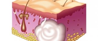
Epidermoid cyst
Epidermoid (epidermal) cyst is a skin cyst, the inner surface of which is lined with stratified squamous epithelium, and the contents are represented by horny scales. It is considered as a developmental defect that occurs as a result of lacing of the epidermis during embryogenesis. Epidermoid cysts occur at any age equally often in men and women, and are localized mainly on the head, trunk and upper extremities.
Microscopic examination reveals a thinned epidermis without skin appendages on the inner surface of the epidermoid cyst, represented by all layers of normal epidermis; the cyst cavity is filled with horny scales. The epithelium lining it, in rare cases, grows, forming outgrowths into the cavity of the cyst in the form of papillae. The contents may be subject to calcification. When the walls of the cyst are destroyed, its contents can penetrate into the dermis, which leads to the development of chronic nonspecific inflammation with the presence of giant foreign body cells in the granulation tissue. Clinically, an epidermoid cyst is a tumor-like formation of a round shape, soft consistency, ranging in size from 5 to 40 mm or more. The skin over it is usually not changed, but if a secondary infection occurs, it may have a reddish tint. The epidermoid cyst is mobile along with the surrounding tissues, painless and grows relatively quickly. Due to the penetration of pathogenic microflora into the cavity of the epidermoid cyst (usually due to microtrauma or blood flow), an inflammatory process often develops in it, up to suppuration with the formation of an abscess.
The diagnosis is made based on the clinical picture and histological examination. Differential diagnosis is carried out with a cystic form of basal cell skin cancer, atheroma, dermoid (see section on soft tissue neoplasms).
Treatment is surgical. The indication for surgery is a cosmetic defect , as well as purulent complications (suppuration and abscess formation). For cosmetic purposes, under local anesthesia, the cyst is excised along with the walls. In cases of suppuration of the epidermoid cyst, it is opened, the contents are evacuated and the cavity is drained. Excision of the cyst walls is performed after the inflammatory phenomena have subsided.
Malignancy of an epidermoid cyst is extremely rare. The prognosis is usually favorable.
Leiomyoma
Cutaneous leiomyoma is a benign tumor that develops from the muscles that raise the hair. It is a smooth yellow-pink or brown plaque, which consists of several fused subcutaneous nodules less than 1 cm in diameter. The tumor is firmly fused to the skin; painful; localized on the face, trunk, limbs. often multiple. Surgical treatment is excision of the tumor.
Dermatofibroma
Dermatofibroma, or histiocytoma, is a benign skin neoplasm represented by fibrous tissue containing a large number of histiocytes and fibroblasts. It occurs mainly in adult women and is localized on the lower extremities, looks like a painless dense subcutaneous node with a diameter of 3-10 mm, which over time usually acquires a reddish-brown color. If you lightly squeeze the skin on the sides of the tumor with your thumb and forefinger, the node seems to fall inward.
Dermatofibroma usually does not require treatment. If the tumor is localized in a site of permanent trauma, its excision under local anesthesia is indicated. Given the deep location of the tumor, minimally invasive techniques (cryodestruction with liquid nitrogen) turn out to be ineffective. because They destroy only the upper pole of the tumor and it can recur.
Milium
Milium, or whiteheads, is a retention cyst of the epidermis with a predominant localization on the face. It is a white, round, dense nodule with a diameter of 1-2 mm. Treatment. The only method of surgical treatment is to open the apex with a scalpel and remove the contents of the cyst. Healing without scar formation.
Keratoacanthoma
Keratoacanthoma is a rapidly growing dense hemispherical node, the size of which can exceed 5 cm. In the center of the tumor there is a crater-shaped depression filled with horny masses. Keratoacanthoma usually occurs on exposed areas of the body in people over 50 years of age. There are indications of a viral nature, the effects of prolonged insolation, trauma, exposure to tar and resins. After 2-3 months, the tumor resolves spontaneously, and a scar may appear in its place. Differential diagnosis is made with squamous cell skin cancer. Keratoacanthoma can be externally indistinguishable from squamous cell skin cancer, so this tumor is classified as both benign and facultative precancerous diseases.
Treatment. Usually resort to excision of the tumor. If the diagnosis is reliably established, electrocoagulation and cryodestruction with liquid nitrogen can be used instead of surgery.
Neurofibroma
Neurofibroma is a benign tumor originating from nerve endings, a soft pinkish-brownish tumor of various sizes, often pedunculated. Occurs on any part of the body except the soles and palms. Multiple neurofibromas are a sign of neurofibromatosis (Recklinghausen's disease). This disease is inherited in an autosomal dominant manner and occurs in adolescence. Over the years, the number and size of neurofibromas increase. Patients often complain of itching. If the tumor bothers the patient, is painful, impedes movement, or leads to disruption of certain functions, its excision is indicated.
Nevus of the sebaceous glands
Nevus of the sebaceous glands is a common developmental defect. It is usually an elongated, hairless neoplasm with a smooth surface, which becomes warty or nodular during puberty. Localized on the face or scalp. In approximately 15% of patients in adulthood, various benign tumors of the skin appendages and basal cell skin cancer develop from the nevus of the sebaceous glands. Differential diagnosis is carried out with linear epidermal nevus. Excision of the nevus is recommended in adolescence.
Fibrous nasal papule
Fibrous nasal papule, or involutive nevus, is a small dome-shaped elevation at the tip of the nose, almost indistinguishable in color from the surrounding skin. Occurs in older people. Differential diagnosis is carried out with basal cell skin cancer, neoplasms of skin appendages.
Treatment. For cosmetic purposes, as well as in case of difficulties in making a differential diagnosis, the tumor is cut off with a sharp scalpel at the skin level and sent for histological examination. The wound heals by secondary intention.
Treatment at the Yusupov Hospital
Doctors at the Yusupov Hospital examine patients with ear cysts, differential diagnoses with benign tumors of adipose tissue (lipomas) and other neoplasms. The key goal of treating a cyst in the ear is to prevent the formation of an ulcer or cellulitis.
Surgeons remove large ear cysts (atheromas) under local anesthesia. During the procedure, a small incision is made on the skin, through which the cystic capsule along with its contents is removed. Complete removal of the cyst walls prevents relapse of the disease. In case of suppuration of an ear cyst after surgery, antibiotics are prescribed.
Small cysts in the ear are removed using a diode laser with a wavelength of 690-1270 nm. The laser produces a gentle temperature effect, after which the tissue structure is restored as completely as possible without the formation of keloid scars. As an alternative to laser, a radio wave method is used to remove ear cysts.