Is there such a thing as ear cancer? This is a question that worries many people who closely monitor their health. Unfortunately, ear cancer is considered a real disease. Ear cancer is a malignant pathology that affects the outer and middle ear. This cancer is rare in Russia.
Of all cancer patients in Russia, only 2% of patients suffer from ear cancer. Pathology develops with approximately the same frequency in women and men. Cases of middle ear cancer developing in children under the age of 10 are not uncommon. During adolescence, pathology practically does not develop. Ear cancer is most often diagnosed in older patients.
Causes
The etiology of ear cancer is studied by specialists from all over the world to this day. Scientists have put forward a theory that the oncological process develops under the influence of cancer viruses, which lead to DNA mutation. However, this assumption was only partially confirmed. Researchers have named the main factors that contribute to the formation of squamous cell ear cancer:
- the presence of chronic pathologies (otitis media, eczema);
- genetic predisposition;
- polyps;
- regular contact with aggressive carcinogens (alkalis, acids, heavy metals);
- inflammation of connective tissue.
It has been established that people who work in enterprises with radioactive radiation are most often exposed to the development of ear cancer.
Classification of ear oncology
Based on the location of the malignant neoplasm, ear cancer is divided into two types: tumor of the outer and middle ear. Depending on the reasons of origin, the oncological process can be primary and secondary. Depending on the nature and intensity of tumor growth, the pathology of ear cancer can be exophytic and endophytic. Based on the type of histological structure, tumors are divided into the following types:
- spinocellular epithelioma. A distinctive feature of the pathology is the accelerated growth of a malignant tumor. As it progresses, the patient develops a growth in the area of the auricle that regularly bleeds and darkens. In later stages, the tumor affects the entire ear canal;
- basalioma. Squamous cell carcinoma of the ear develops from epithelial cells. The pathology is accompanied by the appearance of an ulcerative formation in the area of the ear cavity, which periodically erupts and causes discomfort to the patient. Ear skin cancer grows slowly and metastasizes only in the last stages;
- sarcoma. Develops from connective tissue. One of the most dangerous types of cancer. When the tumor is localized in the area of the auricle, the progression of the disease occurs in a slow manner. If a cancerous tumor appears near the ear canal, the pathology quickly progresses to stages 3 and 4. Symptoms of ear cancer in this case manifest themselves in the form of pain, impaired jaw mobility, and paralysis of the facial muscles.
In rare cases, melanoma (cancer of the skin of the ear) forms in the ear cavity. Signs of ear cancer in this case manifest themselves in the form of a weeping ulcer, which over time metastasizes to neighboring lymph nodes.
Stages
According to its prevalence, ear cancer occurs in several stages:
- Stage 1. The cancer affects the skin or mucous membrane of the outer ear. In this case, bone and cartilage tissue are not damaged;
- Stage 2. The cancerous tumor penetrates the bone and cartilage tissue, the patient begins to show the first signs of ear cancer in the form of bleeding or purulent lesions;
- Stage 3. The cancer metastasizes to neighboring lymph nodes, the general condition of the sick person worsens;
- Stage 4. Squamous cell carcinoma of the auricle penetrates into the deep cervical lymph nodes, conglomerates and hematogenous metastases form in the patient’s body.
If stage 1 or 2 of the disease is detected, the patient can expect recovery in 90% of cases. At a later stage, the prognosis worsens, and survival rate is reduced by almost 2 times. When ear cancer metastasizes, it is fatal.
Middle ear tumors
Benign tumors are represented by fibromas, angiomas, endotheliomas, and osteomas. Treatment of tumors is surgical, angiomas are treated with electrocoagulation, osteomas are treated with x-ray therapy.
True cholesteatomas are rarely found in the area of the temporal bone scales, in the mastoid process, sometimes spreading to the tympanic cavity and cranial fossae. In recent years, glomus tumor has often been observed. This tumor ranks first among middle ear tumors in frequency. It develops from glomus (glomeruli), often found formations along the tympanic nerve, the auricular branch of the vagus nerve, and less commonly the superior petrosal nerve. Glomus have a capsule, consist of numerous capillaries and special glomus cells, and are innervated by parasympathetic nerves (glossopharyngeal and vagus). The tumor is characterized by slow growth, but is infiltrative in nature and often produces bleeding. Treatment is radiation therapy or surgery followed by radiation.
Malignant tumors of the middle ear (sarcoma) are extremely rare, most often in children. Treatment is surgical.
Much more common is middle ear cancer, which, as a rule, develops due to chronic suppurative otitis media, and is therefore diagnosed late. Most often, the tumor originates from the attic-antral region or the area of the tympanic ring. This type of tumor is characterized by rapid infiltrative growth, especially in young people, with spread to the parotid gland, lower jaw joint, inner ear, cranial cavity, and is characterized by early metastasis to regional lymph nodes.
Symptoms and signs of ear cancer
At an early stage of development, ear cancer does not cause severe symptoms, like any cancer. As the disease progresses, the patient develops a small tumor that resembles a wart in appearance. Over time, the tumor thickens and increases in size. Pigmented neoplasm is rarely accompanied by negative signs. Usually it does not hurt, does not burn or cause discomfort.
Many patients pay attention to the growth that appears only because of an aesthetic defect. Ear cancer is discovered when visiting a beauty salon to remove a wart. In later stages of the disease, a person may notice the following negative symptoms of ear cancer:
- regular increase in body temperature;
- profuse sweating;
- general weakness of the body;
- pain in the affected area;
- hearing impairment;
- pressure changes, tachycardia;
- purulent and bloody manifestations from the formed ulcers.
When ear cancer reaches stages 3 and 4, tumor growth accelerates significantly.
Outer ear cancer
Ear cancer at the initial stage of development is asymptomatic. During the transition to stage 2, the sick person experiences severe itching and pain in the area of the ear cavity. Squamous cell carcinoma of the auricle is often accompanied by ringing or noise in the ears.
Upon examination, the doctor may detect a small ulcer or granulation in the area of the external auditory canal. Over time, the ulcers begin to bleed, burn and cause severe discomfort to the patient. In addition to bloody ulcers, the patient may detect serous and purulent discharge.
At stages 3 and 4, ear cancer metastasizes to neighboring lymph nodes. Cancer of the lymph node behind the ear is accompanied by severe pain, hearing loss, deformation of the facial nerves, and deterioration of the general condition.
Diagnostics
An accurate diagnosis is made after the following studies:
- otoscopy. A thorough examination of the ear cavity, eardrum, and ear canal. Otoscopy helps detect tumors in the external parts;
- histological examination. A diagnostic procedure to detect cancer cells in excised skin samples;
- MRI. One of the most accurate methods for determining the presence of cancer in the body. MRI allows you to determine ear cancer at stages 1 and 2 of development. In this case, the procedure allows you to find out more detailed information about the size, location and type of tumor;
- CT. An X-ray method for examining bone tissue for the presence of metastases.
In additional cases, audiometry may be required. This procedure helps to provide more accurate data on the state of the auditory analyzer.
Treatment
In the early stages of ear cancer development, the patient is prescribed courses of radiotherapy. After this, electroexcision is additionally performed. At stage 2, ear cancer is treated with surgical excision and subsequent radiotherapy. At later stages of development, radiation therapy with complete excision of the affected lymph nodes is performed to treat ear cancer. In case of multiple metastases, a Krail operation is performed.
Treatment of ear tumor
The most effective combination treatment is extensive surgery followed by radiation therapy. At stage 4, only radiation or chemotherapy is indicated.
Acoustic neuroma is also classified as an ear tumor. This structurally benign tumor, but with a clinically unfavorable course, located in the pontocerebellar space, can originate from the Schwann sheath of the nerve along its entire length from the bottom of the internal auditory canal to its entrance into the medulla oblongata. At the 1st stage of tumor development, there is noise in the ear on the affected side, decreased hearing, sometimes slight pain in the ear, rarely dizziness, and mild ataxia. In the 2nd stage, hearing loss progresses, spontaneous nystagmus and other vestibular disorders appear, paresis of the 5,6,7 cranial nerves, and headache in the occipital region. In the 3rd stage, severe hearing loss and loss of vestibular function are accompanied by symptoms of cerebellar damage and severe hypertension. Subsequently, bulbar disorders, lesions of other cranial nerves, visual impairment, etc. develop. Treatment is surgical, the prognosis is favorable for stages 2-3 of the disease.
Inner ear cancer
In the first stages of progression, the symptoms of ear cancer resemble chronic otitis media. A sick person develops minor hearing problems, congestion, and purulent lesions. As ear cancer develops, it begins to be accompanied by severe pain and discomfort; patients complain of discomfort in the neck, cheekbone, and temporal zone.
At stages 3 and 4 of auricle cancer, the patient experiences paralysis of facial muscles, impaired swallowing, and immobility of the lower jaw. If the tumor reaches the carotid artery, heavy bleeding may occur. When the tumor spreads into the meninga, carcinomatous meningitis occurs.
Diagnostics
To make a diagnosis, the patient is referred for otoscopy, skull radiography, and histological examination. To clarify the volume of the cancer-affected area, an examination is carried out by a neurologist. MRI and CT are used to determine the characteristics of a malignant tumor, its size and type.
Treatment
Combination therapy is used to treat ear cancer. It usually consists of a subtotal resection of the temporal region with postoperative telegammatherapy. When the tumor spreads to nearby lymph nodes, their excision or Krail operation is performed. At stages 3 and 4 of development, radiotherapy is prescribed. Chemotherapy in this case will be less effective.
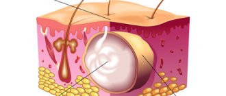
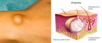
Treatment of earlobe atheroma
This pathology must be treated in a clinic. Self-medication in such a situation will not only not help, but will also provoke complications. There is no conservative treatment for such a tumor. In practice, the following treatment methods are used:
- surgery (Removal during surgery is the most reliable way to cure the ear. To carry out the removal, the parts of the ear where the cyst has arisen are subjected to local anesthesia. To perform the operation, you do not need to go to the hospital. It is performed on an outpatient basis.);
- laser removal (this method is used for small lesions);
- radio wave method (during this method, the tissues of the lobe are heated, and the cyst cells seem to evaporate. The procedure is also carried out under local anesthesia).
There are situations when atheroma becomes inflamed and suppuration occurs. In this case, it is necessary to urgently contact an ENT doctor to open the tumor and prevent inflammation of nearby tissues. An otolaryngologist will open the atheroma, empty its contents and prescribe a course of antibacterial therapy. About a month after the autopsy, after the inflammatory process has subsided, it will be necessary to perform an operation to directly remove the formation. If repeated surgery is not performed, a relapse may develop and the cyst will appear again.