Otitis is a disease accompanied by severe (both shooting, pulsating or aching) pain in the ears. Pain from otitis media can radiate to the teeth, temple, the corresponding side of the head and the back of the head. The patient experiences weakness, insomnia, and loss of appetite.
Depending on the nature of the disease, otitis media can occur in acute and chronic forms.
Acute otitis media is severe and characterized by severe pain.
1
Audiometry in MedicCity
2 Flushing the ear canals
3 Diagnosis of otitis in MedicCity
Acute otitis is a signal for the patient that it is necessary to urgently consult a doctor! Acute ear pain cannot be tolerated; it can cause deafness! Chronic otitis of the ear is less pronounced, but also very dangerous! Otitis media does not go away on its own; after otitis media, the patient may lose hearing forever, so at the first signs of the disease, you should urgently contact a specialist.
Types of otitis
Depending on the direction of the pain, it is customary to distinguish 3 types of otitis: external, middle and internal otitis.
Otitis externa most often appears as a result of mechanical damage to the auricle or external auditory canal. The following symptoms are characteristic of external otitis of the ear: aching, dull pain, swelling of the ear, and a slight increase in temperature.
Otitis of the middle ear is an inflammatory disease of the air cavities of the middle ear: the tympanic cavity, the auditory tube and the mastoid process.
Internal otitis is untreated otitis media of the middle ear. With internal otitis, inflammation of the inner ear occurs and the entire vestibular apparatus is damaged.
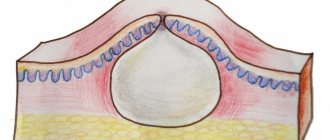
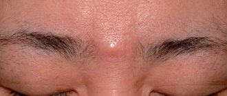
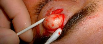
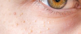
What does atheroma behind the ear look like?
The reasons for the development of atheroma include the following factors:
- Disturbance of metabolic processes in the body.
- Harmful factors of external and internal ecology.
- Excessive sweating (hyperhidrosis).
- Diseases of the endocrine system and hormonal imbalance.
- Increased production of sebum by the glands and oily skin.
- Infection of the sebaceous gland duct (during ear piercing, for example).
- Presence of acne and seborrhea of the scalp.
Factors that can trigger the appearance of atheroma include improper and insufficient hygienic care and hypothermia of the body.
Acute otitis media
According to statistics, acute forms of otitis media account for 30% of the total number of ENT diseases. Most often it occurs in preschool children.
Symptoms of acute otitis media
The disease is characterized by an acute onset with the appearance of the following symptoms:
- earache;
- ear congestion or hearing loss;
- increased body temperature;
- anxiety;
- disturbance of appetite, sleep;
- headache and toothache.
Causes of development of acute otitis media
In most cases, the disease can be caused by various pathogenic microorganisms - viruses, microbes, fungi, etc. In exudate obtained from the middle ear, respiratory viruses are found in 30-50% of cases. The most common causes of otitis are parainfluenza viruses , influenza viruses, rhinoviruses, adenoviruses, enteroviruses, respiratory syncytial viruses, etc.
In 50-70% of patients with acute otitis media, bacteria are detected in the exudate from the middle ear (most often Streptococcus pneumoniae, Haemophilus influenzae, Moraxella catarrhalis).
Often the cause of otitis is a mixed (viral-bacterial) infection.
When making a diagnosis, a differential diagnosis is made with myringitis (inflammation of the eardrum) and exudative otitis media.
The occurrence of otitis media is directly related to the condition of the nose and nasopharynx: rhinitis and tonsillitis often provoke inflammation of the middle ear.
Otitis often occurs against the background of decreased immunity and immunodeficiency states.
1 Diagnosis of otitis in MedicCity
2 Diagnosis of otitis in MedicCity
3 Diagnosis of otitis in MedicCity
Routes of infection
The most common route of infection into the middle ear is through the auditory tube during rhinitis and sinusitis.
It is possible that infection can penetrate through the blood during influenza, scarlet fever and other infectious diseases.
In rare cases, the infection enters the middle ear through the ear canal due to injury (rupture) of the eardrum.
Stages of acute otitis
There are 5 stages of the disease:
- stage of acute eustachitis: feeling of congestion, noise in the ear, normal body temperature (if there is an infection, it may increase);
- stage of acute catarrhal inflammation in the middle ear: sharp pain in the ear, low-grade fever, inflammation of the mucous membrane of the middle ear, increasing noise and congestion in the ear;
- pre-perforative stage of acute purulent inflammation in the middle ear: sharp unbearable pain in the ear, which radiates to the eye, teeth, neck, pharynx, increased noise in the ear and decreased hearing, increased body temperature to 38-39 degrees, the blood picture becomes inflammatory in nature;
- post-perforation stage of acute purulent inflammation in the middle ear: pain in the ear becomes weaker, suppuration appears from the ear, noise in the ear and hearing loss do not go away, body temperature becomes normal;
- reparative stage : inflammation is stopped, perforation is closed with a scar.
Treatment of otitis media
If you have otitis media, treatment can only be prescribed by an otolaryngologist. Treatment of otitis media depends on the stage of the disease and the patient’s condition.
In acute eustachitis, treatment of otitis media is aimed at restoring the functions of the auditory tube. Sanitation of the paranasal sinuses, nose and nasopharynx is carried out in order to eliminate infection - rhinitis, sinuitis, etc.).
Vasoconstrictor nasal drops (otrivin, nazivin, etc.) are prescribed; in case of excessive mucous discharge from the nose, drugs with an astringent effect (collargol, protargol) are prescribed. Catheterization of the auditory tube is carried out using aqueous solutions of corticosteroids, and pneumomassage of the eardrums.
In the stage of acute catarrhal otitis media, catheterization of the auditory tube is carried out with the introduction of aqueous solutions of corticosteroids and antibiotics (penicillins, cephalosporins) into the cavity of the middle ear. Local anesthesia is prescribed (otipax drops, Anauran, Otinum). An intra-ear endaural microcompress according to Tsytovich is carried out: a cotton or gauze turunda soaked in a drug with an analgesic and dehydrating effect is inserted into the external auditory canal. Painkillers with an antipyretic effect (nurofen, solpadeine, etc.) are also prescribed. If there is no effect from symptomatic therapy, antibiotic therapy is prescribed within 48-72 hours.
Purulent otitis in the pre-perforated acute stage requires the same set of procedures as in the second stage, but supplemented with the following measures:
- prescription of penicillin antibiotics (amoxicillin, etc.), cephalosporins or macrolides;
- paracentesis (incision of the eardrum) when the eardrum appears to bulge.
It is important to prevent complications of the disease at this stage. After spontaneous opening of the eardrum or paracentesis, the disease progresses to the next stage.
The post-perforation stage of acute purulent otitis media involves the following treatment regimen:
- started antibacterial therapy continues;
- catheterization of the auditory tube is performed with the introduction of corticosteroids and antibiotics;
- a thorough toilet of the external auditory canal is carried out daily - cleaning it from purulent contents;
- transtympanic infusion of drops with an antibacterial and anti-edematous effect is prescribed (alcohol-based drops (otipax, 3% boric acid solution) are not used in this case).
In the scarring stage of AOM, spontaneous restoration of the integrity of the membrane occurs, and all functions of the ear are completely restored. However, this period requires mandatory observation by an otolaryngologist: there is a danger of chronic inflammation in the middle ear, its transition to a purulent form, or the development of an adhesive scar process in the tympanic cavity. It is also possible to develop mastoiditis.
1 Audiometry in MedicCity
2 Audiometry in MedicCity
3 Audiometry in MedicCity
In case of acute otitis media, timely contact with an otorhinolaryngologist is very important. The only measure to prevent complications is correct and timely diagnostic and treatment measures for otitis media. Sometimes the consequences of acute otitis media are adhesions in the tympanic cavity (adhesive otitis media), dry perforation in the eardrum (dry perforated otitis media), purulent perforation (chronic suppurative otitis media), etc. In addition, AOM can lead to such complications as such as mastoiditis, labyrinthitis, petrositis, meningitis, sepsis, venous sinus thrombosis, brain abscess and other life-threatening diseases of the patient.
Treatment of otitis media during pregnancy
If you experience ear pain during pregnancy, you should urgently see an ENT doctor. Remember that in this case you cannot apply heating pads or warm compresses to the sore spot! This can be very dangerous if purulent inflammation begins in the ear.
If the pain increases and greatly bothers a pregnant woman, and there is no way to see a doctor in the near future, you can take several independent steps. For example, you should put vasoconstrictor drops into your nose.
Reasons for the development of atheroma
Atheroma develops in those places where there is an accumulation of a large number of sebaceous glands - in the area around the nose, on the eyelids, neck, scalp, chest, back and earlobe. Atheroma behind the ear is the most common of all types of atheroma, and, as a rule, affects both men and women equally.
The occurrence of atheroma of the earlobe is characterized by an unpredictable course and frequency of occurrence due to constant rubbing of the tumor with foreign objects - various hats, scarves, headphones, collars of shirts and blouses. In medical practice, cases of degeneration of a benign tumor into a malignant one are described, which occur in cases where proper surgical treatment is not carried out.
What is prohibited for otitis media
- Under no circumstances should foreign bodies be introduced into the ear (geranium leaves, ear phyto-candles). This will make diagnosis difficult and may lead to a worsening of the condition (for example, leaves that have not been removed begin to rot and become a source of infection).
- If the pain is severe, do not apply a heating pad to your ear or apply warm compresses. This is dangerous if purulent inflammation has begun in the ear. Compresses can only help at stages 1-2 of the disease.
- You should not put melted oil in your ear: if there is a perforation, the oil will end up in the tympanic cavity.
- You should not put camphor oil or camphor alcohol into your ear - it can burn the walls of the ear canal and irritate the eardrum, which will increase ear pain.
At MedicCity you will be denied professional help for otitis media and other ENT diseases. Our otolaryngologists will conduct a comprehensive examination of the patient and prescribe a treatment regimen, depending on the cause and stage of the disease. However, the success of treatment depends no less on the patient himself: the sooner he consults a doctor, the more effective the result will be and the lower the likelihood of complications. It is also important to follow preventive measures. So, in the cold season, to prevent otitis media, it is important to wear a hat, protect your ears from drafts, and of course, boost your immunity!
Treatment and prevention of atheroma
Treatment of atheroma is predominantly surgical, with removal of the cavity along with its contents. To do this, a small skin incision is made on the body, through which access to the atheroma is provided. After removing the contents, the cavity is washed, and if necessary, a turunda is inserted for several days to facilitate the release of blood, pus and fat.
The operation is performed not in a hospital setting, but on an outpatient basis. Local anesthesia is used for pain relief. For small atheromas, sutures are not applied, since the skin incision heals on its own after 5-6 days. For large atheromas, cosmetic sutures are applied, which are regularly processed after 1-2 days. The operation itself is painless for the patient, but the danger is that without eliminating the causes that caused the appearance of atheroma, there is a high probability of relapses (almost 50% of all cases). That is why it is so important not only to eliminate the cause of atheroma, but also to take measures to prevent the disease. These include personal hygiene measures and early consultation with a doctor. It is not recommended to treat atheroma on your own.
