- Viral skin diseases: causes, classification
- Herpes
- Shingles (herpes zoster)
| |
Vulgar or simple (ordinary) warts are caused by the human papillomavirus-2 (HSV-2), which selectively affects the epidermis. Vulgar warts on the hands make up 70% of all warts and are localized benign hyperplasia of the epidermis.
Infection with warts occurs through contact—by contact with the affected skin. The penetration of the virus is facilitated by minor injuries, leading to disruption of the integrity of the stratum corneum of the epidermis, dry skin, acrocyanosis, hyperhidrosis, and a decrease in the pH of the water-lipid mantle.
Routes of infection with human papillomavirus, which causes the development of warts
The virus can reach a healthy person through direct contact - a handshake, for example, or through infected objects: toys, handrails on public transport. Two to three hours of virus life in the external environment is enough for someone to become infected with warts: the infection is quite common.
Cosmetic defect is the main complaint of patients with warts. There is usually no pain. Moreover, many medical procedures, such as cryodestruction, cause more suffering than the warts themselves.
Equipment
In the case of laser removal of plantar warts, a special medical laser device is used that emits rays that are absorbed by water or blood in the tissues. Thanks to this specific absorption, water quickly evaporates from the tissues, they dry out with minimal blood loss. In case of removal of plantar warts using the radio wave method, appropriate devices (Surgitron or other brands) are used. In them, electromagnetic radiation with a very high frequency evenly and bloodlessly separates tissues without burning them.
Clinical picture of warts
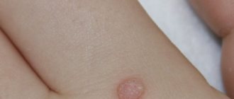
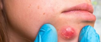
Externally, warts are dense nodules with a diameter of 1 to 10 mm (sometimes more), round or polycyclic in shape. Their surface is covered with cracks, horny layers, and vegetations. The color of warts often does not differ from the healthy skin surrounding them, but under a magnifying glass you can see black-brown dots, which are capillaries filled with blood clots.
The rash can be represented by either one element or multiple, randomly located isolated nodules. After a wart is removed, new ones (daughter ones) may appear around it, arranged in a circle. Warts are localized in easily injured areas: hands, fingers, knees. Localization of warts on the fingers and palms leads to changes in the skin pattern. In this situation, the recovery of fingerprints is a sign of recovery.
There are several types of warts.
Common warts
Dense, dry, limited, painless keratinized elevations with an uneven villous surface, the size of a pinhead to a pea. They can merge to form large plaques. Most often located on the hands.
Plantar warts
They are a type of common warts - they appear in places where shoe pressure is applied, especially in those who sweat a lot. Very dense, keratinized, gray-dirty plantar warts are characterized by severe pain that prevents walking; sometimes cause temporary disability.
Flat or juvenile warts
They usually occur in children and young people. They look like round or irregularly shaped flat nodules that are located on the back of the hands, as well as on the skin of the face. The appearance of flat warts is promoted by skin irritation (they often occur along the course of scratches, cuts, etc.).
Mosaic plantar warts
Mosaic warts are a special type of neoplasm. They are plaques, so-called clusters, formed as a result of the fusion of many small plantar warts tightly pressed together. The arrangement of the plaques resembles a mosaic (hence their name).
This formation is usually observed in a small and localized area. It can reach a diameter of about 6-7 cm. In the early stages of development, mosaic warts look like small black punctures. As they develop, they take on the appearance of a white, yellowish or light brown cauliflower, with dark spots in the middle. These spots are formed due to thrombosis of blood vessels.
This type of wart is quite rare. They usually affect the hands or soles of the feet, and are especially common under the toes. Unlike simple plantar warts, mosaic warts cause little or no pain when walking because they are flatter and more superficial.
Mosaic warts are highly contagious. They are difficult to treat due to the multiplicity of foci of viral infection. The success of treatment is facilitated by its timely initiation. As a rule, mosaic growths are prone to recurrence even after surgical removal.
Basic methods for removing warts
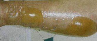
The two most commonly used methods for removing warts are cryotherapy and removal using a radio wave scalpel. If cryotherapy is performed, warts that appear on the patient's skin are treated with liquid nitrogen, a substance that allows objects to be cooled to very low temperatures. Namely, up to -187 degrees. After such a strong freezing of the wart, a characteristic bubble forms in its place after some time. Soon it opens, a wound forms in its place, which heals within 2-3 weeks, then a scar forms in this place. This method of removing warts is almost painless (there is only mild discomfort). The disadvantage of this method is that it is difficult to control the depth of removal, and therefore the remaining scar may be quite large or the wart may not be completely removed.
Removal using a “radio wave scalpel” involves exposing the wart to radio wave energy. This method is effective and safe. It allows you to accurately control the depth and size of the impact.
Are warts always dangerous?
Most warts are completely harmless and can theoretically disappear in a few weeks or at most a month. In this case, patients are more likely to be concerned about a serious cosmetic defect, which causes psychological discomfort and interferes with leading a full lifestyle.
Warts are often painless unless they are on the soles of the feet or another part of the body that is subject to shock or constant contact. But there are cases of itching and discomfort in the affected area.
But as mentioned above, warts are viral in nature, so you cannot expect that the neoplasm will go away on its own or will not bother you for the rest of your life. Any wart should be shown to a dermatologist, and if he deems it necessary, it should be removed using one of the safe methods.
Removing warts using radio waves
1. A 1-2% solution of local anesthetic (for example, lidocaine) is injected into the area around the wart or under it.
2. Using a radio wave scalpel, the wart is excised within healthy skin.
Immediately after removal, a fairly deep wound is formed at the site of the wart (to the entire depth of the removed wart).
Treatment of warts in St. Petersburg
Treating warts requires a lot of patience. Warts may appear and disappear for no apparent reason or for reasons that are difficult to identify. High infectivity and autoculation of warts are arguments in favor of removal.
There are many different treatment methods. Therapeutic choices differ depending on the type of wart, its location, depth, number and extent of the affected area of skin.
Treatment for warts can produce very different results. Some warts respond to treatment while others do not, even if it is given to the same person.
Treatment often requires repetition over several weeks, months and in extreme cases even years before success is achieved. But in any case, wart removal should be performed in a clinic by a dermatologist. Self-medication is highly discouraged, as the consequences may be irreparable.
The most gentle and universal ways to get rid of warts of all types are laser and radio wave therapy.
After wart removal
Immediately after removing the wart, I apply a sterile napkin or a special sterile gel to the wound, and the patient goes home. At home, the patient takes care of the wound independently. Since the wart is removed quite deeply, it may take quite a long time for the wound to heal. 6-10 days after the wart removal procedure, a crust usually forms at the site of the wound. After the crust comes off, a small pink scar usually remains. After 3-4 months, the scar will begin to turn white.
After the final formation of the scar, usually after 6-12 months, it becomes almost invisible.
There are other methods for removing warts - experienced doctors will select the right method for treating warts at our Center for Medical Cosmetology.
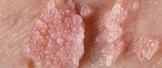
Filiform (acrochord) warts
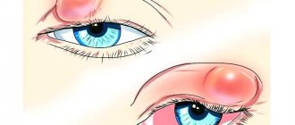
Filiform warts, also known as facial warts, are the most unusual type of these growths. They are thin, long, racem-like shoots that are usually found on the eyelids and surrounding areas, on the neck, near the lips and nose, and less commonly on the legs, in the groin folds, under the mammary glands and in the armpits.
The typical color of acrochords is flesh-colored, which is why people do not immediately notice them. Sometimes they may turn yellow, brown or pink. Usually they reach a length of 5 to 10 mm, extremely rarely - several centimeters. Depending on the severity of the virus, acrochords form singly or in multiple clusters. This distinctive type of filamentous wart is usually diagnosed visually.
Filiform warts form when a strain of the human papillomavirus causes the top layer of skin to grow too quickly. At the inception stage, the growth looks like a yellowish bump. As it grows, it stretches out, transforming into an elongated formation on a stalk. To the touch, the wart has an elastic and dense structure.
People of absolutely any age can become a “target”, but often elderly patients suffer from this disease. According to statistics, about 50% of the world's population over 50 years of age have facial warts.
Infection with the virus often occurs through cracks and abrasions on the face, so people with dry skin are at high risk. Also, the appearance, growth and spread of facial warts is facilitated by various hormonal changes (pregnancy, obesity, menopause, ovarian dysfunction, diabetes, etc.).
Although highly contagious (infectious) and unattractive in appearance, this type of wart is benign, painless, and often responds well to treatment.
When they appear in sensitive areas, such as skin folds, or areas often subject to pressure and injury, some symptoms may occur:
- itching;
- bleeding;
- soreness;
- irritation.
This type of wart almost never develops into a malignant form. However, if the acrochord is injured, there is a high risk of developing an inflammatory process. Unlike many other similar neoplasms, the filamentous type does not disappear on its own. When a wart falls off, a new one grows in its place. Sometimes there is keratinization of the growth and its transformation into a cutaneous horn.
Facial papillomas are contagious and can be spread by sharing towels or facial cosmetics. Touching the acrochords puts a person at risk of spreading them to other parts of the body. Warts will increase in size and number if they are not removed.
Their location and ugly appearance make facial growths a cause of emotional stress and embarrassment for many people, sometimes affecting their self-esteem and self-confidence.
Prevention of warts
• Strict adherence to personal hygiene rules.
After visiting public places, wash your hands with soap. • If wounds or cuts occur on the skin, treat them with an alcohol solution, iodine or brilliant green. • When working with cleaning products or skin-damaging factors, use gloves. • Wear shoes made of genuine leather or fabric, avoid wearing synthetic shoes, especially for children and adolescents. • Normalize your diet, eat foods rich in vitamins. Avoid stress, organize your life. • When in contact with a person who already has warts, it is necessary to follow the rules of personal hygiene - wash your hands often with soap, limit the use of shared objects. [/td]
Senile (age-related keratomas, seborrheic keratoses) warts
Senile warts are one of the most common skin lesions that appear in old age as a general sign of skin aging. Despite their name, they are not caused by the human papillomavirus.
Seborrheic keratoses are extremely common. According to statistics, more than 90% of the population over the age of 60 have one or more of them. They are equally common in both men and women. It is not uncommon for the disease to affect people aged 30-40 years, as well as young people under 20 years of age.
Keratomas and keratoses can appear on any part of the body, including the scalp, face and genitals. The exception is the palms, soles of the feet and mucous membranes. It is rare for a person to develop only one growth. Over time, age-related keratomas become more and more numerous. Many people inherit a tendency to develop a very large number of these tumors. Some of them may have hundreds of wart-like growths scattered throughout their body.
In the early stages, aging warts appear as slightly raised light brown spots or papules. They can remain very flat and resemble freckles in appearance, or they can gradually thicken and develop a rough, warty surface, like a tumor on the skin. In most cases, they darken slowly and may eventually turn black.
These color changes are harmless. Many senile warts remain pinkish in color. Typical of these are small keratin plugs that can be seen on the surface of the wart.
Keratoses are usually round or oval in shape. Some seborrheic warts are irregular in shape. Their size can vary from one to several centimeters in diameter.
The cause of age-related keratomas is unknown. They are generally considered to be degenerative in nature and appear in large numbers as the skin ages. It is assumed that ultraviolet radiation increases the likelihood of their development.
There are five traditional forms of age-related warts:
- Spotted, or popularly “death freckles” . They form in numerous clusters on the hands and face. Such growths are round with an uneven contour and a smooth or slightly rough surface. It has several color options: light brown, brown-brown or pinkish-yellow;
- Papular or nodular . Larger growths tend to grow. Their typical color is gray or yellow. The surface of the wart is covered with horny layers;
- Classic keratoma . It is a collection of plaques tightly connected to each other. It is characterized by a jagged outline and a copper or pinkish color. As it grows, the middle part of the wart sinks;
- Cutaneous horn. It is a modification of keratoma. It is expressed as a cluster of dense keratinized dark brown plaques up to 1.5 cm.
Adverse reactions to certain medications and many chemotherapy drugs can contribute to the formation of irritated seborrheic keratoses—inflamed, red, crusty lesions. This leads to the development of eczematous dermatitis around the growth. Dermatitis can also cause new seborrheic keratoses to appear.
Age-related keratomas are always benign. This means that they do not spread and do not degenerate into a malignant form. The main problem is a cosmetic defect, especially if they develop on the face.
There are rare cases of skin cancer called melanoma that develops in a seborrheic wart. It is unknown whether this is just a coincidence or represents a true change in the cells in a seborrheic wart. A large number of age-related keratomas may be a sign of cancer of internal organs.
Typically, seborrheic keratoses are treated for cosmetic reasons or because they become itchy and irritating. If the growths, especially large and warty ones, are injured (rubbed against clothing, touched by something), they may bleed or become inflamed.
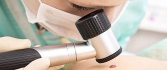
Diagnosis of the disease is usually made through a clinical examination. This type of wart is difficult to distinguish from skin cancer without histological examination. Therefore, very dark lesions that have changed in some way or that are growing rapidly require a biopsy to confirm the diagnosis and rule out the possibility of cancer. Darker lesions should also be checked by a doctor to make sure they are not melanoma.