Causes of mycoses
Mycosis is an infectious dermatological disease caused by filamentous fungi. The source of infection is a sick person; in addition, fungal spores can be found on personal items, the use of which leads to infection.
The main methods of transmission of mycoses are:
- Close contact with an infected person.
- Penetration of microbes through microcracks, abrasions, abrasions and wounds on the body.
- The use of towels, manicure items, shoes, and combs contaminated with fungal microorganisms.
Contact with the skin and subcutaneous layers of pathogenic fungi does not always lead to the development of mycosis. If a person has a strong immune system, then the body itself destroys pathogens. The likelihood of developing the disease increases under the influence of provoking factors, these include:
- Violation of the normal functioning of the immune system.
- Long-term drug treatment.
- Lack of personal hygiene.
- Poor nutrition.
- Chronic diseases.
- Increased body sweating.
Mycoses most often affect the area of the feet and large folds. Sometimes the cause of the disease is one’s own fungi; when immunity decreases, their normal number may increase, which disrupts the skin microflora and leads to the formation of foci of infection.
Skin mycosis: types, symptoms and treatment
20.10.2021
Mycosis of the skin is one of the most popular dermatological diseases. It is very easy to get infected, and you need to be patient during treatment. The treatment process is lengthy and at the initial stage often does not bring the expected results.
Types of skin mycoses
Classification of mycoses by origin:
- Anthropophilic mycoses - transmitted from person to person
- Zoophilic mycoses - the source of which are animals
- Geophilic mycoses - caused by saprophytes found in the soil
Division of mycoses of the skin according to the place of origin:
- Superficial epidermal mycoses - affect the epidermis (for example, pityriasis )
- Mycoses of the skin - concern the dermis and nails (for example, mycoses of the feet, onychomycosis, mycoses of the groin)
Classification of mycoses according to the type of fungus causing the infection:
- Dermatophytoses - are caused by dermatophytes (Microsporum, Trichophyton, Epidermophyton), which can be infected from humans, animals or as a result of contact with contaminated soil. Most often, dermatophytes affect keratinized structures - epidermis, hair, nails.
- Candidiasis (yeast) - caused mainly by fungi of the genus Candida (most often Candida albicans), less often by fungi of the genus Cryptococcus, Malassezia or Geotrichum. Candidiasis most often affects the mucous membranes, but can also cause changes in skin folds.
- Mold - most often caused by fungi of the genus Aspergillus or Scopulariopsis. Most often, mold fungi affect nails or skin areas ( ulcers , burns, erosions).
Symptoms and causes
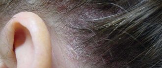
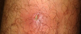
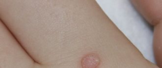
The characteristic symptoms of mycosis of the skin are round or oval erythematous formations with very reddened edges, on which lumps, pimples or blisters may appear. The central part of fungal spots is paler and looks like healing skin. Most often, fungal infections appear on the face , neck , back of the head or hands.
Dermatophytosis usually occurs through direct contact with animals or infected people. The most common causes of cutaneous mycoses are:
- improper (including excessive) personal hygiene
- sharing personal items with other people, such as towels, combs, shoes, clothes
- being in a warm and humid room
- walking barefoot in public places, such as a sauna or swimming pool
- use of public toilets or showers
Factors promoting development
Ringworm can affect anyone, but there are many factors that increase the risk of infection .
People at increased risk of developing athlete's foot include:
- people with chronic metabolic diseases (such as diabetes )
- people with a weakened immune system - people living with HIV or cancer
- people suffering from venous circulation disorders
- people with hormonal imbalances
- obese people
- people who take long-term antibiotics (especially broad-spectrum antibiotics), corticosteroids, or immunosuppressants
- children, elderly and pregnant women
Treatment of mycosis of the skin
Treatment of mycosis of the skin should begin with a visit to a dermatologist , who will rule out other skin diseases and conduct a mycological examination, thanks to which he will recognize the type of pathogenic fungus and select the appropriate treatment. Treatment of mycosis usually begins with the use of topical antifungal drugs - in the form of ointments, creams, sprays or powders, which not only suppress the development of fungi, but also reduce the symptoms accompanying the disease (itching, dry skin, burning).
If local treatment is unsuccessful, a dermatologist may prescribe systemic oral medications. Cured mycosis tends to recur, so after defeating the disease, appropriate prophylaxis should be taken to prevent re-infection.
Published in Dermatology Premium Clinic
Classification of mycoses
Mycoses are usually divided according to the degree of involvement of the skin, mucous membranes and appendages of the skin in the pathological process:
- Keratomycosis
. Fungal microorganisms multiply only in the stratum corneum of the skin; keratomycosis does not cause extensive inflammatory changes on the body. - Dermatophytes
. The changes affect the dermis and epidermis, as well as nails and hair follicles. - Candidiasis
. In addition to the skin, mucous membranes may be involved in the pathological process. - Deep mycoses
. Not only the skin is affected, but also a number of internal organs.
Manifestations and symptoms of mycosis
The main symptom of smooth skin mycoses is the appearance of lesions in the form of red or pink spots covered with epidermal scales. The shape of these lesions can vary from round to oval, their size is usually no more than 1-2 cm. The disease manifests itself after contact with domestic or stray animals, as well as cattle.
It is generally accepted that most often mycoses of smooth skin develop in children who have had close contact with infected animals or their fur. However, under certain conditions, fungal skin infections can also occur in adults, regardless of gender or age.
General symptoms of mycoses
During the incubation period, that is, during the proliferation of fungi, nothing bothers the infected person. The disease begins to manifest itself after the fungi begin to negatively affect the skin.
Depending on the type of fungal microorganism and the location of the infection, mycoses can manifest themselves in different ways, but there are several common symptoms:
- Itching of the skin.
- The appearance of red spots, rashes, blisters.
- Peeling of individual areas of the skin.
When localized on the feet and hands, peeling of the dermis between the fingers is possible.
Mycosis involving the nail plates is called onychomycosis. Nail fungus damage is indicated by a change in their color, delamination, the appearance of dark or yellowish spots, and thickening of the plate.
Squamous-hyperkeratotic form of mycosis of the feet
Also known as the moccasin type of mycosis of the feet (Tinea Pedis Moccasin Type). It is characterized by the presence of peeling on the skin of the interdigital folds, soles, palms, and sometimes with the presence of small surface cracks. In the area of the lateral surfaces of the soles, phenomena of desquamation of the epidermis can also be observed. In later stages, it manifests itself as diffuse or focal thickening of the stratum corneum (hyperkeratosis) of the lateral and plantar surfaces of the feet, which bear the greatest load. Typically, the affected areas of the skin have a mild inflammatory color and are covered with small pityriasis or mealy scales. Peeling in the skin furrows creates an exaggerated pattern, which gives the skin a “powdered” appearance. Subjectively, dry skin, moderate itching, and sometimes pain in the affected areas are noted.
Intertriginous form of mycosis of the feet
Also known as the interdigital type of mycosis of the feet (Tinea Pedis Interdigital Type). This form is characterized by the appearance of maceration and cracks in the interdigital folds, most often between the III and IV, IV and V toes
In the interdigital folds, against the background of edema and hyperemia, a clearly demarcated area of macerated and eroded skin is formed. Rashes in the form of erosions and deep cracks, weeping are often painful, accompanied by itching and burning, have an unpleasant odor due to the addition of a bacterial infection. In some cases, blistering rashes are observed. Subjectively, itching, burning, and pain in the affected areas are noted.
Dyshidrotic form of mycosis of the feet
Also known as the inflammatory type of mycosis of the feet (Tinea Pedis Inflammatory Type). It manifests itself as numerous blisters with a thick tire located on the skin of the arch and inferolateral surface of the foot, as well as in the interdigital folds. The predominant localization is the arches of the feet. The rash can affect large areas of the soles, as well as interdigital folds and skin of the fingers; merging, they form large multi-chamber bubbles, when opened, wet erosions of pink-red color appear. Usually the blisters are located on unchanged skin; as inflammatory phenomena increase, hyperemia and swelling of the skin occur, which gives this type a resemblance to acute dyshidrotic eczema. Subjectively, itching is noted.
Characterized by a chronic undulating course with exacerbations in spring and autumn. Exacerbations are accompanied by severe itching. The dyshidrotic form is often caused by T. mentagrophytes var. interdigitale.
Ulcerative form of mycosis of the feet
It is also known as an acute form of mycosis of the feet and as an ulcerative type of mycosis of the feet (Tinea Pedis Ulcerative Type). Characterized by bright erythema, swelling, severe maceration with the formation of vesicles and blisters, abundant desquamation with the formation of erosions and ulcers. In the acute form, a secondary infection is often associated, accompanied by regional (inguinal-femoral) lymphadenitis and lymphangitis, fever, and malaise. This form of mycosis of the feet is often accompanied by a dermatophyte reaction.
Mycosis of the feet in children
In children, lesions of smooth skin on the feet are characterized by fine-plate peeling on the inner surface of the terminal phalanges of the fingers, often in the 3rd and 4th interdigital folds or under the toes, hyperemia and maceration. On the soles, the skin may not be changed or the skin pattern may be enhanced, and sometimes ring-shaped peeling is observed. The disease is accompanied by itching. In children, more often than in adults, exudative forms of lesions occur not only on the feet, but also on the hands.
Diagnosis of mycoses
A dermatologist makes a preliminary diagnosis during a consultation. The nature of skin lesions during fungal infections often leaves no doubt about the existing pathology. It is possible to accurately and reliably determine the cause of changes in the skin only by studying scrapings taken from the sites of the rash. The resulting material contains pathogens in large quantities.
Staining a smear and examining it under a microscope allows you to identify mycelium threads, which indicate a fungal infection of the skin. Assessment of the morphological structure and properties of the causative agent of the disease makes it possible to determine its exact genus and species, as well as to finally determine which of the fungi caused the disease. All these diagnostic steps are important for choosing effective treatment leading to a quick recovery.
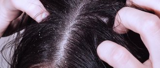
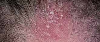
How to cure scalp fungus
It is necessary to start treating scalp fungus at the first symptoms, otherwise it can lead to hair loss, and in severe cases, to complete loss. Treatment should be aimed not only at eliminating aesthetic defects, but also at restoring the natural functioning of the sebaceous glands by normalizing the activity of all body systems.
Treatment and elimination of symptoms of scalp fungus involves the use of local and systemic medications. The most effective are antifungal agents for external use, the action of which is directed directly to the lesion. As a rule, such products include shampoos containing ketoconazole, climbazole, zinc and piroctoneolamine (octopirox). These components attack fungal bacteria, eliminating the cause of inflammation and itching.
Ketoconazole–
has antifungal, fungicidal and antiandrogenic effects, stops the synthesis of substances necessary for the cell membrane, which disrupts the vital activity of fungal bacteria.
Preparations containing ketoconazole are effective against yeast-like fungi of the genus Candida, as well as mold fungi. When applied externally, the substance is practically not absorbed.
Climbazole
– a new generation fungicidal drug that suppresses the active growth of fungi that cause the formation of dandruff and seborrheic dermatitis. Preparations containing climbazole do not disturb the natural bacterial flora of the scalp, providing a calming, regenerating and antiseptic effect on the affected areas.
Zinc
–zinc oxide in the composition of medicinal preparations quickly relieves inflammation and prevents the further spread of fungal infection. On open areas of the skin (temples, neck, area behind the ears), ointments containing zinc are used to relieve itching and flaking. The effectiveness of such drugs is due to their rapid healing and restorative effects.
Piroctone olamine
– blocks the proliferation of fungal bacteria, quickly relieves itching and inflammation of the skin, increases oxygen access to the hair follicles, and has antimicrobial properties. Preparations containing pyroctoneolamine normalize the vital activity of epidermal cells and protect them from the penetration of toxic substances.
When the substance enters the core of pathogenic fungi, it blocks cellular metabolism, which leads to the death of microorganisms.
It is necessary to treat scalp fungus both externally and internally. However, it should be remembered that the fight against the disease is within the competence of a trichologist, and self-medication can only aggravate the problem.
Other treatment measures
- normalization of the activity of the endocrine system and gastrointestinal tract;
- taking anti-inflammatory drugs and keratolytics;
- introduction into the diet of foods high in vitamins A, B, C, zinc, selenium, nicotinic acid, as well as impeccable adherence to the diet prescribed by the attending physician;
- refusal of sweet foods and products containing yeast (baked goods, beer, etc.);
- drug therapy for a systemic disease that causes a disturbance in the microflora of the scalp;
- normalization of sleep and restoration of the nervous system.
How to treat fungal infections?
Treatment of mycoses consists of medicinal use:
- antiseptic solutions;
- antifungal pastes, emulsions, ointments, varnishes;
- combined ointments when a secondary infection occurs, i.e. if fungal and bacterial skin infections are observed at the same time;
- systemic antifungal drugs (in cases of reluctance to external therapy or in severe forms of infection).
Basically, mycoses develop in people suffering from somatic diseases, with reduced immunity and various pathologies. Therefore, in addition to observing personal hygiene rules, it is important to monitor your overall health, strengthen your immune system, maintain a balanced diet and lead an active lifestyle.
Treatment and prognosis
Treatment of fungal infections (mycoses) takes from several weeks in the case of skin mycoses to months in the case of nail mycoses. Smooth skin (without hair) is usually completely restored after treatment, scarring at the site of hair loss is rare and, as a rule, hair growth is restored. A complete cure for nails affected by fungi is not always possible, and nail restoration can take a long time.
Unfortunately, there is no immunity after a fungal infection, so infection can occur again.
The information contained in this publication should not be used as a substitute for medical care or consultation with a physician.
author of the publication Mikhail Sergeevich Betekhtin, PhD, dermatovenerologist, oncologist, cosmetologist.

