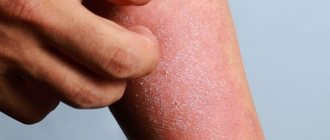
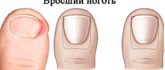
Scratching the skin when itching
It is important to know about the vicious circle “itching – scratching – itching”.
This means that when the skin is damaged, followed by inflammation, itching is provoked. In the skin, inflammatory cells release itch mediators, which again increase inflammation. The itching signal is transmitted to the brain, causing a person to need to scratch. Scratching leads to disruption of the skin (excoriation), which again increases inflammation. The skin becomes more vulnerable to internal and external irritants. This is a vicious circle that is difficult to stop. The vicious circle of “itching – scratching – itching” disrupts sleep, reduces concentration and perception processes.
Theses
In adulthood, itching appears with the formation of a well-defined plaque or polymorphic dermatoses in the same area.
When itchy skin is accompanied by dermatitis and dry skin, treatment of the affected skin should be a priority. When itching appears initially, the causes and time of onset of itching should be analyzed from the medical history. Itching often intensifies in the evening, when the activity of the sympathetic nervous system decreases. Treatment should be carried out taking into account the fact that itching can be caused by increased sensitivity to various external irritants. Heat and sweating are thought to particularly aggravate the itching. Factors that cause itching include cytokines and chemical signaling molecules, which cause itching primarily by stimulating the nervous system. Scratches (scratching) further aggravate existing dermatitis. In atopic dermatitis (AD), there is sometimes hypersensitivity of the skin, in which other sensations such as pain and heat are experienced as itching. Abnormal hypersensitivity of the nerve roots in the epidermis, as well as sensitization of the peripheral/central nervous systems, are possible causes of hypersensitivity leading to itchy skin. To control itchy skin caused by environmental factors such as heat, treating dermatitis is a priority. Sweating is exacerbated by itching, so attention should be paid to the negative effects of sweat on skin homeostasis due to 1) excess sweat on the skin and 2) heat retention due to insufficient sweating. Excess sweat on the skin must be properly eliminated, thus controlling the dermatitis as an appropriate amount of sweat will be released onto the skin. Not only stimulation from the surface of the skin, but also visual and auditory stimulation can cause a new attack of itching. The so-called “contagious itch” can be observed in patients with Alzheimer's disease. This review article describes the causes of worsening itching and provides information on how to treat these causes. Abbreviations: AD (atopic dermatitis), TSLP (thymic stromal lymphopoietin), TEWL (transepidermal water loss)
Clinical features of itching in atopic dermatitis (AD)
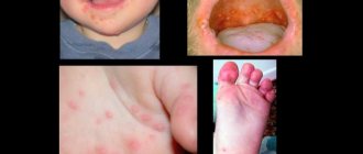
The clinical characteristics of blood pressure change with age, as well as with the duration of the disease. Sulzberger, who first named AD, described patients with Alzheimer's disease according to the characteristic clinical picture at each age. Clinical manifestations of AD in childhood are located on eczematous skin with the presence of serous papules (Fig. 1). Eczema or exudative papules are accompanied by severe itching, excoriation and new papules develop as a result of scratching the skin (Fig. 1). Further, as AD progresses from childhood to adulthood, the clinical course of the disease begins to change (Fig. 1).
The clinical manifestations of atopic dermatitis during this period are more varied, and were historically called disseminated neurodermatitis or Beznier's prurigo. Disseminated neurodermatitis was proposed by Brock and Yacuet and its characteristics include: 1) emotional nervousness, 2) continuous itching against the background of visible skin pathology, 3) the appearance of excoriative scales in almost the same place where the itchy skin first appeared, 4) localization of skin lesions, similar to dermatoses of nervous origin, 5) dry skin, 6) hypertrophy of the papillary layer of the skin and pigmented areas of the skin, and 7) chronicity.
Prurigo Besnier was proposed by Besnier, and is defined as a type of "diabetic prurigo". Early infancy is characterized by nonspecific skin lesions that occur after the onset of itching, and adolescence is characterized by “paroxysmal and chronic polymorphic and prurigo-like dermatoses, such as Hebra prurigo.”
A detailed description of the location, etiology, clinical course and response to treatment of the lesions can be used in modern daily clinical practice. AD is characterized by pruritus that precedes dermatitis, and symptoms and response to treatment vary between early infancy and adulthood.
Thus, elucidation of the distinctive mechanism leading to BP-related pruritus in each age range is required. In addition, itching associated with AD is exacerbated by heat, sweating, wool fibers, emotional stress, certain foods, alcohol, and general cooling. The first three factors are known to be particularly associated with factors that clinically aggravate pruritus. To successfully control blood pressure, it is necessary to eliminate these precipitating factors.
Figure 1. Clinical characteristics of BP vary with age and disease duration. In early childhood, the clinical manifestations are characterized by grouped exudative papules or small blisters, so-called eczematous changes, accompanied by severe itching (left). These clinical signs undergo age-related changes. In adulthood, the cutaneous manifestations develop into a symmetrically localized widespread dermatitis with lichenification (right). Often this chronic skin condition occurs in the area where itching first appeared and is called neurodermatitis.
Transmission and processing of the skin itch signal in AD
Skin itch impulses are transmitted to the brain using electrical impulses and waves passing through peripheral nerve fibers. The brain receives the information and induces a skin response. Recently, it has been shown that astrogliosis in the spinal dorsal horn influences chronic itch in animals with AD-like rashes and that the processing of the itch sensation characteristic of AD occurs in the central nervous system. Further investigation into the mechanism of itch in AD in humans is required. Below is a general picture of the neuronal transmission of itch. The itch associated with cutaneous stimulation is received by a receptor located in the free nerve endings of the skin, and then converted into electrical current by an ion channel and transmitted to the spinal cord. Many receptors and ligands associated with itch have been previously reported (Table 1).
| Table 1 The main factors that induce itching and their receptors in AD. | ||
| Ligands | Receptor | Ref. |
| Histamine | Histamine receptor | 25 |
| Kallikriin tryptases, endogenous/exogenous proteases | PAR-2 | 26, 27 |
| Bradykinin | Bradykinin receptor | 28 |
| Serotonin, 5-HT | 5-HT receptor | 29 |
| Endothelin-1 | Endothelin receptor (ETA) | 30, 31 |
| IL-31 | IL-31 receptor A | 32 |
| Thymic stromal lymphopoietin | Thymic stromal lymphopoietin receptor | 33 |
| Substance P | NK-1R | 34, 35 |
| Platelet activating factor | platelet activating factor receptor- | 36 |
| Leukotriene B4 | Leukotriene B4 receptor | 37 |
| Electrophilmyst, oxidative, pro-inflammatory agents | TRPA1 | 38, 39 |
| 12-HPETE | TRPV1 | 40 |
| Artemin | GFRα3 | 41 |
| IL-2 | IL-2 receptor | 42 |
| Gastrin releasing peptide | Gastrin releasing peptide receptor | 43, 44 |
| β-endorphins | μ opioid receptor | 45, 46 |
| Acetylcholine | Acetylcholine receptor | 47 |
| Calcitonin gene peptide | Gene calcitonin peptide receptor | 48 |
PAR, protease activation receptor; HT, hydroxytryptamine; TSLP, thymic stromal lymphopoietin; NK-1R, neurokinin-1 receptor; PAF, platelet activating factor; LTB4, leukotriene B4; GFR, glial-derived neurotrophic factor; 12-HPETE, 12-hydroxyperoxyeicosatetraenoic acid; GRP, Gastrin releasing peptide; CGRP, Calcitonin gene peptide.
Pruritus ligands (pruritogens) activate voltage-gated ion channels upon stimulation of TRPV1 (histaminergic) or TRPA1 (non-histaminergic), and then the increased action potential leads to the sensation of pruritus. In recent years, TRPV1 and TRPA1 have been the focus of attention as targets for the development of antipruritic drugs, as they are key in the peripheral transmission of itch. Inhibition of TRPV1 or TRPA1 may attenuate pruritus in AD-like animal models. The electrical impulse passing through the free nerve endings is transmitted to the spinal cord. The electrical impulse from the peripheral innervation of the skin is transmitted to neurons, which move to the central nervous system through interneurons in the spinal cord. The intensity of electrical impulses is regulated in the spinal cord and sorted into interneurons.
The functions of inhibitory interneurons are of particular interest. Scratching the skin, thermal stimulation, as well as mechanical stimulation on the surface of the skin can influence the intensity of itching. These stimuli increase the inhibitory activity of interneurons in the spinal cord through the peripheral innervation of the skin, which transmits the sensation of pain from the skin surface.
Neurotransmission inhibitors - gamma-aminobutyric acid (GABA), glycine and dynorphins are released from the nerve endings of inhibitory interneurons, and control neuron activity in the central nervous system, as a result of itching will subside. Controlling inhibitory interneurons is a promising option as a strategy for treating pruritus. This processed skin itch signal is transmitted from the spinal cord through neurons along the ascending pathways to the amygdala through the thalamus or medulla oblongata of the central nervous system. The itch signal is analyzed in the central nervous system, and converted into information about location, quality, scratching, shock, pain and pleasure.
It has been established that the processing of skin itching signals in the central nervous system in patients with AD differs from healthy ones. Although the central nervous system of itch has not been well studied, it is known that the central nervous system may contribute to the control of itch. The descending noradrenergic system suppresses itch signaling through its action on alpha-adrenergic receptors.
Norepinephrine, which is secreted by the descending nerve impulse system due to the excitability of the sympathetic nervous system or midbrain, may inhibit itch. Therefore, the sensation of itching is controlled to a large extent by the sympathetic nervous system, primarily during the daytime, or when a person is concentrating on something.
In addition, in patients with AD, itching can be controlled when the patient's attention is paid to something important. Further study of the mechanism of blocking the transmission of the itch impulse in the nervous system is expected to contribute to the successful treatment of itch in AD.
Itchy skin caused by dry skin or inflammation due to AD
When skin symptoms such as eczema and dry skin are present, various substances that cause itching (pruritogens) such as cytokines and chemical agents are released from the damaged areas (Table 1, Fig. 2). Pruritogens cause itching primarily by affecting nerve endings, and the affected area then itches. However, the skin lesions cannot be removed from the skin and such scratching of the skin aggravates the dermatitis. This vicious cycle is called the “itch and scratch cycle.” With chronic inflammation, such as AD, skin hyperesthesia is formed.
The cause of skin hyperesthesia occurs due to increased sensitivity of the nerve roots in the stratum corneum of the epidermis due to dehydration and inflammation of the skin (Fig. 2). Growth factor, semaphorin 3A, IL-31 and artemin are associated with abnormal nerve sensitivity and lead to the development of AD.
Histamine, produced by mast cell degranulation, and substance P, which is released from nerve endings, act directly or indirectly on the aberrantly increased sensitivity of the nerve and cause itching. Recently, the role of inflammatory mediators, such as thymic stromal lymphopoietin (TSLP), formed from epidermal keratinocytes and IL-31, derived from lymphocytes, on the development of skin itching has been actively discussed. It was found that TSLP and IL-31 induce skin itching by acting directly on nerve fibers. It has been established that liver pathology and an increase in autotoxins (lysophospholipase) as a result of cholestasis are the cause of skin itching. In addition, a correlation was noted between the concentration of autototoxins in the blood and the intensity of itching in patients with Alzheimer's disease.
Figure 2. The illustration schematically shows the triggers of itching and neural sensitization.
It is hypothesized that memories of itch attacks may promote neuronal plasticity and may also increase susceptibility to factors that aggravate itch.
Sometimes skin itching is induced not as a result of cutaneous stimulation, but as a result of visual or auditory signaling. For example, when a person sees images suggestive of itching, such as mosquito bites, or hears the sound of skin abrasions, they may feel the urge to scratch (Figure 2). This phenomenon is known as “contagious itch.” In patients with AD, it has been proven that “contagious itching” is more intense than in healthy people. It is also believed that psychological and mental factors influence itching. Although the actual conditions remain unclear, insomnia is also thought to contribute to the onset and exacerbation of itching. The influence of insufficient sleep and the influence of the circadian rhythm on itch due to BP will be described below.
Temperature and itching
Patients with AD often complain that “the itching increases when it receives heat.” At that time, “heat” was known to be the main inducer of skin itching in AD. There are still many uncertainties in its mechanism. In addition, itching caused by a thermal factor has a poor response to treatment. We may experience a relationship between temperature and itching in everyday life. For example, itching can usually be caused by taking a hot shower or cooling the itchy area with ice. This phenomenon has been scientifically proven in healthy skin, and suppression of histamine, which causes itchy skin, can be achieved by warming or cooling the areas.
However, itching in AD may be suppressed by cold stimulation but increased by heat stimulation. In other words, BP may contribute to abnormal hyperesthesia by causing patients to experience thermal stimulation as itching. This phenomenon is believed to occur due to sensitization of the peripheral or central nervous systems, and its molecular mechanism has been slowly elucidated.
Cutaneous peripheral nerve fibers, as a rule, are located in the dermis, abundantly surrounded by fibroblasts. It is hypothesized that nerve fibers may be activated by factors secreted by skin fibroblasts and sensitized to cause itching.
In summary, we analyzed genes isolated comprehensively from skin fibroblasts that were stimulated by factors associated with allergic inflammation and itch. As a result, we focused on artemin, a neurotrophic factor produced by dermal fibroblasts that were stimulated to produce substance P. Although artemin is thought to be important primarily in the development of sympathetic innervation, its function in the skin was unknown. Artemin production is not observed in normal human skin, but accumulates abundantly in skin lesions in AD and nummular eczema.
In mice administered artemin, an increase in the number of peripheral sensory nerves in the skin was noted with the development of thermal hypersensitivity of the skin. In addition, mice injected with artemin into the skin of the back exhibit abnormal behavior, such as increased skin temperature in the area under warm temperature conditions at 38°C. This phenomenon was not observed in artemin receptors (GFRα3) isolated from mice.
It is believed that abnormal accumulation of artemin in a localized area of the skin may cause systemic heat-induced itch. Now, the question remains as to why local accumulation of artemin in the skin affects sensations throughout the body. Capsacepin, a selective TRPV1 temperature receptor antagonist, does not suppress the abnormal behavior induced by artemin in a warm environment. Thus, it is currently believed that this reaction is caused by a mechanism independent of TRPV1.
With blood pressure, even the temperature of the water or air should be taken into account. In fact, everyday life and clinical practice provide us with the opportunity to consider the importance of temperature changes in the formation of skin itching. For example, patients with AD experience itching immediately after taking a hot bath, or patients scratch their body immediately after removing clothes. These phenomena are believed to be associated with rapid changes in skin surface temperature.
In fact, rapid changes in skin surface temperature have been experimentally demonstrated to increase skin itching. Fab et al found that pruritus increased as a result of rapid changes in skin surface temperature in response to repeated alternations of warm (32°C) and relatively cold (25°C) temperatures at 20-second intervals. These results indicate that even within the temperature range familiar in our daily lives, rapid changes in temperature can increase itching. Patients with Alzheimer's disease should be advised to avoid extreme temperatures for both air conditioning and water. The Japanese BP manual recommends the appropriate water temperature: 38-40 °C.
Sweating and itchy skin
Regulating body temperature activity: Warm-blooded mammals have a stable body temperature. As body temperature rises, thermal control mechanisms such as sweating are depleted and heat exchange processes are subsequently activated. Humans have exocrine sweat glands located throughout the body and sweat to effectively regulate body temperature. Sweating is an important physiological function of regulating human body temperature.
Sweat contains components to maintain skin homeostasis, such as natural moisture-retaining factors (urea and sodium lactate), bactericidal peptides (dermicidin, cathelicidin, β-defensin) and secretory IgA for protection against infections [64]. In addition, sweat has an inhibitory effect on the activity of cysteine and serine proteases, and also helps prevent the proinflammatory response against allergens with the activation of cysteine protease, and also promotes the formation of a mature stratum corneum of the epidermis. In addition, sweating has been reported to be decreased experimentally as a result of the activation of pruritus.
On the other hand, sweat is known to be an aggravating factor in blood pressure. Evidence linking the mechanism of exacerbation of blood pressure and sweating can be divided into three categories, 1), the presence of excess sweat on the skin, 2) an abnormality in the components of sweat, 3) an abnormal decrease in sweating. Sweating itself has been seen as an aggravating factor, as a result of the fact that many patients with AD consider sweating as an aggravating factor, as shown in many large-scale studies using self-administered questionnaires. In addition, in a report indicating the existence of "sweat allergy", basophils isolated from patients showed a positive reaction in a histamine release test in response to semi-purified antigens isolated from the sweat of healthy individuals.
On the other hand, Adachi et al. reported that no positive reaction was detected using the patch pot test. Based on the data above, sweat itself is not considered to be associated with a delayed allergic reaction, although it may be involved in the development of an acute phase allergic reaction. It has been reported that an antigen derived from Malassezia globosa, a saprophytic skin fungus, can be found in high levels in contaminated sweat and may be associated with “sweat allergy.” Because fungal antigens are not major components of sweat, the role of sweat in AD should be carefully discussed. On the other hand, the ability to reduce antimicrobial components in sweat is associated with a decrease in the ability to protect against the occurrence of AD.
In addition, an abnormal decrease in the ability to sweat is noted in hypertension. Its mechanisms are considered: A) sweating is produced, but does not spread over the surface of the skin due to obstacles, and B) an anomaly occurs in the production and secretion of sweat itself, etc. One theory about the cause of obstruction is the formation of a horny plug in the pore. However, it is not conclusive whether this is due to accompanying keratosis inflammation or dehydration due to decreased sweating. Shiohara et al confirmed the hypothesis of a deficiency of dermicidin, a bactericidal peptide contained in sweat, in the area surrounding the sweat duct in the area of AD lesions. As sweat leaks out of the sweat duct, the volume of sweat is discharged and reduced to the surface of the skin.
Further, in AD, the decrease in sweat secretion was confirmed by a sweat test using acetylcholine load and temperature stress. Regarding the mechanism, the influence of autonomic dystonia and anxious personality type can be considered. In addition, we have recently discovered that histamine inhibits sweating by acting on sweat ligands. In other words, allergic inflammation may be the cause of decreased sweating in blood pressure. Reduction of sweating in AD can be improved by reducing skin symptoms. Thus, normalization of abnormal sweating should be an important therapeutic step in the management of blood pressure.
Excess sweat left on the surface of the skin contributes to exacerbation of AD symptoms. One of the factors that contribute to higher skin surface pH levels is excessive sweating. As a rule, the pH of normal sweat remains quite low, since the reabsorption of sodium bicarbonate and alkali metal ions from the sweat duct is reduced in sweat.
However, as the amount of sweat increases with body temperature, the reabsorption of sodium bicarbonate from the sweat ducts is impaired, resulting in an increase in the pH of sweat. If excess sweat with a higher pH value remains on the skin, it can easily lead to detachment of the stratum corneum of the epidermis and increased susceptibility to inflammation. Contamination of bacteria or antigens (antigens derived from Malassezia, etc.) contained in sweat on the surface of the skin can cause itching.
In addition, protective antipruritic components of sweat, such as inhibitory proteinases, etc., may be lost as sweating increases. Therefore, in case of itchy skin accompanied by sweating, excess sweat should be washed off or wiped off the surface of the skin with a damp towel. The fact that sweat is an aggravating factor for blood pressure can easily lead to the misconception that “patients with blood pressure do not sweat.” However, there is no evidence that skin symptoms improve when the skin does not sweat. Thus, as mentioned above, improving sweating dysfunction may be the best treatment goal.
Lifestyle and skin itching
An unhealthy lifestyle leads to fluctuations in skin homeostasis. Patients with AD often complain that “the itching gets worse at night.” In fact, as 24-hour monitoring shows, core body temperature increases at night and drops with dilatation of the peripheral blood vessels of the skin during sleep. As a result, if the skin temperature rises during sleep, itchy skin can easily appear. Melatonin is synthesized in the thymus and is associated with a decrease in body temperature after sleep.
The habit of going to bed late reduces the production of melatonin and inhibits the decrease in core body temperature (Fig. 2). Transepidermal water loss (TEWL), which is a measure of skin barrier function, also increases at night. As a result of the action of factors with high temperature and high humidity TEWL, the skin surface is susceptible to itching. In addition, there was a decrease in the amount of cortisol and an increase in the synthesis of IL-2, which is associated with inflammation, during the night period. Poor lifestyle choices also cause an imbalance of μ-opioid receptor agonists and κ-opioid receptors, which contributes to the development of an imbalance towards susceptibility to itchy skin.
Conclusion
Recent data on the factors associated with the exacerbation of itching, the mechanisms of its intensification, as well as its control, have contributed to the development of blood pressure control. AD can be aggravated by natural human physiological phenomena and our lifestyle. Thus, choosing a management strategy for a patient can sometimes be difficult. Some problems still remain unresolved, but we hope that the recently accumulated data can be used as often as possible in patient management.
Types of skin itching
At the appointment, doctors usually clarify with the patient the following characteristics of itching due to dermatitis:
- Intensity: weak, moderate, intense;
- constant or increasing at certain times of the day, for example, in the evening and at night;
- permanent or periodic;
- common – itchy skin all over the body;
These criteria allow you to more accurately determine the causes of itchy skin and prescribe the necessary treatment.
It should be remembered that scratching itchy areas of the skin is dangerous due to possible microbial infection - the so-called secondary infection, which can significantly worsen the condition of the skin and complicate treatment.
Symptoms of itchy dermatitis
- Simple prurigo is characterized by the appearance of symmetrically located areas with intense itching. There, over time, small dome-shaped flesh-colored bumps (papules) form, which itch intensely. Sometimes they transform into blisters containing clear liquid. To a greater extent, the disease affects the outer surfaces of the arms and buttocks, but can also be located in other areas of the head, neck, torso and limbs. This type of prurigo can affect people of any age; it occurs in both children and adults. Over time, the papules that appeared at the beginning of the disease are scratched off and disappear, but new ones are formed. This condition leads to a significant deterioration in quality of life, the itching worsens at night and interferes with sleep.
- Pigmented prurigo is a simple variant when pigment accumulates in papules and they change color and become darker than the underlying skin.
- With nodular prurigo, dense bumps and pimples appear on the skin, similar to warts, which bring a lot of discomfort. They may be darker than the skin surrounding these lesions. Their surface is rough, it is noticeably thickened, and many scratches and scratches are visible. The nodules are usually no more than a centimeter in diameter, they are very dense to the touch, can be sensitive, ulcerate, crust over and itch. The arms and legs are the areas most commonly affected, but the back, buttocks, shoulders and chest can also be affected by the disease. Scars and cicatrices are noticeable, differing in color from the underlying epithelium, which form at the site of scratching. Nodular prurigo is more common in young and middle age (30 - 60 years), equally common in men and women, and in representatives of all nationalities. The number of nodules gradually increases. This variant of prurigo is difficult to treat.
For diagnosis, a doctor usually only needs to examine the patient. The very appearance of skin rashes in combination with itching indicates itchy dermatitis. In some cases, a piece of the rash is taken and a biopsy is performed to clarify the diagnosis.
Types of dermatitis and diagnosis
Pruritic dermatoses and dermatitis are a group of diseases that are completely different in nature, which are united by a common symptom - itchy skin. It is not always easy to distinguish them even for an experienced specialist.
Table of itchy skin diseases, diagnosis
| Disease | Itching | Localization | Nature of the rash | Age of onset |
| Allergic contact dermatitis | Pronounced or strong, burning sensation | Any where the skin comes into contact with the allergen | Bubbles against the background of redness of the skin, clear boundaries of the lesion | Any |
| Atopic dermatitis | Pronounced or strong | Face, limbs; depends on the patient's age | Bubbles against a background of redness, weeping, crusts, cracks | Most often children's, can be any |
| Psoriasis | From weak to strong depending on the stage of the process | Scalp, natural skin folds or friction points | Psoriatic plaques with gray scales | Any |
| Seborrheic dermatitis | Weak to moderate, may be absent | Scalp, nasolabial folds, upper third of the body | Areas of redness with accumulation of yellow, greasy scales | Teenage, adult |
| Scabies | Strong, unbearable, worse at night | Between the fingers, stomach, groin, buttocks | Red spots or papules arranged in pairs | Anyone, usually children |
| Herpetic infection | Severe, burning skin | Most often the face (lips), along the nerves (shingles) | Small tense bubbles | Anyone, for shingles most often an adult |
| Microbial eczema | Moderate, burning, soreness | Most often limbs, asymmetrically, legs | Bright red spots 1-3 cm with clear boundaries, weeping, blisters and crusts | Anyone, usually an adult |
Causes of itchy dermatitis
It is not known exactly what causes prurigo, or why the skin reacts the way it does. Once itching occurs, rubbing and scratching makes the nerve endings in the dermis thicker and even more susceptible to irritation. This leads to more itching, thus making the condition worse.
In half of the patients, the exact cause cannot be detected, but there are some combinations of diseases that may be related.
- Up to 80% of patients with pruritic dermatitis are also prone to atopy, and may have conditions such as asthma, eczema and seasonal pollen allergies.
- Prurigo nodosa also develops in people with venous eczema (a condition where skin lesions occur due to venous blood pooling in the legs), and may be associated with nummular eczema (which is characterized by a well-defined, coin-shaped rash).
- In some people, nodular prurigo may begin after an insect bite,
- Several diseases may be associated with nodular prurigo, which must be excluded when pruritic dermatitis appears. This is bullous pemphigoid, in which blisters filled with fluid appear on the skin, like after a burn. Dermatitis herpetiformis is characterized by small, round blisters and is a symptom of gluten intolerance.
In 30% of patients, pruritic dermatitis develops as a consequence of a systemic disease, and metabolic and biochemical causes play a role.
- Nutrient deficiency. Here, iron deficiency comes to the fore, which may be associated with a lack of this microelement in food, increased losses (in women with menstruation), or poor absorption in the intestine (with gluten intolerance, after removal of a section of the intestine).
- liver disease,
- Uremia (a condition that develops with kidney failure),
- Severe diabetes mellitus.
Less common causes include thyroid disease, stroke, calcium metabolism disorders, and lymphoma (blood disease).
For the remaining 20%, psychological factors such as depression, anxiety and psychosis play a role. But even ordinary emotional stress can become a trigger. When under stress, people are more prone to scratching the epithelium. Thus, stress worsens the course of prurigo.
How to get rid of itchy skin
Before relieving the itching of dermatitis, it is necessary to establish its cause, since in most cases it will be difficult to get rid of the itching without eliminating the cause.
Regardless of the causes of itching, treatment usually uses remedies aimed at alleviating the patient’s condition, relieving the urge to scratch, and preventing the development of secondary infection in areas of scratching.
Among skin diseases, common causes of itching are atopic dermatitis, eczema, neurodermatitis, seborrheic dermatitis, and psoriasis.
Allergic contact dermatitis
This is a delayed-type hypersensitivity reaction in sensitized individuals who have specific immune cells - T-lymphocytes in the epidermis, which cause an atypical allergic reaction to seemingly harmless substances.
Sensitizing agents may include:
- Adhesives for cosmetics, wigs;
- Some topical antibiotics;
- Some resins;
- Clothes;
- Fragrances used in perfumes, soaps or cosmetics;
- Other cosmetics, including nail polish or hair products;
- Nickel, chromium or other metals;
- Poisonous plants such as poison ivy or poison sumac;
- Products made of rubber, latex (gloves or shoes).
Contact between immune cells of the skin and specific antigens provokes an allergic reaction. The reaction goes through two phases as shown below: Sensitization. E
This is the phase when immune cells react to a hapten (a molecule with an antigenic determinant) in a certain way.
At this stage, nonspecific immune cells assemble the hapten-carrier protein complex and migrate to local lymph nodes. There they activate antigen-specific lymphocytes to produce a proliferative clone of T cells that enter the bloodstream and enter the dermis. The sensitization phase is now complete and new T cells are ready to respond to the antigen if they encounter it. The whole process takes about 10-14 days. Selection.
This phase accompanies re-exposure to the antigen. The prepared T cells create an immune challenge that causes an allergic reaction.
Increasing long-term use of a sensitizing, irritant, agent can lead to persistent, severe and more rapid reactions, which are called anamnesis.
Treatment of itchy skin with medications
Considering that itching can be provoked not only by mechanical and chemical irritants, but also by emotional factors, treatment should be comprehensive and aimed at eliminating the cause.
Medical recommendations for the treatment of skin itching due to dermatitis highlight the following means to reduce the intensity of itching:
- Systemic medications for skin itching in dermatitis: antihistamines, mast cell membrane stabilizers, systemic hormonal agents, antidepressants, sedatives, immunosuppressants.
- External medications, which include external anesthetics, hormonal agents, tacrolimus, zinc, menthol and camphor.
Irritant contact dermatitis
The content of the article
Irritant contact dermatitis is more common than the allergic type of dermatitis. Its appearance is the result of irritation and disruption of the skin surface by the substance. For this reason, the nature and extent of the rash depends on the degree of presence of the irritant and the duration of exposure.
The type of reaction does not have a characteristic time interval between visual manifestations and the onset of dermatitis. Irritants act through wet or dry skin and extreme climatic conditions. Typical irritants include:
- Chemicals such as acids or alkalis that burn the skin, including soaps and detergents;
- Fabric treatment chemicals such as softeners, solvents;
- Shampoos;
- Cement;
- Hair Dye;
- Moisture, for example from prolonged use of diapers;
- Pesticides;
- Rubber gloves.
The resulting rash is a skin reaction to irritation caused by a chemical.
Skin cap for itchy skin due to dermatitis
To combat skin itching in atopic, seborrheic dermatitis, eczema and psoriasis in adults and children from 1 year of age, external medications based on zinc pyriton activated SKIN-CAP have been successfully used, which:
- can help reduce the severity of skin itching within 5-7 days [1] after the start of use;
- reduce the need to use hormonal [2] and antihistamines [1];
- help prevent the development of secondary infections when scratching, thanks to the antibacterial and antifungal activity of activated zinc pyrithione;
- have a high level of safety, unlike, for example, hormonal ointments, since they have only one contraindication in the form of individual intolerance to the components of the drug [2].
Anti-itching cream for dermatitis and Skin-cap aerosol can be used in children from the age of 1 year.
Treatment of atopic dermatitis
A feature of the treatment of atopic dermatitis is a stepwise approach from simpler methods of external therapy and skin care to complex innovative techniques.
Remedies and methods for treating atopic dermatitis
When treating atopic dermatitis, the following are used:
- Softening and moisturizing creams and ointments are the basis for the treatment of atopic dermatitis. These products help keep your skin hydrated and soft.
- Steroid creams and ointments are applied to the skin and help relieve redness and itching. In severe cases, steroid tablets or injections may be required, but your doctor will keep the course of treatment as short as possible, since the high effectiveness of injections comes with a high risk of side effects.
- Medicines that affect the immune system are highly effective, but have side effects and may be prescribed when safer treatments do not produce the desired results.
- Antihistamines may be used in patients who report decreased itching.
- To increase the effectiveness of therapy, it is possible to perform wet wraps (Wet Dressing, Wet Wrap Therapy).
Wet wraps (Wet Dressing, Wet Wrap Therapy)
Treatment with Wet Wrap Therapy helps to quickly clear skin of rashes and can be used both in a medical facility or hospital, as well as at home. The effect is associated with improved penetration of the drug used, deeper and longer-lasting hydration, and reduced water loss by the skin. The dressings provide protection from scratching, which leads to the formation of an itch-scratch cycle, preventing scratches from occurring, which further intensify the itching, which makes the skin heal better. Cooling the surface of inflamed skin by evaporating water from the dressings helps reduce inflammation, itching and soreness.
For severe eczema, wet wraps are prescribed in a medical facility. They can also be used at home to maintain good health or at the first sign of deterioration and can reduce the need to seek medical help and the likelihood of hospitalization.
There are various modifications of Wet Wrap Therapy; the essence of the method is to use external medications (emollients or steroids) under two layers of bandages. The bottom layer is warm and moist, on top of which a second, dry layer is made. For the bandage, you can use a regular bandage, special tubular bandages, or special clothing. The bottom layer must be periodically moistened with ordinary warm water, preventing it from drying out. The use of topical steroids under a wet dressing can increase the effectiveness of treatment. The procedure can be performed 1-2 times a day, every day, during an exacerbation. As the exacerbation decreases, the procedure can be done less frequently, performed 1-2 times a week during the period of remission. If well tolerated, the bandage with the medicine can be left overnight, remembering to periodically moisten the bottom layer of tissue.
The procedure takes quite a long time, and in the first stages (especially in patients with severe skin damage) may require the participation of medical personnel. After training and improvement, the procedures can be continued at home.
Types of dermatological diseases
In medicine, there are more than a dozen varieties of this disease; let’s look at the most common forms of its manifestation. These include: contact, dry, atopic (or allergic, toxicoderma, eczema, urticaria), seborrheic dermatitis and the price of their treatment depends on the type of lesion.
Dry dermatitis
Symptoms of dry dermatitis appear only in the cold season, due to prolonged exposure to cold or, conversely, too dry air indoors. There is a decrease in immunity and a deterioration in the general condition of the body. A distinctive feature is the duration of the course and the seasonal manifestation of exacerbations.
Contact dermatitis
Foci of simple contact dermatitis form at the site of exposure to the primary irritant. It is distinguished by the absence of manifestations of an acute reaction and the further spread of the inflammation process after the exclusion of the irritating element. For example, when they often wear clothes made from synthetic fabrics, to the fibers of which the body is highly sensitive.
Atopic dermatitis
By atopic dermatitis, clinic doctors mean a chronic, often hereditary allergic disease with recurrent periods and significant complications. In the first stages (in children from 3 months to 2 years, and at 4-7 years), the development of the disease manifests itself in the form of redness with copious discharge and the formation of small blisters. And in the last stages, severe dryness and pallor of the skin predominates.
Toxidermy
Toxicoderma is accompanied by significant damage to the skin from toxic substances. It differs in that the allergen enters the body through the blood, respiratory system and oral cavity. This is a very serious and dangerous disease and at the first manifestations (shortness of breath, swelling of the mucous membranes, stomach pain), you must urgently contact specialists who will professionally provide first aid. It is impossible to remove toxic substances from the body on your own.
Methods of infection
Sometimes we ourselves, without noticing it, put ourselves in danger, without thinking about what consequences allergic dermatitis can lead to when postponing a visit to the clinic. Ways of human contact with allergens that can cause disease:
- medicinal – manifests itself after the introduction of drugs into the body. For example, antibiotics, vitamins, analgesics, gamma globulins;
- food - allergens enter our body along with food;
- professional - observed in people who are constantly in contact with allergens, for example in production with a high concentration of cobalt, nickel, chromium;
- autointoxication – characterized by the accumulation of autoallergens, for example, in patients with pathologies of the gastrointestinal tract.
Eczema
Eczema is also a chronic disease and manifests itself in the form of acute inflammation of the skin and subcutaneous tissue under the influence of internal or external irritants. This allergy is manifested by areas of dry, flaky skin, cracks, redness, swelling and weeping lesions. In severe cases, even damage to the entire surface of the epithelium. Most often, workers in metallurgical plants, pharmaceutical and food industries suffer.
Seborrheic dermatitis
Seborrheic dermatitis is a disease caused by a certain fungus. Very often, the causes of its appearance are stress conditions, hormonal disorders and diseases of the nervous system.