The epidemiology (prevalence) of tongue cancer is on average 5 cases per 100 thousand population.
Among the recorded cases of oral tumors, it accounts for up to 60%. Despite the relatively simple diagnosis of this disease, there are advanced cases - people often do not notice the signs or ignore the symptoms of tongue cancer.
Etiology - causes of tongue cancer
The main cause of tumors on the tongue, like any other cancer, is a genetic malfunction in the cells.
In this case, these are epithelial cells - the tissue that forms the mucosa. Several main factors contributing to this process have been identified:
- Exposure to carcinogens. A huge amount of harmful substances is contained in cigarette smoke and chewing mixtures (nasvay, betel). It is in smokers and nasal users that cancer of this localization is most often diagnosed, and in men it is detected 3 times more often than in women. Alcohol increases the impact of carcinogens.
- Occupational hazards. The incidence of tongue cancer in people employed in hazardous industries is much higher. Salts of heavy metals (mercury, lead), asbestos, and petroleum products can also be classified as carcinogens by their nature.
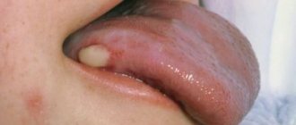
- Photo: neoplasm on the tongue
Impact of viruses. Recent studies have proven a direct link between chronic viral infection and the incidence of cancer. Human papillomavirus, herpes simplex virus and HIV are capable of transforming the genome of cells, turning them into cancerous ones. According to statistics, up to 70% of women are carriers of HPV. The mechanism of oncogenic effects of viruses is associated with their ability to suppress antitumor genes.
- Chronic oral injuries. They may be associated with improper installation of dentures, improper treatment of fillings, or chronic biting of the mucous membrane.
Long-term exposure to these factors on the mucosa is accompanied by damage to the DNA structure of epithelial cells. As a result, papillary hyperplasia (excessive growth) develops, which looks like a thickening at the sides, or dysplasia (improper development) of the mucous membrane.
Further exposure to these causes leads to the development of precancerous conditions: leukoplakia, Bowen's disease, hyperkeratosis and papillomas. Subsequently, these conditions transform into oncology.
Stages of tongue cancer
There are several stages of the disease, each of which has its own distinctive features.
Initial stage of tongue cancer (first)
Photo: this is what the initial stage of tongue cancer looks like
It is characterized by an asymptomatic course - practically nothing bothers patients. It appears as whitish spots on the mucous membrane, the so-called papillary growths. They are often mistaken for plaque, which is located on the lateral surface of the muscular organ.
When examined, doctors often mistake these formations for manifestations of other diseases: glossitis or stomatitis. There is no pain at this stage.
Stage of clinical manifestations (second)
As the disease progresses, the “plaque” gradually turns into a lump on the tongue, which, if left untreated, transforms into a tumor on the tongue. At this stage, a pain syndrome appears; the pain is diffuse or local in nature, very often radiating to nearby organs (neck, temple, ears).
Patients at this stage often complain of bad breath, which is caused by infection and suppuration of the tumor. The clinical picture is accompanied by difficulty swallowing, articulation disorder, swelling and numbness of part of the tongue. Metastasis to surrounding tissues and nearby lymph nodes - submandibular, cervical - is possible.
Advanced degree (third)
It is possible to bring the disease to this stage if you completely ignore the symptoms of the initial and second stages of tongue cancer. It manifests itself as aggressive invasive (penetrating into the thickness of the organ and surrounding tissues) growth, accompanied by tissue decay.
Terminal stage (fourth)
In this phase, distant metastases occur - in the bones, liver, lungs. Treatment of advanced tongue cancer is not very successful and the prognosis is very doubtful. Patients who progress the disease to the terminal stage rarely live more than a year.
Treatment is only palliative - the fight against pain and cancer intoxication.
What does plaque color tell you?
White
As we have already said, a thin white mucous coating on the tongue after sleep is not a deviation from the norm. A white coating of increased density indicates constipation, and a cheesy coating on the tongue indicates the unhealthy activity of yeast-like fungi of the genus Candida.
Yellow
A bright yellow coating on the tip of the tongue indicates hepatitis A (Botkin's disease). If there are problems in the functioning of the gallbladder, a yellowish coating and cracks appear on the tongue.
Dark
A dark coating on the tongue is a sign that something is wrong with the lungs. You don’t often see a completely black plaque: for example, in advanced stages of cholera due to dehydration of the body or in Crohn’s disease.
Types of tongue cancer
The disease is classified by localization - the location of the tumor on the tongue:
- Cancer of the tongue body accounts for more than 2/3 of all cases of this pathology.
- Cancer of the root of the tongue - 1/5 of all cases.
- Lower surface cancer – all remaining cases (about 10%).
Photo: squamous cell carcinoma of the tongue
Based on the principle of tumor growth, the following clinical forms of the disease are distinguished:
- Exophytic – the tumor grows mainly in the oral cavity. Sometimes this form of the disease is called papillary.
- Endophytic (infiltrative) – growth is directed into the thickness of the organ. Endophytic growth is a characteristic symptom of tongue root cancer. Very often, this form of the disease leads to swelling and difficulty in moving the tongue or to its complete immobility.
Tongue cancer is classified as squamous cell in 95% of cases, and only in 5% is it another histological form: carcinoma or basal cell carcinoma.
Based on the appearance of the tumor, papillary and ulcerative forms are distinguished. Papillary cancer is the most common type of disease.
It looks like a dense growth, somewhat elevated above the mucous membrane and covered with “warts”.
In the ulcerative form of the disease, an ulceration appears on the surface of the tongue on the side or back, surrounded by a ridge. A characteristic property of an ulcer at an early stage is its complete painlessness. As the ulcer grows, pain appears and bleeding is noted. The addition of an inflammatory component masks cancer and complicates diagnosis.
Photo of tongue cancer
Pictured is papillary tongue cancer
The photo shows a tumor on the side of the tongue
The photo shows an ulcerative form of cancer
Pictured is tongue root cancer
Anatomical structure of the pharynx
The pharynx, another name is the pharynx.
It starts at the back of the mouth and continues down the neck. The wider part is located at the base of the skull for strength. The narrow lower part connects to the larynx. The outer part of the pharynx continues with the outer part of the mouth - it has quite a lot of glands that produce mucus and help moisten the throat during speech or eating. When studying the anatomy of the pharynx, it is important to determine its type, structure, functions and risks of disease. As mentioned earlier, the pharynx is shaped like a cone. The narrowed part merges with the laryngopharynx, and the wide side continues the oral cavity. There are glands that produce mucus and help moisten the throat during communication and eating. From the front side it connects to the larynx, from above it adjoins the nasal cavity, on the sides it adjoins the cavities of the middle ear through the Eustachian canal, and from below it connects with the esophagus.
The larynx is located as follows:
- opposite 4 - 6 cervical vertebrae;
- behind - the laryngeal part of the pharynx;
- in front - formed due to the group of hyoid muscles;
- above - hyoid bone;
- lateral - adjacent to the thyroid gland with its lateral parts.
The structure of a child's pharynx has its own differences. Tonsils in newborns are underdeveloped and do not function at all. Their full development is achieved by two years.
The larynx includes in its structure a skeleton, which contains paired and unpaired cartilages connected by joints, ligaments and muscles:
- unpaired consist of: cricoid, epiglottis, thyroid.
- paired ones consist of: corniculate, arytenoid, wedge-shaped.
The muscles of the larynx are divided into three groups and consist of:
- thyroarytenoid, cricoarytenoid, oblique arytenoid and transverse muscles - those that narrow the glottis;
- posterior cricoarytenoid muscle - is paired and expands the glottis;
- vocal and cricothyroid - strain the vocal cords.
Entrance to the larynx:
- behind the entrance there are arytenoid cartilages, which consist of cornuform tubercles, and are located on the side of the mucous membrane;
- in front - epiglottis;
- on the sides there are aryepiglottic folds, which consist of wedge-shaped tubercles.
The laryngeal cavity is also divided into 3 parts:
- The vestibule tends to stretch from the vestibular folds to the epiglottis.
- Interventricular section - stretches from the inferior ligaments to the superior ligaments of the vestibule.
- Subglottic region - located at the bottom of the glottis, when it expands, the trachea begins.
The larynx has 3 membranes:
- mucous membrane - consists of multinucleated prismatic epithelium;
- fibrocartilaginous membrane - consists of elastic and hyaline cartilages;
- connective tissue - connects part of the larynx and other formations of the neck.
Pharynx: nasopharynx, oropharynx, swallowing department
The anatomy of the pharynx is divided into several sections.
- How is swallowing done?
Each of them has its own specific purpose:
- The nasopharynx is the most important section, which covers and merges with special openings into the back of the nasal cavity. The function of the nasopharynx is to moisturize, warm, clean the inhaled air from pathogenic microflora and recognize odor. The nasopharynx is an integral part of the respiratory tract.
- The oropharynx includes the tonsils and uvula. They border the palate and the hyoid bone and are connected by the tongue. The main function of the oropharynx is to protect the body from infections. It is the tonsils that prevent the penetration of germs and viruses inside. The oropharynx performs a combined action. Without its participation, the functioning of the respiratory and digestive systems is not possible.
- Swallowing department (hyopharynx). The function of the swallowing department is to carry out swallowing movements. The laryngopharynx is related to the digestive system.
There are two types of muscles surrounding the pharynx:
- stylopharyngeal;
- muscles are compressors.
Their functional action is based on pushing food towards the esophagus. The swallowing reflex occurs automatically when muscles tense and relax.
The process looks like this:
- In the oral cavity, food is moistened with saliva and crushed. The resulting lump moves towards the root of the tongue.
- Further, the receptors, irritating them, cause muscle contraction. As a result, the sky rises. At this second, a curtain closes between the pharynx and nasopharynx, which prevents food from entering the nasal passages. The lump of food moves deep into the throat without any problems.
- Chewed food is pushed down the throat.
- Food passes to the esophagus.
Since the pharynx is an integral part of the respiratory and digestive system, it is able to regulate the functions assigned to it. It prevents food from entering the respiratory tract during swallowing.
What functions does the pharynx perform?
The structure of the pharynx makes it possible to carry out serious processes necessary for human existence.
Functions of the pharynx:
- Voice-forming. Cartilage in the pharynx takes control of the movement of the vocal cords. The space between the ligaments is constantly subject to change. This process regulates the volume of the voice. The shorter the vocal cords, the higher the pitch of the sound produced.
- Protective. The tonsils produce immunoglobulin, which prevents a person from becoming infected with viral and antibacterial diseases. At the moment of inhalation, the air entering through the nasopharynx is warmed and cleared of pathogens.
- Respiratory. The air inhaled by a person penetrates the nasopharynx, then the larynx, pharynx, and trachea. The villi located on the surface of the epithelium prevent foreign bodies from entering the respiratory tract.
- Esophageal. The function ensures the functioning of swallowing and sucking reflexes.
The diagram of the pharynx can be seen in the next photo.
- The structure of the human throat and larynx: photo
Diseases affecting the throat and pharynx
Diseases of the ENT organs can be triggered by an attack of a viral or bacterial infection. But pathology is also caused by fungal infections, the development of various tumors, and allergies.
Pharynx diseases manifest themselves:
- ARVI;
- sore throat;
- tonsillitis;
- pharyngitis;
- laryngitis;
- paratonsillitis.
Only a doctor can determine an accurate diagnosis after a thorough examination and based on the results of laboratory tests.
Possible injuries
The pharynx can be injured as a result of internal, external, closed, open, penetrating, blind and through injuries. Possible complication - blood loss, suffocation, development of a retropharyngeal abscess, etc.
First aid:
- in case of injury to the mucous membrane in the oropharynx area, the damaged area is treated with silver nitrate;
- deep injury requires the administration of tetanus toxoid, analgesic, antibiotic;
- severe arterial bleeding is stopped by finger pressure.
Specialized medical care includes tracheostomy and pharyngeal tamponade.
Diagnostics
On your own, you can only suspect the first symptoms of oncology in a given localization. Strangely enough, the doctors who most often encounter this pathology are not oncologists, but dentists. This is due to the specifics of their activity - in the course of their professional activities, they may be the first to notice signs of an early stage of cancer in the form of a lump on the side of the tongue or at the root.
The similarity of the first symptoms of tongue cancer with other diseases of the oral cavity leads to its late diagnosis - most often at the second stage, when a malignant neoplasm of the mucous membrane takes the form of an ulcer or a pronounced tumor.
Specific diagnostics consists of the following examinations:
- Examination of a fingerprint smear to detect signs of squamous cell carcinoma.
- Tumor biopsy followed by histological examination under a microscope.
- Ultrasound of the tongue and soft tissues of the lower jaw, as well as ultrasound of the neck to search for metastases.
- X-ray of the skull or computed tomography - if there is a suspicion of tumor growth into the bone.
In order to exclude distant metastases, it is necessary to do an X-ray of the lungs, CT (MRI) of the brain and ultrasound of the liver.
Despite the fact that squamous cell carcinoma of the tongue is less common in women, the frequency of its detection at an early stage is much higher in them. This is due to the more sensitive attitude of women to the condition of the oral cavity.
What to do at home if a growth appears - folk methods
In addition to traditional treatment of growths under the tongue, many patients use traditional recipes. It is impossible to replace treatment with a completely unconventional method, but nothing prevents you from trying it.
Popular folk recipes for growths under the tongue:
- daily rubbing of the formations with garlic juice or applying cloves of spice;
- lubricating newly appeared growths with egg white;
- treating affected areas with castor oil (using cotton swabs);
- rinse the mouth twice a day with decoctions of medicinal herbs (medicinal chamomile, string, sage, echinacea, etc.);
- ingesting rosehip decoction, freshly squeezed potato or carrot juice.
Any infections and viruses developing in the body signal a decrease in immunity, so during the treatment period you should take care of taking fresh vegetables and fruits. A good vitamin complex is also suitable for these purposes.
Before using an unconventional approach, you should consult your doctor to rule out contraindications.
Treatment methods for tongue cancer
Regardless of the cause of tongue cancer, combination therapy is used to treat it, including the following methods:
- Surgery. The operation is aimed at radical removal of a malignant tumor: either partial resection (excision) of the tongue or its complete removal (glossectomy) is performed. In advanced cases, when the tumor has grown into the surrounding tissues, they are resected down to the bones of the lower jaw.
- Radiation therapy. There are remote therapy, when the tumor is irradiated at a distance with X-rays, and contact therapy (brachytherapy), when the radiation source (radioactive isotopes) is placed deep within the organ. Radiotherapy is performed both before and after surgery. The doctor determines how many sessions are needed.
- Polychemotherapy. It is used in advanced cases in the presence of distant metastases, when other methods cannot be used, or their effect is insufficient. Patients are treated with Cisplatin, Methotrexate and other drugs.
Surgeries in the later stages of the disease are often mutilating in nature - in some cases it is necessary to remove almost the entire lower jaw. After surgery, patients live with some restrictions. In order to create a satisfactory quality of life, they undergo reconstructive medical operations.
Therapeutic exercises
There are alternative methods that allow you to do without surgery, for example, a set of special exercises for developing the frenulum of the tongue.
- Reach alternately to the upper and lower lips.
- Extend your tongue and make pendulum movements from cheek to cheek.
- Suck your tongue to the roof of your mouth and let go, imitating a horse clopping its hooves.
- Place your tongue on each cheek with your mouth closed.
- Smile with your mouth open.
- Stretch your lips.
- Pull your lips into a tube, pretending to kiss.
To achieve results, you need to perform the exercises every day for 5 - 10 minutes. It is important to remember that this method is suitable for correcting mild disorders; more severe pathologies require surgical intervention.
Prevention methods, prognosis of tongue cancer
You can reduce the likelihood of developing the disease by giving up bad habits: smoking, drinking alcohol and chewing tobacco. If there is dental pathology that contributes to chronic injury to the mucous membrane, the oral cavity is sanitized: dentures are reinstalled, fillings are treated, and the bite is corrected.
Maintaining basic oral hygiene can significantly reduce the risk of developing cancer. In terms of prognosis, regular preventive examination by a dentist is important, during which the first signs of cancer can be noticed, which allows a person to be referred to an oncologist in a timely manner.
With radical combined treatment of tongue cancer in the early stages, the five-year survival rate approaches 90%; in advanced cases it drops to 60%. If treatment is started at the metastatic stage, the five-year survival rate is less than 35%, even in Moscow.
What parents should pay attention to and be wary of
- Breathing through the mouth if the child does not have a runny nose.
- No gaps between teeth by 4-5 years.
Baby teeth are small and there is usually always room for them, even if the jaw has not developed enough. As the child grows up and prepares for a mixed dentition, so-called trema should appear between the baby teeth - small but distinct distances. When baby teeth fall out, permanent teeth will erupt, which are much wider than baby teeth, and they should have enough space for normal growth and the formation of an even, beautiful dentition.
- Observe the child’s swallowing of saliva; normally, swallowing occurs imperceptibly, without tension in the facial muscles.
Usually, during an appointment, a pediatric dentist draws the attention of parents to the emerging problems of forming a correct bite and refers such children to an orthodontist.
If this does not happen, it is necessary to show the child at the age of 4-5 years to a pediatric orthodontist, preferably one who takes into account the functional approach in his work.
