Atheromas rarely degenerate into malignant neoplasms. They do not cause physical discomfort, so many people seek medical help only when the tumor becomes large and unsightly. But the larger the atheroma, the more difficult it is to remove it, after which no noticeable scars remain.
Atheroma occurs in 5–10% of the population. It is diagnosed in men half as often as in women, and in middle-aged people - more often than in young people¹.
Symptoms of atheroma
Atheromas occur on any part of the body where there are sebaceous glands: on the upper and lower extremities, on the head, torso, in the popliteal fossae and armpits. The exception is the area of the feet and palms.
Sebaceous gland cysts look like a round, dense and painless subcutaneous formation, on the surface of which in some cases a clogged duct is visible. A sebaceous secretion accumulates inside the atheroma capsule. It is a thick, yellowish liquid that often has an unpleasant odor.
When palpating (feeling) large cysts of the sebaceous glands, fluctuation (fluctuation) is often detected. The occurrence of this symptom is associated with the ability of the fluid to transmit pressure in all directions. To identify fluctuations, the doctor uses the fingers of one hand to sharply but lightly press on the area under study, while using the fingers of the other hand he feels the wave shocks.
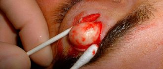
Atheroma occurs in places where sebaceous glands accumulate. Photo: Archives of plastic surgery / Open-i (CC BY-NC 3.0)
Atheromas themselves do not cause discomfort, but if the cystic formation is traumatized as a result of scratching or rubbing with clothing, it can become inflamed.
Important!
When inflamed, atheroma causes pain. The skin over it turns red and becomes hot to the touch. If these symptoms appear, you should immediately consult a surgeon. If treatment is not started in a timely manner, the inflammatory process can spread and cause an abscess or phlegmon - purulent melting of soft tissue.
Atheromas on the scalp are called trichodermal cysts. They can be either single or multiple. These formations are usually quite large and reach a diameter of 5–6 cm or more. Above the surface of the formations, the hair thins greatly, and in many cases falls out completely.
With oily seborrhea, atheromas on the face or neck gradually become denser, and the skin above them acquires a bluish tint. Minor pain may occur upon palpation.
Treatment of atheroma
Retention cysts are removed surgically. Conservative treatment of atheromas is appropriate only at the preoperative stage for patients with an inflammatory process in the area of formation. The doctor may prescribe treating the skin with an antiseptic solution and applying ointments with antibiotics and anti-inflammatory components to it.
Removal of atheromas is carried out using various methods:
- Cystectomy. During this operation, the surgeon, through a small incision in the skin, removes the cyst along with its capsule. After which the wound is sutured and covered with a sterile bandage.
- Cystotomy. Removing the contents of atheroma through a skin puncture². This procedure gives a good cosmetic result. Suitable for face and neck. After healing, an inconspicuous round scar remains in the area of the former atheroma, the diameter of which does not exceed 3–4 mm.
- Laser removal. The laser beam has coagulating and disinfecting properties. Thanks to this, the operation is almost bloodless, and the wound heals quickly.
Laser removal of atheroma.
Photo: Ioannis Dontas / YouTube When suppuration occurs, the atheroma is removed in two stages. First, an incision is made and the purulent contents of the cyst are evacuated through it, and the cavity is washed with antiseptic solutions. After the inflammatory process has completely subsided, the second stage of surgical treatment is performed - removal of the capsule.
What should I do?
If signs of inflammation appear, you should not hesitate - you must urgently go to the surgeon. But what you definitely can’t do is squeeze out or pick at the formation, trying to make a hole in it. If a breakthrough occurs and the pus is outside, the wound needs to be treated and wait until it heals. After this, the capsule and contents can be removed. The cyst should not be heated or steamed at any stage - this can cause infection.
At an emergency consultation with a surgeon, it may be recommended to relieve inflammation with levomekol or ichthyol ointment. The product is applied to a clean bandage and applied to the cyst, and then secured with a bandage. The compress is applied three times a day until the process subsides.
Diagnostics
Most benign tumors of the skin and subcutaneous tissue have a similar clinical picture. Therefore, only a dermatologist can make a correct diagnosis based on research results.
A general examination reveals signs of a cavity neoplasm. It has clear boundaries and can easily move along with the skin in relation to the deeper soft tissues. Other characteristic signs are the slow growth of the swelling and the presence on its surface of a clogged opening of the sebaceous gland duct.
Epidermoid requires differential diagnosis with other diseases:
- lipomas are benign tumors from adipose tissue cells;
- hygromas are synovial cysts that look like a dense “ball” under the skin and are usually localized in the joint area, but can appear in other parts of the body;
- Fibromas are benign tumors made from connective tissue fibers.
A diagnosis of “atheroma” can be made with high accuracy after ultrasound examination of soft tissues, which allows visualization of the capsule and cavity with liquid contents.
After surgical removal, the contents of the sebaceous gland cyst are sent for histological examination. Studying the cellular structure of the neoplasm allows not only to confirm the preoperative diagnosis, but also to exclude the possible malignant nature of the atheroma.
Atheroma and lipoma are different diagnoses!
There is a misconception that atheroma and lipoma are different names for the same benign neoplasm. In fact, these are two independent diseases. Atheroma is a capsule in which sebaceous secretions accumulate, and lipoma is a tumor that is formed due to the proliferation of adipose tissue.
General information
Atheroma is also called a sebaceous cyst. Its appearance is typical for young people 20–30 years old. According to statistics, the problem is more often found in women.
The location is the zones that most actively produce sebaceous secretions and are prone to clogging pores. Cysts can be either single or multiple. They slowly increase in size over several years. Large atheromas can compress nearby tissues and blood vessels.
As a rule, cysts do not hurt, itch or fester. The formation itself does not pose a health hazard, but with prolonged existence, atheromas often become inflamed and suppurate, and can also become a chronic source of infection. This often occurs due to mechanical pressure or irritation, which facilitates the penetration of a bacterial infection. Timely qualified assistance will help avoid such complications.
Attempts to independently open the atheroma in the hope of squeezing out its contents do not lead to the expected effect. Self-medication can lead to infection and other unpredictable consequences. Even in the case of a favorable outcome, after an independent autopsy, in one hundred percent of cases there will be a scar. In addition, what you think is a cyst may turn out to be a formation of a completely different nature and even malignant.
A surgeon can correctly diagnose and safely remove a sebaceous gland tumor. Don't be afraid of going to the doctor. The procedure for removing atheroma is completely safe and painless. [3.5]
Causes of atheroma
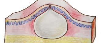
The formation of atheroma is associated with a violation of the outflow of sebaceous gland secretions due to blockage of its duct. The sebaceous secretion accumulates and puts pressure on the walls of the duct, which leads to the formation of a cavity. Over time, connective tissue fibers begin to grow in its walls, forming a fairly dense capsule.
Figure 1. Formation of atheroma. Image: rob3000/Depositphotos
The skin above the growing cyst rises, as a result of which a compaction with a soft-elastic consistency and rounded shape becomes noticeable. The growing atheroma does not compress the nerve endings. Therefore, the neoplasm is not accompanied by itching or pain.
The reasons for the formation of epidermoid are different. A combination of the two is often observed. Most often, the formation of atheromas is caused by:
- Changes in the quantity and nature of the secretion. During puberty, age-related decline of sexual function, and pregnancy, hormonal levels change. This is accompanied by an increase in the viscosity of the secretion secreted by the sebaceous glands. As a result, the outflow worsens, which leads to blockage of the duct.
- Features of the anatomical structure of the sebaceous glands. Some genetic defects cause underdevelopment or even complete absence of the excretory ducts of the sebaceous glands. In this case, atheromas occur in the child during the period of intrauterine development. They can be found on the baby’s body from the first days of life.
- Mechanical skin injuries. As wounds heal, scar tissue forms, which compresses the ducts of the sebaceous glands. The outflow of secretions worsens, and a cystic formation appears.
Some factors also increase the risk of retention cysts³:
- hyperhidrosis - increased sweating of the skin;
- acne - acne;
- oily seborrhea is a chronic inflammatory process that affects skin areas in the area where the sebaceous glands are located;
- boils - acute purulent inflammation of sebaceous jelly or hair follicles.
Atheromas are more often observed in open areas of the body. According to some researchers, this is due to the negative impact of environmental factors on the condition of the skin: radiation, ultraviolet radiation, high and low temperatures.
Postoperative period
In the postoperative period, it is important to monitor the condition of the wound. In the first days, dressings after atheroma are carried out every day or every other day, under the supervision of the attending surgeon. If the cyst was inflamed, the rubber releases are changed daily and the tissues are treated with an antiseptic.
On average, the healing process lasts about 2 weeks. The patient is undergoing outpatient treatment; only patients with severe forms of cysts are admitted to the hospital. The sutures are removed after the formation of good connective tissue “bridges” between the edges of the wounds. This procedure is painless, does not require local anesthesia and takes 3-5 minutes.
Classification
The cellular structure of atheromas is different. Since this does not manifest itself clinically, the histological classification is practically not used and is of interest only to researchers. The more common classification is based on the mechanism of formation of sebaceous gland cysts:
- True or primary. These are congenital formations that are formed at the stage of intrauterine development of the fetus. It is usually diagnosed in children from the moment of birth. Often the formations are multiple in nature and rarely exceed 5 mm in diameter. Mostly located in the perineal area.
- False or secondary. They occur mainly in adults as a result of blockage of the duct of unchanged sebaceous glands. Over time, the cyst gradually increases and can reach 10 cm in diameter. Localization of false atheromas is most often observed on the head, neck and upper back.
Prognosis and prevention
After removal of the atheroma, the wound heals easily. An inconspicuous scar remains. To prevent the formation of new sebaceous gland cysts, you need to consult a dermatologist and select skin care products. In case of multiple atheromas, consultation with an endocrinologist is necessary to exclude hormonal imbalances or correct them.
Consultation with a dermatologist is an important part of preventing atheroma. Photo: IgorVetushko / Depositphotos
It is important to regularly and promptly treat skin diseases - hyperhidrosis, seborrhea, acne. Avoiding aggressive depilation methods, such as wax or sugar paste, can reduce the likelihood of trichodermal cysts. You should also limit the percentage of fats and carbohydrates in your diet.
Photos "before" - "after"
Removal of a large lipoma. There is no need to wait and grow a lipoma to a large size; a small lipoma is easier to remove and the operation is less traumatic. Performed by surgeon: Gladysheva Vladislava.
Surgical removal of back atheroma. “Before” and on the 8th day after surgery: the suture is removed. Surgeon - Vasiliev Maxim.
Sources
- Kisilev V. A., Murzagareeva M. N., Vasilyeva N. A. Experience in surgical treatment of atheromas. Consultative and diagnostic clinic of the Federal State Institution “1477 Naval Clinical Hospital” of the Ministry of Defense of the Russian Federation. Vladivostok. year 2014.
- Grigorieva T. S. Results of surgical treatment of facial atheroma using a gentle method (cystotomy). Crimean State Medical University named after. S.I. Georgievsky. Simferopol.
- Skin atheroma. Wikipedia