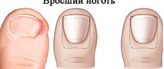
Dermatophytes are microscopic fungi belonging to the genera Trichophyton, Microsporum and Epidermophyton. Dermatophytes live in the keratin of the skin, hair and nails, where their metabolism occurs.
These microscopic fungi cause the most common fungal infections of the skin - foot fungus (Tinea pedis), foot fungus (Tinea cruris), scalp fungus (Tinea capitis), nail fungus (Tinea unguium), body fungus (Tinea unguium). Tinea corporis). Body fungus appears on the groin, face, scalp or beard.
Most often, a fungal infection of dermatophytes affects the superficial layer of the skin, reaching only the epidermis. Sometimes dermatophytes also affect the hair follicles and dermis, then this infection is called Majocchi granuloma. A fungal infection of the scalp and beard damages the hair, but not the hair follicles.
Fungal infection of the head
Fungal infections caused by dermatophytes can be suspected based on clinical signs, but diagnostic tests are also recommended to confirm the diagnosis, as other skin diseases can cause very similar symptoms.
The infection is diagnosed using potassium hydroxide (KOH), fungal culture, or a dermatophyte test. If a cutaneous dermatophyte infection is not recognized and corticosteroid ointment is given for initial treatment, clinical symptoms change. Then it can be difficult to recognize a fungal infection, and the disease itself continues to progress and can reach deeper layers of the skin, dermis, and hair follicles. Patients develop erythema, dandruff, and the rash disappears.
Fungal infections of dermatophytes can predominate in multiple sites at the same time (eg, athlete's foot and groin, athlete's foot and nails). The patient must be thoroughly examined from head to toe, skin, hair and nails. Some patients with fungal skin infections caused by dermatophytes may experience a secondary skin reaction away from the site of infection. This leads to an immunological reaction to the fungal infection.
For treatment, local or systemic antifungal drugs are used:
- Dermatophytic skin infections that affect only the superficial layer of the skin are treated with topical antifungals such as butenafine, ciclopirox, tolnaftate, and allylamine.
- Nystatin, which is effective in treating fungal infections caused by Candida, is not suitable or effective in treating fungal infections caused by dermatophytes.
- Systemic drugs (flucanozole, griseofulvin, itraconazole or terbinafine) are used in cases where the fungal infection is recurrent, although superficial, or when deeper layers (hair follicles, dermis) and nails are affected.
- Patients should not be prescribed the systemic drug ketoconazole due to possible liver damage, adrenal insufficiency and interactions with other drugs.
Griseofulvin
Concomitant use of antifungals with moderate to severe corticosteroids may be effective and may relieve symptoms of superficial dermatophyte skin infection, but is not recommended because corticosteroids are not necessary for successful treatment of the disease and may cause skin atrophy (2). This treatment (antifungal drugs combined with corticosteroids) has been reported to be completely ineffective (3–5).
Immunosuppressive conditions are one of the risk factors for the development of fungal infections caused by dermatophytes, which makes them difficult to treat and recurrence of infection after treatment. A patient with an intractable dermatophyte infection should be evaluated for immunodeficiency disorders.
In this article, we will discuss the most common dermatophyte skin infections, their symptoms, diagnosis, and treatment.
Foot fungus: diagnosis and treatment
The content of the article
Foot fungus (Tinea pedis) is the most common dermatophyte infection. Foot fungus can occur between the toes, develop with hyperkeratosis or vesicobulosis, and in very rare cases can cause ulcerative skin lesions.
Typically, foot fungus predominates between the toes, and often nail and foot (groin) fungus develop together. Foot fungus is most often diagnosed in adults (especially young men). It is estimated that 70% will develop foot fungus throughout their lives. Human. Foot fungus is caused by the dermatophytes Trichophyton rubrum, Trichophyton interdigitale and Epidermophyton floccosum. Infection most often occurs when walking barefoot in changing cabins, saunas or swimming pools and through direct contact with pathogens.
Patients with athlete's foot between the toes usually complain of severe itching, dandruff between the toes, and redness of the skin. The fungus most often spreads under the sole, less often the back of the foot is damaged. In particular, cracks occur between the third and fourth toes, which can be very painful. A less common form of the disease is toenail fungus along with ulcers, where a secondary bacterial infection should be suspected.
Hyperkeratosis foot fungus (also called moccasin foot fungus) appears as diffuse hyperkeratosis of the soles and lateral and medial surfaces of the foot. The skin is scaly and erythema is always present.
Hyperkeratosis foot fungus
Inflammatory or blister foot fungus is painful, causing severe itching, blistering and blistering of the skin that may burst. These lesions are accompanied by severe erythema. The medial side of the foot is most often injured.
The main diagnostic methods are the examination of scratches on the skin after staining with KOH. Microscopic examination of KOH-stained skin scratches showing segmented hyphae of filamentous fungi helps differentiate athlete's foot from nonfungal foot disease, and in the case of Candida infection, a clustered fungus with pseudohyphya is visible.
In case of vesicular disease, vesicles are used for microbiological studies (inoculation on a special medium). If ulcers or cracks form on the skin, a swab should be taken and the culture examined for a secondary bacterial infection.
Taking a smear for foot fungus
In clinical practice, foot fungus can be suspected solely on the basis of signs and type of lesion, but it is recommended to confirm the diagnosis with clinical trials, since the symptoms of a fungal infection can be similar to other dermatological diseases - candidal infection of the fingers, atopic dermatitis.
The main goals of treatment are to relieve symptoms (itching and pain), reduce the risk of secondary bacteria, and stop the further spread of the fungal infection to both the patient and others.
- First-line drugs are topical antifungals, and systemic drugs are reserve drugs prescribed in cases where topical drugs are ineffective or ineffective on their own.
- Topical antifungals include allylamine, butenafine, ciclopirox, and tolnaftate. Data from a 2005 meta-analysis support the effectiveness of topical treatment for tinea pedis.
Local antifungal drugs are prescribed 1-2 times a day and treatment is continued for at least 4 weeks. Patients requiring systemic therapy receive itraconazole (250 mg twice daily for 1 week) and terbinafine (250 mg once daily for 1 week) or fluconazole (150 mg once daily for 2 weeks). -6 weeks).
Topical antifungal drugs
Griseofulvin can also be used to treat athlete's foot, but it is not as effective as other systemic antifungals and requires longer treatment (1000 mg/day for 4–8 weeks or 660 or 750 mg/day for 4–8 weeks ).
In addition to topical antifungal drugs, patients with hyperkeratosis can be prescribed weak solutions of salicylic acid (1). A bandage soaked in this product should be applied to the affected areas and held for 2-0 minutes, repeating the procedure 2-3 times a day.
In children
The main sources of infection of children with zoophilic fungi are stray cats and kittens. The latter are more susceptible to the disease because they have an imperfect immune system and a developed delicate undercoat, which is a breeding ground for microsporum. Microsporia in children is called “feline” lichen. The main contingent consists of children aged 5 to 14 years. The peak incidence occurs in August-October. Microsporia spreads quickly when the pathogen is introduced into schools, sports clubs and kindergartens. Infection from a sick child is possible through household items, a shared towel. The incubation period of zoonotic microsporia is 5-8 days. Anthropophilic pathogens of microsporia (M. ferrugineum) are transmitted through household items and from person to person.
A fungus on a child’s face manifests itself in small, coin-shaped, multiple lesions 1-2 cm in size. They tend to merge. Visually, these foci are difficult to distinguish from foci of trichophytosis . The difference is that with microsporia there are a larger number of foci, vellus hair, eyebrows and eyelashes are more often affected. Microsporidae can also occur - these are allergic rashes, like nodules.
Manifestations of anthroponotic microsporia are similar to superficial trichophytosis. The lesions have clear boundaries and merge, forming bizarre outlines. The primary elements in the lesions are bubbles or nodules, and the secondary elements are crusts. In the classic version, two lesions are formed in the shape of a “ring within a ring”.
Photo of a fungus on a child’s face
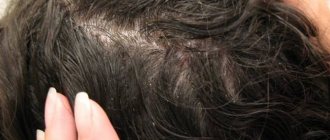
With microsporia and trichophytosis, lesions are often found on the head. These are large, round, scaly patches of baldness. The hair within the lesion is broken off and, with microsporia, protrudes several millimeters above the skin; with trichophytosis, it breaks off at the level of the skin and is visible as a black dot (“stump”). Inflammatory phenomena on the scalp are not expressed. Only bestial pathogens in some cases can cause an infiltrative-suppurative form, in which the lesion protrudes above the skin and is covered with purulent crusts. In case of complications, the child may be hospitalized. If the diagnosis of microsporia and trichophytosis is confirmed, the patient is isolated for 35 days. Contact persons during this period are examined by a doctor. The fireplace is disinfected.
Body fungus: diagnosis and treatment
Tinea corporis is a dermatophytic skin infection. The most common body fungus is caused by T. rubrum. Other pathogens may include Trichophyton tonsurans, Microsporum canis, T. interdigitale, Microsporum gypseum, Trichophyton violaceum and Microsporum audouinii.
Tinea corporis is a dermatophytic skin infection
The infection most often occurs after direct skin contact with a diseased body fungus, can be transmitted from animals, or the infection can spread to the patient himself from other places if he has fungus on the head, legs or other areas. Adults can become infected with the body fungus T. tonsurans from children with a capitis fungus, which is often caused by this pathogen. M. canis infection is transmitted from sick cats or dogs. Outbreaks of body fungus are possible in young athletes who have a lot of contact with other athletes (basketball players, wrestlers, etc.),
Clinical signs of fungus on the body are itchy, round or oval, erythematous, scaly plaques that spread eccentrically. As the disease progresses, the middle of the rash becomes slightly lighter and the edges protrude forward. Round annular plates are formed.
These plaques may coalesce and form blisters. Infections in animals, especially young kittens and puppies, are more severe and predominantly inflammatory. If the rash is widespread, the possibility of an immunodeficiency disorder (eg, HIV infection) or diabetes should be considered.
The main diagnostic method is to examine skin scratches using a KOH solution. Dermatophytes are segmented filamentous hyphae. For analysis, it is necessary to take skin sections from the most active focus of the disease - the edge of the round plate.
A number of dermatological skin diseases must be distinguished between body fungi. The suspicion that this is not a fungus of the body should be caused by the fact that there is no dandruff, treatment is ineffective, a negative response to the KOH test, and the rash is very common. Fungus on the body must be differentiated from other skin diseases that manifest themselves as a ring rash. These diseases include subacute cutaneous lupus erythematosus, granuloma orbicularis, and erythema orbicularis.
The cause of subacute lupus erythematosus is idiopathic and may be associated with systemic lupus erythematosus or the use of certain medications. The disease manifests itself on sun-exposed areas of the body in the form of circular erythematous plaques of dandruff.
Circular granuloma is a benign inflammatory dermatosis. Its clinical features are one or more erythematous annular plaques on the extremities, but no dandruff. Centrifugal circular erythema is an inflammatory skin disease of unknown cause, also manifested by ring-shaped erythematous plaques.
Centrifugal circular erythema
Subacute lupus erythematosus
Circular granuloma
Tinea corporis is effectively treated with topical antifungals such as azoles, allylamine, butenafine, ciclopirox, and tolnaftate (2). Research shows that treating body and foot fungus with two antifungal drugs is very effective (terbinafine and naftifine were studied) (2). Nystatin is not effective in treating dermatophyte infections. Local medications are administered 1-2 times/day. 3 weeks. Treatment is stopped after all clinical signs disappear.
Systemic treatment is indicated for patients who have failed treatment with topical antifungals or whose disease is very advanced. The most common treatment options are terbinafine (250 mg 1 day/day for 1-2 weeks) and itraconazole (200 mg/day for 1 week).
Terbinafine
General information
Fungal diseases are infectious diseases caused by pathogenic and opportunistic fungi.
Currently, the number of fungal infections is growing, which is associated with the expansion of risk groups and climate change. Risk groups include people with allergic conditions, malignant neoplasms, those who have undergone organ and stem cell transplants, patients with immunological diseases, those who have undergone complex operations and who have received massive antibacterial therapy. Superficial skin lesions can be caused by dermatophytes, molds and yeast-like fungi. Dermatophytes rank first in the list of causative agents of skin diseases, and they are grouped into three large genera: Trichophyton, Microsporum and Epidermophyton. Mycosis on the skin of the face (Tinea faciei) is considered a variant of mycosis of smooth skin, but is less common than mycoses of other localizations.
Most often, facial damage is caused by T. rubrum (rubromycosis disease) and T. mentagrophytes, less commonly by microsporum fungi: M. Canis and M. audouinii. Dermatophytosis of the face can be detected as an independent primary lesion, or be secondary in nature when the process spreads from primary lesions to the trunk, folds, neck or limbs. This is facilitated by concomitant pathology of internal organs, diabetes mellitus , pathology of the thyroid gland, as well as the lack of treatment for mycotic lesions. Facial mycosis caused by dermatophytes occupies a separate place in the classification - ICD-10 code B35.0. The most common yeasts that affect the skin of the face and scalp are Candida and Pityrosporum ovale yeast.
Majocchi granuloma: diagnosis and treatment
Majocchi granuloma is a rarer fungal dermatophyte infection that affects the deeper layers of the skin - the dermis, subcutaneous tissue and hair follicles. The most common pathogen is T. rubrum, but infection can also be caused by other dermatophytes. The development of Majocchi granuloma can be caused by skin trauma or occlusion of hair follicles.
Shaving legs is one of the factors in the development of Majocchi granulomas. Immunocompromised and immunocompromised patients are at increased risk of developing Majocchi granuloma. Corticosteroid therapy for dermatophytic superficial infections is not recommended as it may lead to local immunosuppression and the development of Majocchi granuloma.
Clinical signs of infection occur in a localized area of the body where erythematous, perifolicular papules or small nodules or pustules are visible. In patients with immunosuppressive conditions, nodules and abscesses may form subcutaneously. In rare cases, it is also possible for the disease to spread.
Majocchi granuloma
The disease is diagnosed based on the patient's medical history and clinical signs, the diagnosis is confirmed by a skin biopsy, and histopathological examination confirms the presence of fungal forms in the dermis. The response to a culture survey helps identify the exact pathogen. The KOH test may be negative because the fungal infection in this case develops in a deeper layer of the skin.
Topical antifungals are ineffective for treating Majocchi granulomas because they do not penetrate the dermis and subcutaneously. Treatment with systemic antifungal drugs is recommended. The usual recommendation is terbinafine 250 mg once a day. 2–6 weeks, duration of treatment depends on response to treatment. Treatment is continued until all clinical signs disappear.
Pathogenesis
The main location of pathogenic fungi is keratin-containing tissues (the stratum corneum of the epidermis, nails and hair), since they feed on keratin and actively multiply in these tissues. Dermatophytes penetrate the keratin of the skin using keratinolytic proteases. They also produce multiple proteases that destroy other proteins - fibrin , collagen , elastin .
The introduction of a fungus into the skin does not always cause the development of an infectious process. Thus, in the absence of skin damage, long-term asymptomatic carriage of pathogenic fungi occurs. For the development of the disease, certain conditions are necessary: viability, the number of fungi on the skin, the body’s ability to resist pathogens. Mycotic infection is resisted by the barrier function of the skin and immune . Patients with weakened immune systems are susceptible to fungal infections. Elimination of pathogens depends on the condition and function of phagocytic cells - macrophages and neutrophils. The former provide antifungal protection at the initial stages of infection, and neutrophils eliminate fungal invasion.
Having penetrated the skin, the fungus forms threads of mycelium, which penetrate into healthy areas of the skin, causing characteristic damage. If the rate of exfoliation of the stratum corneum (desquamation) is low, then the rate of growth of the fungus prevails and the infectious process spreads quickly. If the rate of desquamation exceeds that of the fungus, then the infection does not spread, since the fungus is eliminated with dead scales of the stratum corneum.
Summary
Fungal skin infections are most often caused by dermatophytes from the genera Trichophyton, Epidermophyton and Microsporum. These dermatophytes metabolize keratin and can be found in various parts of the body - legs, groin, head, nails, beard hair, and can damage the deeper layer of skin, dermis and hair follicles.
Fungal infections caused by dermatophytes can often be suspected by clinical signs, but the diagnosis must also be confirmed by testing. In cases of misdiagnosis, the patient may be prescribed corticosteroids, which complicates both the confirmation of the dermatophyte diagnosis and the healing process itself.
Most skin infections caused by dermatophytes are treatable with topical antifungals such as azole, allylamine, ciclopirox, butenafine, and tolnaftate.
Classification
In the classification of all mycoses, the following lesions are distinguished:
- Superficial ( dermatomycosis ).
- Deep subcutaneous lesions.
- Visceral mycoses (damage to internal organs).
Superficial mycoses are classified according to the localization of the process:
- Mycosis of the feet.
- Smooth skin of the body.
- Inguinal mycosis.
- The scalp.
- Mycosis of the hands.
- Onychomycosis (damage to nails).
- Mycosis of the face.
Facial dermatophytosis is most often found in countries with warm climates. There are two forms: classic and with deep inflammatory lesions. In the first form, the lesion is represented by flat scaly papules, the borders of which are raised and enlarged, turning into a ring-shaped lesion. The desquamation border contains elevated papules and vesicles. The merged elements occupy large areas.
Deep lesions are caused by the fungus Trichophyton verrucosum, which is transmitted from cattle. Large inflammatory foci with a raised surface and pustules appear on the skin of the face. A secondary bacterial (staphylococcal) infection is often associated. The process resolves with hyperpigmentation and scarring .
Sources
- Adam O Goldstein, et al. Dermatophyte (dermatophyte) infections. 2022 UpToDate.
- El-Gohary M, et al. Topical antifungals for herpes zoster and body rash. Cochrane Database Syst Rev 2014.
- Greenberg H.L. et al. Clotrimazole/betamethasone dipropionate: a review of costs and complications in the treatment of common cutaneous fungal infections. Pediatric Dermatol 2002.
- Rosen T., Elewski B.E. Ineffectiveness of clotrimazole-betamethasone dipropionate cream in the treatment of Microsporum canis infections. J Am Acad Dermatol 1995.
- Goldstein A.O. etc. Fungal infections. Effective treatment of diseases of the skin, hair and nails. Geriatrics. 2000.
- Crawford F, Hollis S. Topical treatments for fungal infections of the skin and toenails. Cochrane Database Syst Rev 2007.
- Gupta A. K., Cooper E. A. Update in antifungal therapy for dermatophytosis. Mycopathology 2008.
ONLINE REGISTRATION at the DIANA clinic
You can sign up by calling the toll-free phone number 8-800-707-15-60 or filling out the contact form. In this case, we will contact you ourselves.
Prevention of fungal infection
Prevention of fungal infections is to comply with the following rules:
- Do not wear excessively narrow shoes;
- Make sure your underwear is fresh and change your socks daily;
- Avoid wearing wet clothing (sportswear and swimsuits);
- Dry your hands and feet after bathing;
- Follow the rules of personal hygiene, do not use other people's bed linen, towels, shoes and clothes;
- Don't go barefoot in swimming pools and gyms;
- Use antibiotics only if prescribed by your doctor.
Causes of candidiasis in the mouth
Candidiasis is a mycosis that affects the oral mucosa (gums, tongue, palate, inner cheeks). The disease occurs for a number of reasons that are associated with impaired immunity and the creation of favorable conditions for the development of fungal microflora. Before treating oral candidiasis, you need to determine the cause in order to act directly on it.
The main reasons for the development of fungal disease:
- taking antibacterial drugs:
- lack of hygiene;
- pathologies that provoke a decrease in immunity;
- nervous tension, stress;
- poor diet, excessive consumption of alcoholic beverages.
Dry seborrhea
If there is a lack of activity in the sebaceous glands, dry seborrhea can occur. The reason is not only low sebum production, but also:
- stress, anxiety, psychological shocks;
- hereditary factors;
- hormonal disruptions (puberty or abnormalities in the functioning of the glands);
- use of incorrectly selected cosmetics, as well as cosmetic paints with ammonia;
- poor hygiene, excessively frequent hair washing and blow-drying;
- lack of vitamins (this affects the general health and functioning of the glands).
A deficiency of sebum contributes to the appearance of inflammatory processes on the scalp, which provoke the proliferation of Malassezia fungi. Symptoms of dry seborrhea:
- discoloration, dryness and brittleness of hair;
- formation of cracks in the epidermis;
- the appearance of large scales that quickly peel off.
If measures are not taken, dry seborrhea moves into the next stage. In addition to dandruff, a person is bothered by redness of the skin and seborrheic spots. Sometimes dry seborrhea is an advanced form of oily seborrhea, in other cases the problem arises on its own.