Seborrheic dermatitis is chronic, with periods of improvement and worsening that persist for decades.
The disease usually worsens in cold and dry weather, in situations of increased fatigue or emotional stress.
After consuming fatty foods or alcoholic drinks, in people who smoke or after hot baths.
The development of seborrheic dermatitis is based on the increased activity of a fungus called Pityrosporum (Malassezia).
Malassezia is a yeast that is part of the normal skin flora in more than 90% of adults.
As part of the normal microflora, the fungus is usually apathogenic (not capable of causing disease).
But under the influence of certain factors it is considered as a trigger for pityriasis versicolor, seborrheic eczema and neonatal cephalic pustulosis.
The fungus is characterized by an oval, cylindrical or round cellular structure.
Belongs to the class of imperfect fungi.
Protozoa range in size from 1.5 to 5.5 microns.
They consist of round or oval fungal cells that reproduce by asexual spores.
Malassezia has lipophilic properties and can dissolve fats and oils well.
The fungus feeds on sebum, which is produced on the human epidermis and consists of monounsaturated fatty acids.
Therefore, its main localization is on the scalp, face, occipital area, and back.
A weakened immune system and infectious diseases are considered risk factors for overcolonization of Malassezia yeast.
Most often, fungal infections of the skin occur on the face (almost 80% of cases) and on the scalp (more than 70% of cases).
According to many specialists and patients, the greatest difficulty is treating seborrhea on the head.
It appears more often in men aged 20-40 years.
In the absence of various pathological processes and provoking factors, the body regulates the functioning of the fungus.
And it does not give them the opportunity to multiply excessively.
Normally, the concentration of Pityrosporum on the epidermis of the head varies from 30 to 45-50%.
Against the background of various factors that are triggers in the development of seborrhea, excessive proliferation of the fungus occurs.
The concentration can reach 95%.
These include:
- negative changes in the functioning of the endocrine system and hormonal levels
- chronic pathologies of the autonomic system and central nervous system
- conditions caused by immunodeficiency
- digestive diseases
- prolonged exposure to stressful situations
- therapy with certain groups of medications
What is eczema
Eczema is an inflammatory skin disease that manifests itself as a rash, itching, and redness. The rashes can take a wide variety of forms, affecting different parts of the body. The disease is not contagious. Eczema is often recurrent.
The mechanism of inflammation developing in the dermis and epidermis is that a certain group of cells, when irritated, release substances that cause inflammation. The latter in turn causes itching. The patient itches - inflammation intensifies, microbes penetrate, which together leads to systemic disorders.
Eczema is a polyetiological disease, that is, it is not possible to name a reliable root cause of this pathology. As a rule, this is a combination of exogenous and endogenous factors, the main of which are:
- weakness of the immune system;
- tendency to allergic reactions;
- nervous disorders;
- metabolic disorders in the body;
- genetic predisposition.
According to statistics, this type of dermatitis most often occurs in women aged 30–40 years. This is probably due to high emotional lability (women are more often nervous), as well as frequent contacts with potential allergens: cleaning products, detergents, cosmetics.
Causes of the disease
Today, the main cause of seborrheic eczema is considered to be an allergic reaction to an infection. Another reason is fungus.
There are certain risk factors that contribute to the formation of eczema
:
- excessive production of fat by the sebaceous glands;
- pathologies of the gastrointestinal tract;
- liver disorders;
- hormonal imbalances;
- VSD (vegetative-vascular dystonia);
- chronic infectious pathologies;
- weak immunity.
The main types of eczema that can occur on the scalp
There are wet and dry forms of eczema. The latter most often manifests itself on the hands and fingers, and the ingress of water onto the affected areas significantly aggravates the condition. The weeping form is accompanied by a large amount of serous discharge with the resolution of small vesicular rashes, papules, and pustules.
The following types of eczema can be localized on the scalp:
- seborrheic;
- mycotic;
- true;
- microbial;
- sycosiform.
Let's take a closer look at what each variety is and how it manifests itself.
Make an appointment with NEARMEDIC
Doctors provide effective comprehensive treatment using the latest discoveries in the field of dermatology and long-proven classical methods that are still relevant.
Treatment at NEARMEDIC has a number of advantages:
- complexity - drug therapy, individually developed diet, vitamin intake;
- modern techniques - physiotherapy, ozone therapy, innovative equipment;
- our own laboratory, which allows us to quickly obtain tests for fungi and bacteriological contamination;
- professional doctors with work experience and internships in the best foreign clinics.
Treatment tactics depend on the diagnosed type of disease, laboratory and clinical research data. The program is compiled individually each time. Since the disease is sometimes provoked by problems with the gastrointestinal tract, blood vessels and heart, consultation with an additional specialized specialist may be required. Complex treatment using laser irradiation, ointments, and glucocorticoids relieves inflammation, after which the disease enters a long-term stage of remission.
In the initial stages, when not advanced, eczema can be treated at minimal cost. If primary symptoms appear, do not self-medicate, but immediately seek advice from a dermatovenerologist (specialist, candidate or doctor of medical sciences) in the selected department of the NEARMEDIC clinic network.
True eczema
It is developing rapidly. Small scattered bubbles appear, which soon, sometimes within several hours, open. A serous secretion is released, foci of weeping are formed, crusts, peeling, peeling of crusts, and open wounds appear. Each of the stages of eczema is rapidly replaced by the next: erythematous - papulovesicular - weeping - cortical.
In parallel, fresh vesicles and papules with serous content may appear in another area, since the process is rarely limited to one area. Characteristic is the symmetry of the rashes and the tendency of the rash from the scalp to the skin of the upper, lower extremities, and torso. There are no clearly defined lesions.
True eczema is accompanied by severe itching, which can cause neurosis and insomnia.
General approaches to the treatment of eczema in the practice of an internist
Eczema is a common skin disease, represented by a polymorphism of morphological elements, which is formed as a result of a complex set of etiological and pathogenetic factors. The disease appears at any age, often occurs acutely, less often there are chronic forms. This dermatosis accounts for 30–40% of all skin pathology. Eczema is a polyetiological disease caused by a combination of exogenous and endogenous factors, and is also associated with allergic, metabolic, neurogenic processes, endocrine, and gastrointestinal disorders. Exogenous are chemical, biological, bacterial, physical factors. Drugs, food products, cosmetics and household chemicals are important for the development of the disease.
Eczema is determined by the presence of grouped vesicular elements, when opened, serous “wells” are formed, resembling “bubbles of boiling water.” Dermatosis has been known since the 2nd century BC. e.
Allergic reactivity is of greatest importance in the development of this dermatosis and represents monovalent and polyvalent sensitization. Often the allergic reaction develops as a delayed type, sometimes it occurs as an immediate-delayed one. For the development of an antigen-antibody reaction, a certain humoral environment is required, a change in homeostasis, the presence of immune shifts, a change in the function of prostaglandins and cyclic nucleotides (A. A. Kubanova). Changes in the immune response are determined by the regulation of prostaglandins and cyclic nucleotides and contribute to the development of allergic reactivity and the formation of an eczematous process. This process characterizes the degree and severity of the clinical manifestations of the disease.
The formation of prerequisites for the occurrence of eczema is determined by the presence of a genetic predisposition and factors contributing to the development of an immediate-delayed type reaction. An increase in the content of the thyroid hormone, thyrocalcitonin, has been proven, stimulating the activity of prostaglandins and cyclic nucleotides, which is a compensatory reaction of the body (A. A. Kubanova). In the presence of eczema, there are disorders of the central nervous system, with the activity of the parasympathetic nervous system predominating over the sympathetic, unconditioned reflexes over conditioned ones, and increased sensitivity of skin receptors. Increased permeability of vascular walls is promoted by pituitary-adrenal insufficiency and increased sensitivity of smooth muscle cells. The presence of bacterial and microbial antigens, immune deficiency, a combination of exogenous and endogenous factors influence the development of chronic inflammation in the dermis and epidermis and the formation of immune complexes and the appearance of autoantibodies. Pathological changes in various organs and systems, chronic inflammation in the dermoepidermal area stimulate the development of eczema.
Currently, there is no uniform classification of eczema. For example, one of the classifications represents the following forms of eczema: acute, subacute, chronic.
Yu. K. Skripkin proposed the following classification:
- true eczema, which includes pruriginous and dyshidrotic;
- microbial eczema, which includes nummular, varicose, paratraumatic, sycosiform, nipple eczema, seborrheic, childhood, occupational, mycotic, tilotic eczema.
True eczema is determined by the presence of erythema, against which vesicles and microvesicles, papules, and pustules develop. This eczema is characterized by a polymorphism of rashes, accompanied by an acute inflammatory reaction, infiltration of skin areas, excoriations and the presence of severe itching. When the elements are opened, areas of weeping with exudative crusts, maceration of the skin, scales, fragments of the epidermis and horny layers are formed.
The transition of acute to chronic eczema is determined by the presence of pronounced tissue infiltration, the transition of active hyperemia to chronic, the formation of lichenification, and constant itching.
Pruriginous eczema is characterized by the presence of papulovesicular elements on a compacted base on the extensor surfaces of the limbs, elbows, popliteal areas, face, and inguinal folds. The elements do not open and do not become wet. The disease is chronic and accompanied by severe itching. In this case, dryness, lichenification, cracks, thickening, peeling, and pigmentation of the skin appear.
Pruriginous eczema is accompanied by changes in the nervous system (sleep disturbance, severe neurotic reactions). In childhood, the disease is combined with bronchial asthma and has persistent white dermographism due to the involvement of the parasympathetic nervous system.
Dyshidrotic eczema is determined by the appearance in the palms, soles, and on the lateral surfaces of the fingers of small vesicular elements, dense on palpation, grouped, characterized by the presence of peeling skin, cracks in the palms. The disease is accompanied by severe itching, weeping, the presence of hemorrhagic and yellowish crusts, and has clear boundaries.
Microbial eczema - develops as sensitization to a microbial antigen (streptococcus, staphylococcus) against the background of changes in the neuroendocrine and immune systems, dysfunction of the gastrointestinal tract.
The disease is an asymmetrical process on the skin of the legs, the dorsum of the hands, the lateral surfaces of the torso, and the scalp. The lesions have a clear boundary and are represented by microvesicles, pustules against a background of erythema, infiltration, purulent and yellowish crusts, and peeling. The rashes often spread to all the skin, where serous-purulent or hemorrhagic crusts appear. When the crusts are removed, the surface presents with erosions, bleeding easily, with serous effusion. The process is accompanied by severe itching and pain on palpation.
Nummular eczema (coin-shaped) - is defined by erythematous scaly elements of a round shape, with pronounced exudation, infiltration, the presence of papules, pustules, micropustules, oozing, yellowish, hemorrhagic crusts on the skin. When the process spreads to healthy areas of the skin, papules, papulovesicles, and erythematous spots are formed, which are accompanied by severe itching.
Often the development of microbial eczema is associated with foci of chronic infection (cholecystitis, adnexitis, ENT pathology), parasitic diseases (helminthiasis, giardiasis, enterobiasis, etc.).
Paratraumatic eczema - develops against the background of vascular pathology and in the presence of a focus of infection, in the area of postoperative scars, with bone fractures, osteosynthesis. In this case, erythematous, infiltrative changes appear in the peri-wound area, more often in the presence of edema, with the release of exudate and the formation of yellowish-hemorrhagic crusts. Superficial sclerosis of the skin and deposition of hemosiderin in tissues are possible.
Hypostatic eczema (varicose) - associated with severe vascular pathology of the lower extremities, varicose veins, trophic changes in tissue, trophic ulcers, edema. Vesicular-pustular elements appear on the skin against the background of erythema, infiltration, and serous-purulent crusts form. The disease is accompanied by itching. Foci of sclerosis also form. Differential diagnosis is carried out with erysipelas, pretibial myxedema.
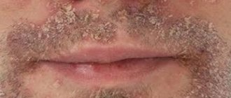
Sycosiform eczema - occurs in the presence of sycosis or with a predisposition to ostiofolliculitis. The process is localized not only on the face, but also in places of increased hair growth (pubic, axillary areas, etc.). Against the background of hyperemia and infiltration, papules and multiple pustules appear, peeling and weeping appear, which leads to the formation of crusts. The disease is chronic, often relapsing.
Eczema of the nipples - with minor infiltration or against the background of severe hyperemia, pustulization develops, erythematous plaques and peeling appear, then, in the presence of weeping, crusts and cracks with bloody-serous discharge form. Differential diagnosis is carried out with Paget's disease, streptostaphyloderma, seborrheic eczema.
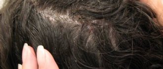
Seborrheic eczema is determined by its location on the scalp, chest, back, interscapular, postauricular area, nasolabial fold. The disease is characterized by the presence of yellowish-pink erythematous spots, with infiltration, fine-plate peeling, and yellow scales. Serous-purulent exudation is possible, sebum secretion is increased, and serous-purulent crusts are formed. In the area of the scalp, the hair is glued together with exudate, there are scales, crusts, and pronounced infiltration. Differential diagnosis is carried out with psoriasis, lichen asbestos, mycotic lesions, pyoderma.
Children's eczema - appears both independently and against the background of atopic dermatitis. The disease develops at an early age. According to statistics, childhood eczema accounts for 13% to 29% of all skin diseases (F.A. Zverkova).
The most common localization is the face, cheek area, scalp, buttocks, hands, legs, and abdomen. Erythematous spots, infiltrative areas with exudative papules, pustules, vesicles and microvesicles form on the skin. The rapid formation of yellowish, brown, hemorrhagic crusts, with oozing, fine-plate and large-plate peeling is possible. When removing or scratching the crusts, eroded areas of the skin appear. In the presence of weeping, erythematous-spotty seborrheic lesions form on the limbs, torso, and face. Purulent-hemorrhagic crusts often appear against the background of severe infiltration, accompanied by biopsied itching.
The disease is a combination of seborrheic, microbial, true eczema. Often a combination of childhood eczema with bronchospasm, bronchitis with an asthmatic component, hay fever, and allergic conjunctivitis.
Most close relatives or parents had allergic diseases. Genetic predisposition depends on the presence of an immune response gene, a positive association of histocompatibility system antigens. At the same time, with an increase in the synthesis of prostaglandins and an imbalance of prostaglandin F2a, a decrease in prostaglandin E, the production of histamine and serotonin is activated, which contribute to the development of an allergic reaction, the appearance of inflammation, and an increase in the permeability of the vascular wall (A. A. Kubanova).
The anamnesis reveals the presence of toxicosis during pregnancy, irrational nutrition, nervous stress, chronic diseases of the mother (pyelonephritis, nephropathy, hepatitis, cholecystitis, diabetes mellitus, foci of chronic infection, diseases of the nervous system). Also important for the spread of the process and its progression are disorders of the gastrointestinal tract (excesses and dyskinesia of the gallbladder, changes in the pancreas, hepatitis). It is necessary to examine children for carriage of helminthic infestations and enterobiasis.
When determining the immune status, immune deficiency and the presence of antibodies to staphylococcal and streptococcal antigens are revealed (F.A. Zverkova).
The most common forms of eczema in childhood are true eczema, seborrheic, microbial, and dyshidrotic. Differential diagnosis must be made with atopic dermatitis, psoriasis, contact dermatitis, streptostaphyloderma, toxicoderma, mycotic skin lesions, prurigo, and Dühring's dermatitis herpetiformis.
Occupational eczema - develops in the presence of industrial allergens (chemical, bacteriological, etc.) and a change in the allergic reactivity of the body.
Professional allergens are amine hardeners, synthetic adhesives, paraphenylenediamine, dinitrochlorobenzene, epoxy resins, phenol-formaldehyde, penicillin and semi-synthetic antibiotics, heavy metal salts, turpentine and its derivatives, mercury compounds, alloys of precious and semi-precious metals, etc.
With occupational eczema, a delayed reaction develops to a substance that is used in production and is an occupational allergen. At the same time, an occupational disease does not develop in all workers of the enterprise, but when the reactivity of the body changes. The inflammatory process manifests itself after a certain period of time with constant contact with substances. Cross-sensitization is possible when exposed to multiple occupational allergens. Workers in metallurgical plants, chemical plants, pharmaceutical and food industries are most susceptible to developing occupational eczema.
The clinical picture of occupational eczema is quite varied. Erythema with infiltrative tissue changes, edema, papulopustular rashes, serous-exudative crusts, oozing, and erosions develop on the skin. The process is accompanied by severe itching. Occupational eczema is often complicated by the addition of a pyogenic infection, which aggravates the course of the disease, promotes the formation of purulent, hemorrhagic crusts, the spread of pustules, vesicles, which tend to open and form weeping areas with serous effusion, vesicular and cystic elements. In this case, lymphadenitis and lymphangitis develop, and body temperature rises. The clinical picture of the disease depends on the degree of allergic reactivity. When the etiological factor disappears, the disease resolves quite quickly.
Occupational eczema is often accompanied by respiratory changes, bronchospasm, allergic rhinitis, allergic conjunctivitis and is a consequence of allergic dermatitis, toxicoderma. The diagnosis of occupational eczema is not made during the initial examination of the patient, since dynamic observation of the clinical manifestations of the process and additional examination methods are necessary. The most common of them are allergological, immunological, and functional diagnostic methods.
Patients with occupational eczema undergo an examination of their ability to work, and the degree of disability due to the occupational disease is determined.
Treatment
Treatment of eczema is carried out comprehensively, taking into account the form and stage of the disease, as well as the severity of the process. Be sure to take into account the condition of internal organs and systems. Complex therapy for various forms of eczema includes a combination of hyposensitizing therapy, detoxification agents, small doses of steroid drugs, sedatives, drugs for correcting changes in the gastrointestinal tract, B vitamins, antibacterial therapy, immunomodulators and immunocorrective drugs, non-steroidal anti-inflammatory drugs, angioprotectors. It is also necessary to use external means and physiotherapeutic techniques.
Systemic therapy
Desensitizing and hyposensitizing therapy includes calcium preparations (calcium chloride and calcium gluconate), which are used both parenterally and orally; sodium thiosulfate solution intravenously (up to 20 injections) or orally; intravenous hemodez solution 200–400 ml drip (4–8 infusions). Polysorbents include Polyphepan, activated carbon, Enterodes, Enterosgel.
The use of H1-histamine blockers (antihistamines) is mandatory - parenteral administration of clemastine, diphenhydramine, chloropyramine (up to 20 injections), in combination with ingestion of H1-histamine receptor blockers, histamine H1-blockers with antiserotonin activity or mast cell membrane stabilizers, for example , ebastine.
In the most severe cases, corticosteroid drugs are used - betamethasone solution (Diprospan) 1.0 ml (once every 10 days, 4 injections in total), prednisolone IM, IV 30-60 mg or orally 30-35 mg per day until a clinical effect is achieved, followed by a reduction of 0.5 mg 1 time every 3–5 days until complete withdrawal.
It is necessary to prescribe immunomodulatory and immunocorrective agents (solutions of Splenin, Humisol, placenta extract, Plazmol, vitreous body, Imunofan, immunoglobulin, Myelopid, Timalin, Timogen, Taktivin); oral drugs (Licopid, Kemantan, Glyciram, sodium nucleinate, Polyoxidonium, Leukinferon, interferon drugs, Diucifon, Dimocyfon, Avlosulfone, dapsone). To correct IgE and IgG, use Histaglobulin intravenously at 0.25, 0.4, 0.6, 0.8, 1 - up to 2 ml after 2-3 days.
Vitamins B1, B6, B12, B15, A, E, C, calcium pantothenate, folic acid, Essentiale are used.
In the presence of a purulent, microbial process, antibacterial agents are used with preliminary culture of the flora and sensitivity determination - broad-spectrum antibiotics (strengthened and antistaphylococcal penicillins, cephalosporins of the first and second generations, aminoglycosides, macrolides, fluoroquinolones, etc.).
Among sedatives, it is advisable to use tranquilizers and antidepressants, especially tricyclic antidepressants with antihistamine activity (doxepin, etc.).
Drugs that influence the synthesis of prostaglandins are used - non-steroidal anti-inflammatory drugs (ibuprofen, indomethacin, diclofenac, etc.) and angioprotectors - xanthinol nicotinate, pentoxifylline, dipyridamole, etc.
The literature (Yu. K. Skripkin) indicates the use of Prodigiosan and Pyrogenal, but they should be used with great caution.
In some cases, it is advisable to prescribe gastrointestinal enzymes; in the presence of dysbacteriosis, eubiotics are used accordingly. Among the biostimulating agents used are tinctures of Eleutherococcus, aloe, ginseng, aralia, pantocrine, etc.
Local treatment
External treatment is determined by the clinical picture of eczema.
If there is weeping, lotions are prescribed from a solution of silver nitrate, a solution of tannin, in addition, it is rational to use quickly drying gels - dimethindene (Fenistil gel also has a local anesthetic effect), solutions of Furacilin, Dioxidin, methylene blue, a solution of potassium permanganate, chlorhexidine (Gibitan) , 0.5% Resorcinol, 2% boric acid.
For excoriation, peels are applied with aniline dyes (Fukortsin, brilliant green).
For exudative changes, aerosols of Polcortolone, Oxycort, Oxycyclosol, Panthenol, Aekol, Levovinizol are used. To form crusts, it is possible to use the following pastes: 2% boron/5% tar, 2% boron/5% (up to 10–20%) naphthalan, 5% ASD, zinc, as well as pastes with the addition of antibiotics, 2–5% ichthyol, 2–5% sulfur, salicylic acid.
Steroid ointments are used: Celestoderm-B, Lokasalen, Lorinden A, C, Belosalik, Belogent, Beloderm, Diprosalik, Diprogent, Ultralan, Polcortolon (ointment), Prednisolone (ointment), Hydrocortisone (ointment), Betnovate, Dermovate, Cutivate, Lokoid, Elokom, Advantan, Apulein, Flucinar, Fluorocort, Locacorten, Gioksizon, Triderm, etc.
Together with steroid ointments, dimethendene (Fenistil gel) is prescribed for a faster onset of effect, as well as for the purpose of drying or, conversely, moisturizing.
Physiotherapy
Physiotherapy is prescribed in combination with the specified treatment. A helium-neon laser (wavelength 0.632 nm) is used directly on the lesions (also intravenously, navenno) with preliminary application of a 1% solution of methylene blue and oral administration of Anavenol, Redergin.
The use of selective phototherapy, local and general UV irradiation is effective. Treatment with acupuncture is possible.
Resort therapy and balneothalassotherapy are also of great importance, especially in the summer. The most effective stay is on the Black Sea coast of Crimea, the Caucasus, the resorts of the Krasnodar Territory, Transbaikalia, Altai Territory, Azerbaijan, the Mediterranean, and the Dead Sea.
It is important to limit water procedures, adherence to hygiene rules, a rational regimen, the elimination of neuro-emotional overload, adequate physical activity, and good sleep. A hypoallergenic dairy-vegetable and fortified diet is a necessary preventive measure to prevent the development and exacerbation of eczema. It is also necessary to treat concomitant diseases, normalize the function of the gastrointestinal tract, and avoid contact with various allergens, household chemicals, synthetic and woolen items.
Patients with eczema should be monitored by a local dermatologist.
Literature
- Allergic dermatoses in children: Clinic, treatment, organization of clinical observation and prevention: Method, recommendations of the Sverdlovsk NIKVI (author: N.P. Toropova et al.) Sverdlovsk, 1990. 65 p.
- Antonyev A. A. Candidiasis of the skin and mucous membranes // A. A. Antonyev, L. A. Bulwakhter, L. K. Glazkova, I. I. Ilyin M.: Medicine, 1985. 160 pp. 1.l.
- Arzumanyan V. G. Yeast-like fungi on the skin of patients with atonic dermatitis/V. G. Arzumanyan, M. A. Mokronosova, T. M. Samuilova, V. B. Gervazieva // JMEI. 1998. No. 3. p. 10–13.
- Kubanova A. A. State of the immune system in patients with eczema/A. A. Kubanova, L. L. Vasilyeva, L. V. Alekseeva, N. G. Dmitrieva // Vestn. dermatol. Venerol. 1983. No. 8. pp. 16–19.
- Kubanova A. A. The significance of disorders of immunological reactivity, the ratio of the level of cyclic nucleotides and prostaglandins in the pathogenesis and clinic of true eczema and therapeutic correction: Diss.. Dr. med. Sci. M., 1986. 240 p.
- Chalimova R. A. Blood clotting and fibrinolysis in eczema // Vestn. dermatol. Venerol. 1975. No. 4. pp. 54–57.
- Chuchalin A. G. Bronchial asthma. M.: Medicine, 1985. 157 p.
A. A. Danilova, Candidate of Medical Sciences
TsNIKVI Ministry of Health of the Russian Federation, Moscow
Contact information about the author for correspondence
Treatment of scalp eczema
Curing eczema on the scalp is quite difficult. Firstly, eczematous wounds are difficult to treat. Secondly, patients most often consult a doctor when the disease has already taken on a complex form and a secondary infection occurs. The reason for this lies in the same “insidiousness” of the disease, especially when it comes to eczema of the scalp: localized in the crown area, such lesions are difficult to detect. Simply put, the disease goes unnoticed for some time: itching is considered an accident, flaking is considered dandruff.
Mycotic infections can complicate the course. In this case, treatment will not bring results; preliminary antimycotic therapy is necessary. To do this, you need to determine the type of fungus that has settled. If the fungal component of the process was not excluded before starting treatment, then hormonal ointments and antibiotic-based ointments, which are used to combat eczema, will lead to active reproduction and mutation of fungi, if present. Therefore, in the treatment of eczema, you cannot do without consulting a dermatologist or trichologist.
As a rule, complex therapy is prescribed and may include the use of:
- hormonal ointments;
- antibiotic-based ointments;
- antihistamines;
- sedatives;
- desensitizing drugs;
- diet.
During treatment, it is also important to visit a specialist who will monitor the progress of the disease. In no case should you independently extend the prescribed course of treatment - corticosteroid hormones can cause serious side effects if used uncontrolled.
Diagnosis of dermatitis
Which doctor should I contact for help, and what tests should I take?
Although identifying dermatitis is not difficult, it is necessary to determine the root cause and triggering factors.
In addition to collecting the patient’s medical history and general examination, you will need to diagnose hormonal levels, as well as study the functioning of the digestive organs.
Laboratory tests are prescribed to exclude endocrine disorders, in particular diabetes mellitus.
Additionally, a stool examination is carried out to determine the presence/absence of dysbacteriosis.
If the dermatologist deciphers the test results and detects pathological changes, the doctor will redirect the patient to specialized specialists.
Most often such doctors are a gastroenterologist and an endocrinologist.
Tips for those suffering from eczema
If you have a genetic predisposition or have already experienced eczema, you should be extremely careful by following these recommendations:
- avoid wearing clothes made of synthetic, wool or flannel fabric;
- include in your diet more vegetable dishes, boiled meat, fermented milk products, fruits (except for those that can provoke allergies: oranges, strawberries, raspberries, tangerines);
- avoid gastronomic experiments, alcohol, too spicy, salty and fatty foods, marinades;
- take vitamin complexes, in particular vitamins A and B are important;
- avoid contact with suspected allergens in everyday life (powders, detergents, etc.);
- in case of a history of varicose veins, wear rubber stockings and bandage the legs with medicinal bandages;
- be as nervous as possible;
- Visit a dermatologist periodically.
At the MediLife clinic in St. Petersburg, highly qualified experienced dermatologists and trichologists receive treatment. You can make an appointment online. Prices for services are presented on the website. You can clarify any details by phone. Friendly staff, the ability to choose a convenient time to visit and complete anonymity are pleasant and significant additions to the qualified consultation that will be provided at MediLife.
Bibliography
- Ado A.D. General allergology. M.; "Medicine", 1970.-543 p.
- Results of a study of the effectiveness of non-halogenated corticosteroids in the treatment of chronic eczema / I.M. Korsunskaya, N.A. Lukashova, Z.A. Nevozinskaya, E.E. Agafonova // Clinical. dermatology, venereology. 2008. - No. 4. — P. 101-105.
- Ryzhko, P.P. The use of antihistamines in the treatment of various dermatoses / P.P. Ryzhko // Ukr. magazine dermatology, venereology, cosmetology. 2002. - No. 1. — P. 39-41.
- Skripkin, Yu.K. Allergic dermatoses / Yu.K. Skripkin, B.A. Somov, Yu.S. Butov. M.: Medicine, 1975. - 245 p.
- Tishchenko, A.L. Optimal therapeutic doses of vitamin A in patients with eczema and psoriasis / A.L. Tishchenko // Vestn. dermatology and venereology. 1998. - No. 4. — P. 36-38.
- Turchina, I.P. Complex treatment of patients with eczema and neurodermatitis localized on the lower extremities / I.P. Turchin // Dermatovenerology, cosmetology, sexopathology. 2002. - No. 1-2 (5). - S. 98101.
- Yusupova, L.A. Treatment of patients with eczema / L.A. Yusupova, R.Kh. Kha-fizyanova // Ross. magazine skin diseases. 2005. - No. 6. — P. 20-22.
- Brazzini, B. New and established topical corticosteroids in dermatology: Clinical pharmacology and therapeutic use / B. Brazzini, N. Pimpinelli // Am. J. Clin. Dermatol. 2002. - N3. — P. 47-58.
- Buznikov, GA Serotonin and serotonin-like substances as regulators of early embryogenesis and / GA Buznikov, HW Lanbert, JM Lauder //Cell Tissue Res. 2001.-N 39.-P. 177-186.
- Catania, A. Inhibition of acute inflammation in the periphery by central action of salicylates / A. Catania, J. Arnold, A. Macaluso et al. //Proc. Natl Acad Sci USA. 1991. -Vol.88, N19. -P. 8544-85
Types and stages of the disease
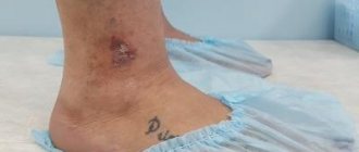
Regardless of the location, weeping eczema goes through several stages of development:
- The appearance of vesicles with a diameter of up to 5 mm, reminiscent of bubbles when water boils.
- Formation of erosions at the site of opened blisters, which burst due to scratching or on their own.
- The formation of wet areas covered with serous fluid, which is released from the burst vesicles.
- Transition of the disease to the dry stage with a decrease in redness and weeping, the formation and rejection of scales.