Itchy skin
Itchy skin:
Occurs in 2% of pregnant women in the third trimester of pregnancy.
Pathogenesis:
Presumably, itching is caused by increased excretion of bile acids under the influence of endogenous hormones (estrogen and progesterone). Its development is also associated with recurrent cholestasis.
Clinical picture:
Usually the itching is localized in the abdominal area, but it can also be generalized. Some patients have pronounced scleral icterus and an increase in the content of alkyl phosphatase in liver tests. After childbirth, the itching disappears, but may reappear during the next pregnancy or use of oral contraceptives.
Differential diagnosis:
- Scabies.
- Toxicoderma.
- Hepatitis.
Prurigo of pregnancy occurs at an earlier stage (from the 25th to the 30th week) and is not urticarial in nature, unlike polymorphic dermatosis. The clinical picture is characterized by disseminated itchy pruriginous rashes in the form of excoriated nodules with a hemorrhagic crust at the top. The rash persists throughout pregnancy and is difficult to treat (see Pruritus); After childbirth it regresses on its own.
Make an appointment for a consultation by phone. +7 (495) 589-85-42, +7 (495) 589-85-43.
Intrahepatic cholestasis of pregnancy
Definition.
Intrahepatic cholestasis of pregnancy (ICP, intrahepatic cholestasis syndrome of pregnancy, obstetric cholestasis, idiopathic jaundice of pregnant women) is one of the specific dermatoses of pregnancy (IDP), which is characterized by an increase in the level of aminotransferases and bile acids (≥10 μmol/l) in the blood serum, the development of intense itching (this intensity is not typical for other SDB) at the end of pregnancy (in 80% of patients - in the II-III trimester), mainly secondary damage to the skin (excoriations occur, etc.), spontaneous and rapid (from 2 days to 3 weeks ) regression of clinical symptoms after childbirth and, finally, not always a favorable outcome for the fetus [1-5].
In 1883, the disease was first described by F. Alfeld [6].
Epidemiology.
ICP is a relatively common cause of liver problems during pregnancy. In Central and Western Europe and North America, the incidence of ICP is 0.4-1.0%, in the Scandinavian and Baltic countries - up to 3%, and in Chile and Bolivia 5-28% (this may be due to the gastronomic preferences of local residents) [7, 8]. Unlike polymorphic rash of pregnancy (EPP), ICP recurs during subsequent pregnancies [1, 8].
ICP is more often observed in the cold season [7].
Etiopathogenesis.
A genetic predisposition to the development of ICP is evidenced by a family history (in 50% of patients), ethnic and geographical differences, the presence of histocompatibility antigens HLA-A31 and HLA-B8 (positive correlation), mutations of the ABC-B4 genes on the 7th chromosome (encodes MDR-3 protein; disruption of the activity of transport proteins, phospholipids, bile formation, etc.), ABC-B11 on the 2nd chromosome (encodes the BSEP protein; changes in the so-called bile salt export pump), ATP-8-B1 on the 18th chromosome (encodes the FIC1 protein; insignificant role) [4, 9–11, 33].
The role of endocrine factors deserves attention. ICP is most often observed in the last trimester of pregnancy (estrogens concentration reaches a maximum), in twin or triple pregnancies (estrogens concentration is higher than in singleton pregnancies), as well as in persons who took hormonal (estrogenic) contraceptives before pregnancy (ICP in in the appropriate context, is considered as a hyperergic response of the body to increased estrogen levels). Subsequently, the cholestatic effects of sex hormones and their metabolites develop. We must also not forget about the role of progesterone (hyperprogesteronemia), selenium (a decrease in selenium concentration leads to cholestasis, and vice versa) and others [1, 4, 12, 13].
Under the influence of the above factors, the content of bile acids in the blood serum of pregnant women increases, they enter the blood of the fetus (pregnant women experience severe itching, uteroplacental blood flow is disrupted - acute oxygen deficiency develops, fetal cardiac activity is inhibited, etc.).
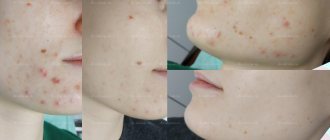
Clinic.
The main symptom of ICP is severe itching of the palms and soles, which usually occurs without any warning signs after 30 weeks of pregnancy, rapidly progresses and becomes unbearable, having a strong psychotraumatic effect and leading to the development of various nosogenies (for example, insomnia, depressive reactions, suicidal behavior) [4, 14].
Over time, the itching spreads to other areas (extensor surfaces of the limbs, buttocks, abdomen) and covers the entire body. It sometimes weakens, but then intensifies and becomes continuous and unbearable, continuing until the end of pregnancy. Usually, the itching disappears immediately after childbirth.
Itching is not caused by a primary skin lesion - it is affected secondarily (excoriation, erythematous-papular elements occur, and the itching worsens) [7].
Only in 10-20% of patients, a few weeks after the onset of the disease, jaundice develops in parallel with itching (sometimes preceding the appearance of itching), but the condition of the patients does not change much because of this, since the bilirubin level does not increase more than 100 μmol/L (not a critical value : complications such as bilirubin encephalopathy and so on do not occur) [14-16].
Sometimes observed malabsorption syndrome and steatorrhea (the result of impaired processes of breakdown and utilization of fats) lead to impaired absorption of various nutrients in the small intestine, for example fat-soluble vitamin K (necessary for the synthesis of prothrombin complex factors), which in turn leads to an increase in prothrombin time (hypocoagulation , intrauterine and postpartum hemorrhage) [1, 14, 17, 18].
Subsequently, women who have had ICP during pregnancy have an increased risk of developing hepatobiliary pathology (for example, cholelithiasis, cholecystitis) [19].
During subsequent pregnancies or when using hormonal contraceptives, the process recurs.
Laboratory diagnostics
.
The most sensitive biochemical indicators
for diagnosing ICP are an increase in the level of bile acids (more than 10-100 times) and aminotransferase activity (2-10 times) in the blood serum [1, 15, 20, 21]. However, it should be taken into account that the level of bile acids in healthy pregnant women at the end of pregnancy somehow increases, exceeding the normal limits (0-6 μmol/l), but not more than 2 times, which is not correctly attributed to pathology (asymptomatic hypercholanemia) [ 1, 22-24].
Pathohistological
changes in ICP are not specific.
The results of immunomorphological
research methods (direct and indirect immunofluorescence reactions) for ICP are negative.
Treatment.
The goal of drug treatment for ICP is to improve the mother’s condition (eliminate itching by reducing the level of bile acids in the blood serum, carry the pregnancy to term, improve the quality of life, etc.) and reduce the risk of developing pathology in the fetus (improving the medical prognosis of the fetus’s health). In this context, ursodeoxycholic acid (USCA, Ursofalk) is the most effective drug (antihistamines, sleeping pills, hormonal and other drugs are less effective). Often they also resort to induction of labor (during the 37-38th week of pregnancy, since stillbirths are often observed precisely at these dates or later) [1, 4, 15, 25-31].
Course and prognosis.
ICP in the DBS group is considered a disease that poses the maximum, although controllable, danger to both the mother and the fetus. However, for ICP, what is considered more important is not the mother’s itching, the possibility of intrauterine and postpartum hemorrhage (especially with jaundice and/or vitamin K deficiency), but possible complications in the fetus. With ICP, premature birth (19-60%), intrauterine hypoxia (22-41%), bradycardia (14%), stillbirth (0.4-4.1%), etc. are very often observed. The likelihood of these complications increases, especially when the concentration of bile acids in the mother’s blood serum exceeds 40 μmol/l [1, 4, 5, 20].
Both in the blood serum of patients with ICP, and in the umbilical cord blood and in the amniotic fluid of the fetus, the concentration of total bile acids can be very high, which leads to pathological changes in the surfactant of the fetal lung tissue with corresponding consequences [32].
There is also a positive correlation between high levels of total bile acids in the blood serum of maternal and newborn rats, the possibility of developing pathological changes in the lungs and a greater risk of stillbirth (experimental dermatology) [3].
Thus, early diagnosis of ICP, prescription of specific treatment and constant obstetric and gynecological monitoring are important for the correct choice of medical tactics and strategies in the management of patients.
Atopic rash of pregnancy
Definition.
Atopic rash of pregnancy (ASB, prurigo of pregnancy, pruritic folliculitis of pregnancy, eczema of pregnancy) is a benign, most common (in 50% of patients) RAS, which occurs in the first months of pregnancy (in 75% of cases - in the first trimester) in persons with predisposition to atopy, manifested by vesicular or papular elements. ASP usually recurs during subsequent pregnancies [34, 35].
Pathogenesis.
Currently, in the pathogenesis of ASP, the dominant version is a violation of the ratio and imbalance between Th1 and Th2 lymphocytes, as a result of which the production of Th1 cytokines (IL-2, γ-interferon, IL-12) is inhibited, and Th2 cytokines (IL-12) are inhibited. 4, IL-10), on the contrary, is stimulated [36].
Clinic.
ASD is divided into two types - intrinsing (internal, IgE-non-associated, non-allergic, atopiform dermatitis) and extrinsing (external, IgE-associated, allergic), but the issue of such a division is disputed [37].
In 80% of patients, ARS first occurs during pregnancy (only in this case the rash is classified as ARS). In 20% of patients, existing atopic dermatitis worsens (perhaps the disease has been in remission since childhood).
The process in 65-70% of cases manifests itself in the form of diffuse eczema (the so-called IgE-associated type of ASD), located in the favorite places of atopic dermatitis (face, neck, décolleté, flexor surfaces of the limbs), and in 30-35% of cases small erythematous (on the body and limbs) and lichenoid (on the legs and palms) papules [38]. Other characteristic stigmas of atopic dermatitis (severe dry skin, dermographism of the palms, follicular keratosis, lichen alba, and so on) are also often found [4, 35, 37, 39].
Laboratory diagnostics.
Pathohistological changes in ICP are not specific.
The results of immunomorphological
research methods (direct and indirect immunofluorescence reactions) for ASP are negative. A high concentration of IgE is detected in the blood of 20-70% of patients [34].
Treatment.
Local moisturizers (containing 3-10% urea) and antipruritic drugs (menthol, polidocanol and others) are often used. To prevent additional skin dryness, you can use mild, non-alkaline synthetic cleansers (skin lipid replacements), oil shower solutions, etc.
Local corticosteroid drugs are very effective. Moderately active corticosteroids are applied to the skin of the face and folds, and “strong” corticosteroids are applied to the rest of the body (extra-strength corticosteroids should be avoided) [40-42].
In severe cases, systemic non-halogen corticosteroid drugs are prescribed. Due to the fact that cortisol, prednisone and prednisolone are deactivated in the placenta (unlike betamethasone and dexamethasone), prednisolone is considered the drug of choice during pregnancy. The dosage depends on the nature of the pregnancy and the severity of ASP. The initial dose of prednisolone is 0.5-2 mg/kg/day (if necessary, the dose is increased, but in the first trimester of pregnancy it should not exceed 10-15 mg/day, since the likelihood of the child developing a cleft lip and cleft palate increases) [ 40-42].
To avoid side effects, the duration of corticosteroid therapy should not exceed 1 month. If the course is prolonged, then fetal growth must be monitored using ultrasound and not forgetting about the possibility of adrenal insufficiency developing in the newborn.
Antihistamines are also prescribed (preferably only in the first trimester of pregnancy) of both the first (dimetindene, clemastine, pheniramine, etc.; extensive experience with use) and second generation (loratadine, cetirizine, etc.; no pronounced sedative effect), phototherapy (UVB -therapy in difficult cases in early pregnancy is an additional, safe and effective tool), etc. [41, 42].
Course and prognosis.
The prognosis for ASP in the mother is favorable, but the disease can recur again during subsequent pregnancies.
The health of the fetus is also out of the danger zone, although in the future children may experience signs of atopic dermatitis.
Polymorphic dermatosis
Polymorphic dermatosis of pregnant women:
Developing in the third trimester of pregnancy, on average at the 36th week of pregnancy, usually within 1-2 weeks. before giving birth; Regresses on its own within a few days after birth.
Frequency:
One case in 120-240 pregnancies.
Pathogenesis:
The disease is associated with an abnormal increase in body weight of the pregnant woman and fetus, and overstretching of the abdominal skin.
Clinical picture:
Red papules, 1-3 mm in size, appear on the skin of the abdomen, buttocks, thighs, occasionally the face, palms and soles, which quickly merge into plaques resembling large blisters, in places forming confluent polycyclic lesions. The rash is accompanied by severe itching. The mucous membranes are not affected.
Diagnostics:
Usually based on the clinical picture.
Differential diagnosis
- Herpes in pregnant women.
- Drug toxicoderma.
- Diffuse neurodermatitis.
Course and prognosis:
All symptoms usually disappear 7-10 days after birth. Most women do not experience relapses in subsequent pregnancies. No adverse effects on the pregnant woman or fetus were noted.
Synonyms:
- Pruritic urticarial-papular and plaque dermatosis of pregnant women.
- Toxic erythema of pregnancy.
Make an appointment for a consultation by phone. +7 (495) 589-85-42, +7 (495) 589-85-43.
Irrigation of the nasal cavity with saline solutions
Irrigation with saline solutions allows you to cleanse the nasal cavity of mucus and various foreign particles [3]. Saline solutions provide hydration to the sinuses.
The use of saline solutions increases the activity of ciliated epithelial cells of the nasal mucosa.
Preparation of the solution:
Prepare a pre-sterilized glass container.
To fill the container, use a large medical syringe (30 cc), a water collector with an irrigation attachment, or a Neti pot. The selected liquid collection device must also be pre-sterilized.
Fill the container with distilled or boiled water.
Add 1 to 1.5 teaspoons of canned salt to the water. The use of table salt is not recommended due to the large number of additives it contains.
Add one spoon of table salt.
Stir the resulting solution.
The solution should be stored at room temperature. Shelf life - 1 week. from the moment of manufacture.
Instructions for use:
Rinse your nose with saline solution 1-2 times a day.
If you have been prescribed medications for intranasal inhalation, use them only after irrigating the nasal cavity with the prepared solution. Preliminary cleansing of the nasal cavity from mucus and foreign particles increases the effectiveness of medications.
First, pour the volume of liquid required for irrigation into a clean container. Liquid collection should be carried out each time with a new, sterile syringe. To prevent contamination of the prepared solution, do not use syringes more than once.
Warm the solution slightly in the microwave before use.
During irrigation, tilt your head forward and gently rinse each nasal passage, directing the stream of liquid towards the back of the head. When properly irrigated, the solution will pass through one nasal passage and out the other.
Some people may experience a burning sensation during the first irrigation, but most often it goes away after the nasal mucosa adapts to the procedure.
Herpes in pregnant women
Herpes in pregnant women:
Dermatosis, manifested by a polymorphic rash and severe itching, occurring during pregnancy and the postpartum period.
Frequency:
One case in 4,000–50,000 pregnancies.
Risk factors:
- Episodes of illness during a previous pregnancy.
- Use of oral contraceptives.
- Tumors from trophoblast.
Etiology and pathogenesis:
An autoimmune disease involving complement-fixing IgG antibodies. In patients, alleles HLA-B8, HLA-DR3 and HLA-DR4 are detected with high frequency.
Clinical picture:
The disease usually begins in the 4th to 7th month of pregnancy, but can occur in the first trimester and in the postpartum period. Manifests itself in the form of papules and vesicles. There are also small and large red blisters, large tense blisters, erosions and crusts. The elements are arranged in groups, which gives rise to the name “herpes of pregnant women.” The process most often begins from the navel area, and then spreads to the skin of the abdomen, thighs, palms and soles, and rarely to the mucous membrane of the mouth.
Differential diagnosis:
- Polymorphic dermatosis of pregnant women.
- Polymorphous exudative erythema.
- Dühring's dermatitis herpetiphora.
- Drug toxicoderma.
Treatment:
For mild cases, corticosteroid ointments are used topically in combination with oral antihistamines. For bullous rashes, prednisolone is prescribed orally 40 mg/day. followed by a reduction to a maintenance dose. During childbirth, a significant increase in the dose is sometimes necessary due to a sharp increase in rashes and itching.
Make an appointment for a consultation by phone. +7 (495) 589-85-42, +7 (495) 589-85-43.
Rhinitis
The main causes of mucous discharge from the nose during pregnancy are rhinitis (vasomotor, allergic, drug) and sinusitis [2]. Regardless of their form, non-drug methods are the basis of first-choice therapy.
Saline sprays
reduce the severity of rhinitis symptoms, they are characterized by an extremely low risk of side effects due to the absence of pharmacological components in them [3]. To irrigate the nasal cavity, nozzles and nebulizers are used, and up to 200 ml of solution should flow into each nasal passage. The spray can be used daily or as needed. According to the results of randomized clinical trials, the use of sprays is recognized as the treatment of choice for nasal symptoms during pregnancy [3].
Regular physical activity
promotes physiological vasoconstriction and reduces symptoms of nasal congestion and rhinorrhea [3, 4].
Nasal dilators
allow you to mechanically widen the nasal passages, thereby facilitating nasal breathing.
The advantages include their high availability, as well as the possibility of use at night. Raising the head end of the bed
by 30–45 degrees greatly facilitates nasal breathing during sleep [5].
Atrophic stripes
Atrophic stripes:
They appear in most pregnant women on the skin of the abdomen, buttocks, and mammary glands.
Pathogenesis:
Apparently, in addition to the mechanical factor (stretching), degeneration of the dermis caused by endocrinopathy, autonomic disorders, etc. is of certain importance.
Clinical picture. Atrophic stripes of skin up to 4-6 mm wide, located symmetrically. Areas of band-like atrophy are sharply defined, with a wrinkled surface of a whitish-lilac hue, slightly sunken.
Treatment:
Use of Mederma or Contratubes creams after childbirth.
Synonym:
Striae of pregnant women.
In addition, during pregnancy, more pronounced general skin pigmentation is observed, especially in brunettes, developing in the area of the nipples of the mammary glands, genitals, and midline of the abdomen. In many women, the number and size of melanocytic nevi increase. The hair thins somewhat, even diffuse alopecia is possible, but after childbirth, over time, the properties of the hair are restored. Sometimes there is some hypertrichosis in combination with acne, usually in the second half of pregnancy. This may be the result of an androgen-secreting tumor, luteoma, luteal cysts, or polycystic ovarian disease and requires careful evaluation. Vascular changes are associated with the action of estrogens; they manifest themselves in 70% of pregnant women with erythema of the palms, which in some cases can be expressed only in the thenar and hypothenar areas. Small hemangiomas may occur, most often on the scalp and neck. Varicose veins of the extremities, hemorrhoids, and, less commonly, deep vein thrombophlebitis are often observed. Swelling of the face, eyelids, feet, and hands is also possible, which occurs in the morning and disappears in the evening. 80% of pregnant women develop gingivitis (swelling and redness of the gums), sometimes accompanied by pain and ulceration. In 2% of cases, changes in the gums are accompanied by the appearance of small vascular formations such as pyogenic granuloma, which bleed when touched (granuloma of pregnancy). These symptoms are somewhat relieved by taking vitamin C.
During pregnancy, there may be an increase in the frequency of infectious lesions, especially candidiasis, genital warts, herpes simplex, bowenoid papulosis. During pregnancy, autoimmune diseases (especially lupus erythematosus), lymphomas, and atopic dermatitis worsen. The effect of pregnancy on psoriasis can be different, and some patients may develop an acute pustular form.
Allergic rhinitis and conjunctivitis
In most cases, allergic conjunctivitis first develops before pregnancy. The leading complaints of patients are sneezing, itchy nose and watery rhinorrhea. In some cases, irritation of the eye mucosa occurs. Allergic rhinitis often develops after contact with allergens such as dust mites, pet dander, mold and pollen [5].
Diagnosis during pregnancy.
One of the most effective methods for diagnosing allergic diseases is skin testing with allergens. However, their use during pregnancy is not recommended due to the risk of developing systemic inflammatory reactions. In this regard, for routine use in clinical practice, it is recommended to study the level of allergen-specific IgE in venous blood [5, 6].
Treatment.
Pregnant women with minor allergy symptoms, including those with a significant allergic history, should undergo preventive measures and/or treatment with non-drug methods. If necessary, second-generation antihistamines are prescribed, such as loratadine (10 mg once a day) or cetirizine (10 mg once a day) [5, 7].
For moderate to severe rhinitis, budesonide, fluticasone or mometasone (in the form of a spray for intranasal inhalation) are recommended as basic therapy. Additionally, it is possible to prescribe second-generation antihistamines [7].
Cromoglicic acid
(in the form for intranasal inhalation) is considered a first-line drug for the treatment of mild allergic rhinitis in pregnant women. This drug penetrates poorly into the systemic bloodstream, has maximum effect at the site of application, and causes virtually no side effects [7]. Clinical studies have not revealed any effect of intranasal inhalation of cromolyn sodium on fetal growth and development [7]. The maximum safe dose is 6 inhalations per day in each nostril. The disadvantage of the drug is the need for frequent inhalations to achieve the effect.
Glucocorticosteroid preparations
(GCS) for intranasal inhalation have a high mucolytic effect, which provides significant relief from congestion and rhinorrhea. GCS are the drugs of choice for the treatment of moderate and severe allergic rhinitis (Table 2).
Before inhaling GCS, it is recommended to rinse the nasal cavity with saline solution. GCS preparations after inhalation should linger in the nasal cavity and not flow down the back wall of the pharynx; this ensures maximum effectiveness of the procedure [8].
As nasal congestion and rhinorrhea decrease, the dosage of the drug used should be reduced until the minimum possible effective dose is reached. The safety assessment of intranasal inhalation of GCS is based on clinical studies of the use of GCS in bronchial asthma [6]. In 2016, a study was conducted that included 140,000 pregnant women who received GCS; in 2502 of them, therapy began in the first trimester [6, 7]. It has been proven that inhaled corticosteroids do not increase the likelihood of fetal growth restriction. An exception is the drug triamcinolone, the use of which may increase the risk of developing respiratory system defects (odds ratio: 2.71; 95% confidence interval: 1.11–6.64).
Many clinicians prefer to start therapy with budesonide due to the fact that it belongs to category B drugs and has a larger evidence base compared to other inhaled corticosteroids used to treat allergic rhinitis [8]. Currently, fluticasone and mometasone are also considered safe for use in the first trimester [9].
Antihistamines for oral administration.
Antihistamines differ from GCS in having a smaller therapeutic effect, but do not affect fetal growth [7]. For the treatment of allergic rhinitis, it is recommended to use second-generation antihistamines, which differ from first-generation drugs in less pronounced sedative and cholinergic effects. Among the second generation drugs, loratadine (10 mg once a day) and cetirizine (10 mg once a day) are preferred. Both drugs belong to category B and are considered safe for use during pregnancy [8]. Levocetirizine (category B) and fexofenadine (category C) are also used.
First generation drugs are characterized by high prevalence and low cost. The drug of choice among them is chlorphenamine, which is associated with its safety even in the first trimester, proven by many preclinical and clinical studies [7]. Chlorphenamine should be taken at a dose of 4 mg every 4–6 hours. Forms of the drug are also available containing 8 mg (take 1 tablet 3 times a day) and 12 mg (take 1 tablet 2 times a day). The daily dose should not exceed 24 mg [7–9].
Antihistamines for intranasal inhalation
(azelastine and olopatodine) should not be used due to the lack of data on their safety [4].
Anticongestants.
These drugs have a pronounced vasoconstrictor effect. There are no data on their ability to penetrate the placenta. Intranasal decongestants are used to relieve severe nasal congestion during pregnancy, but not for more than 3 days in a row. Oxymetazoline is most widely used, but there is evidence of its negative effect on fetal growth processes [8]. Anticongestants for oral administration are not recommended in the first trimester due to the expected risk of developing defects in the fetus [8]. In the second and third trimesters, the use of pseudoephedrine is recommended [8].
Pseudoephedrine should be taken 60 mg no more than 4 times a day or 120 mg no more than 2 times a day. Taking pseudoephedrine in the first trimester is associated with an increased risk of developing gastroschisis in the fetus (approximately 1 case per 10 thousand newborns) or underdevelopment of the limbs [9].
Phenylephrine is not recommended for use during pregnancy due to its low effectiveness and high risk of side effects [9]. To achieve a pronounced therapeutic effect, it is possible to simultaneously use antihistamines with pseudoephedrine (in the second trimester) or with corticosteroids for intranasal inhalation (in the first trimester) [10].
Herbal treatment.
Pregnant women are not recommended to use medicinal herbs due to the lack of study of their safety.
Allergen-specific immunotherapy
(
ASIT
)
.
It is unacceptable to start any form of ASIT for the first time during pregnancy due to the high risk of developing anaphylactic shock or other systemic allergic reactions. If this therapy was started earlier, it can be resumed after pregnancy. Clinical studies have not revealed an increased risk of developing obstetric complications and fetal defects when using ASIT [7].
It is advisable to resume ASIT in pregnant women if:
before pregnancy, ASIT led to a decrease in the severity of symptoms of the disease;
there is no history of systemic allergic reactions;
To achieve a noticeable effect, a maintenance or minimal therapeutic dose is sufficient.
During pregnancy, it is recommended to gradually reduce the dose of the administered allergen to reduce the risk of developing systemic allergic reactions. If such a reaction develops, ASIT is completely stopped.
Drug-induced vasomotor rhinitis
Drug-induced vasomotor rhinitis develops due to excessive exposure of the nasal mucosa to drugs. The most common cause of the disease is the too frequent use of intranasal sprays containing oxymetazoline. The pathogenesis of the disease is based on the adaptation of receptors in the nasal mucosa and vascular endothelium to the constant effect of the drug, which leads to a decrease in its therapeutic effect and a forced increase in doses. Treatment of drug-induced vasomotor rhinitis consists of discontinuing the drug and prescribing intranasal forms of GCS [6, 10].
Autoimmune progesterone dermatitis of pregnancy (APD)
APD is characterized by a papulopustular rash, transient arthritis, and eosinophilia in the tissues and peripheral circulation. With intradermal administration of an aqueous solution of progesterone, patients develop a delayed-type hypersensitivity reaction. The risk of spontaneous miscarriage is increased in this condition [12]. With allergic sensitization to progesterone, urticaria can develop even outside of pregnancy; it usually appears 7–10 days before the onset of menstruation [13–15]. In some cases, progesterone-induced anaphylactic shock may develop.
Urticaria and angioedema
Urticaria (including those not accompanied by angioedema) can develop in pregnant women due to exacerbation of any previously acquired allergic diseases. In cases where urticaria appears for the first time during pregnancy, it is customary to talk about urticaria during pregnancy. This disease tends to recur in the next pregnancy [9, 11]. The pathogenesis of urticaria in pregnant women is not completely clear; there are suggestions about the role of allergic sensitization to placental hormones.
Diagnosis of urticaria in pregnant women is based on identifying characteristic changes in the skin. It is important to differentiate between urticaria and dermatoses of pregnancy, autoimmune progesterone dermatitis, pruritic urticarial papules and plaques of pregnancy, and other pruritic dermatoses of pregnancy (pemphigoid, prurigo, cholestasis) [12].
Treatment
The most effective method of treating acute allergic diseases, manifested by changes in the skin, is to exclude a pregnant woman from contact with allergens. If symptomatic treatment is necessary, H1-histamine receptor blockers are prescribed (20 mg of cetirizine 2 times a day is sufficient). Exacerbations of diseases are usually effectively controlled by oral prednisolone (not recommended in the first trimester). For the treatment of chronic forms with severe symptoms, omalizumab can be used [17].