More than 90% of people living with HIV suffer from skin rashes. An HIV rash takes the form of any skin disease and causes symptoms unusual for skin diseases. Skin irritation is the first sign of infection and appears in the first month after infection. Therapy for rashes is aimed at reducing the intensity and suppressing unpleasant sensations.
What does a rash look like in people with HIV?
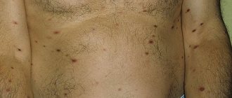
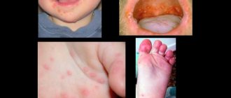
The characteristics of the rash depend on the skin disease that caused it. The only thing that all types of rashes with HIV infection have in common is more pronounced redness and itching, difficulty in treatment, and frequent relapses. In the photo below you can see in more detail the nature of the rashes in various diseases.
Skin diseases in HIV-infected people are more severe than in uninfected people, and are accompanied by fever, enlarged lymph nodes, intestinal upset, body and throat pain, and sweating.
Seborrheic dermatitis
It is expressed in the appearance of red plaques, which become crusty over time. Inflammations appear first on the face, then spread to the scalp, the inside of the knees and elbows.
Plaques characteristic of seborrheic dermatitis form at an early stage of the disease in half of those infected.
Stomatitis
Bubbles, white plaque and ulcers of various sizes, also covered with white plaque, appear on the oral mucosa.
Stomatitis in an HIV-infected person, which occurs against the background of allergies, is accompanied by fever and sore throat. Rashes in the mouth are one of the first external signs of HIV infection.
Allergy
In patients with immunodeficiency, allergic urticaria is noted, which is accompanied by the appearance of red spots and a rash similar to an insect bite.
Kaposi's sarcoma
Characterized by skin spots and lumps in the form of burgundy colored bumps. The rashes are localized on the face, mouth (tongue), genitals, from where they spread to the torso, arms and legs. A year after HIV infection, the disease enters the final stage, in which many malignant neoplasms appear on the patient’s skin and mucous membranes.
Kaposi's sarcoma is more common in HIV-infected men.
Fungus
In case of fungal diseases, such as candidiasis, lichen, rubrophytia, foci of inflammation with pronounced peeling, redness, severe itching, and a white coating appear on the patient’s skin.
Irritation is first localized on the hands, feet and groin area, then quickly spreads throughout the body.
Exanthema
Rashes occur when the body becomes infected with an infection. Exanthema appears as a colorless or bright red papular rash. The location of the rash depends on the pathogen, but most often appears in the torso and groin area.
In HIV-infected people, exanthema is caused by herpes virus, cytomegalovirus, Coxsackie virus, enteroviruses, hepatitis C virus, molluscum contagiosum, and human papillomavirus.
Psoriasis
It appears as red plaques with clear boundaries, covered with gray scales.
The severity of skin lesions depends on the degree of impairment of the immune system.
Scabies
Scabies affects up to 30% of HIV-infected people. The disease is characterized by intense itching, worsening at night.
White or red S-shaped itch burrows appear on the hands, between the fingers, on the mammary glands, under the armpits, and in the groin.
Pyodermatitis
The rash occurs against the background of inflammation of the hair follicles and takes the form of burgundy pimples and pustules, reminiscent of a teenage rash.
Syphilis
With secondary syphilis, a pink or red maculopapular rash appears on the patient’s body, which spreads throughout the body.
Secondary syphilis occurs 3-4 months after infection with Treponema pallidum.
Types of Skin Infections
The content of the article
There are three main groups of viruses:
- Infections caused by the herpetic virus: herpes zoster, herpes, chickenpox.
- The cause of infections of the second group is poxvirus. It causes molluscum contagiosum, vaccinia and false cowpox.
- The third group includes skin diseases that are caused by 3 viruses - polyoma, papilloma and vacuolinating virus. Manifestations of these viruses are genital and plantar condylomas, flat and vulgar warts.
Diagnosis of rashes on the body
With HIV and AIDS, it is difficult to visually determine the type of rash - the nature of skin inflammation in infected people is different.
To determine the type of rash and its cause, the following diagnostic measures are carried out:
- Clinical analysis of blood and urine.
- Blood test for sexually transmitted viruses.
- Skin scraping. The method is used to detect intradermal mites and fungi that cause skin diseases.
- Blood tests for antibodies to allergens and allergy skin tests.
- Hormonal studies.
- Biopsy of skin lesions. They detect the presence of cancer cells, which is important for diagnosing Kaposi's sarcoma.
Treatment of viral skin infections
To choose the right treatment, you first need to determine the type of infection. Diagnostic tests and treatment are performed by a dermatologist. Each individual type of disease has its own treatment regimen.
Herpes and herpes zoster occur when the immune system is weakened, so complex therapy will include, in addition to antiviral agents, immunomodulators, vitamins and physical therapy. For treatment to be effective, it is necessary to examine the blood for the presence of antibodies to the herpes virus - immunoglobulins and assess the general state of the body's immunity.
Condylomas and warts are removed in various ways. The cheapest is electrocoagulation. Modern safe techniques are laser and radio knife. Some clinics still use special solutions - concentrated alkalis or acids that cause necrosis of warts.
But this technique is considered outdated, since it can damage surrounding tissues, causing a burn. Removing molluscum contagiosum involves the use of medications that enhance immunity and the removal of plaques.
For each case, the dermatologist chooses individual treatment, taking into account the location of the tumor and its size. Additionally, immunocorrective agents, restorative treatment and other methods can be used.
Viral eye diseases
They are constantly exposed to attacks by all kinds of microorganisms, so the number of eye diseases is very large, but for some time now it has been viral eye disease
. There are about 150 viruses that can infect the mucous membranes of the organs of vision, but the most common are several diagnoses.
Viral conjunctivitis
An extremely contagious disease, which, in turn, is divided into the following types:
- Adenoviral conjunctivitis is characterized by redness of the membranes of the eyes, swelling of the eyelids, transparent discharge from the eyes and an increase in general body temperature. In the case of the catarrhal type, the external signs of the disease are not expressed significantly; in the case of the filmy type, a thin film is formed on the mucous membrane, and the follicular type of conjunctivitis is characterized by the appearance of small blisters on the mucous membrane of the eyes.
- Herpetic conjunctivitis - as a rule, only one eye is affected by the virus. The course of the disease is sluggish and long-lasting; in the catarrhal form, the redness is not too pronounced, and in the follicular form, blisters also appear in the eyes.
- Epidemic keratoconjunctivitis affects a large number of people at once, first affecting one eye, and then involving the other in the disease. There is both redness of the membranes of the eyes and profuse lacrimation. Plus there is a feeling as if the eyes are clogged, obvious photophobia and a persistent increase in body temperature.
Conjunctivitis of these types occurs equally in both adults and children.
Viral conjunctivitis
Viral keratitis
It is an inflammation of the cornea, which is most susceptible to the elderly and very young children. It can be both superficial and deep, and it can be caused by adenoviruses, herpes infection, smallpox viruses, measles, etc. Viral keratitis can be triggered by severe hypothermia, weak immunity and frequent stress.
The main symptoms of this pathology are redness and swelling of the eyes, as well as rashes of blisters on their mucous membranes and clouding of the cornea. Additionally, there is a decrease in visual acuity and neuralgic pain. In this case, antibiotics are also prescribed along with antiviral drugs.
Viral keratitis
Viral uveitis
This concept hides inflammation of different parts of the choroid of the eyes. In almost half of the cases of inflammatory eye diseases, this diagnosis is made, because many factors can lead to the disease. Most often, it is caused by the common herpes virus, but sometimes it can also be shingles (shingles).
With viral uveitis, the eyes become red, painful, watery eyes and light sensitivity appear, and vision loses its clarity. Often, anti-inflammatory and some symptomatic drugs are sufficient to treat this pathology.
Viral uveitis
Ophthalmoherpes
In some cases, eye infection with the herpes virus leads to this very disease, and it is extremely important that the first symptoms of eye disease
became a reason to consult a doctor, because this diagnosis can even lead to complete loss of vision. Mostly, ophthalmoherpes is caused by the herpes simplex virus type 1 or the chickenpox virus, but in recent days cases of infection with herpes type 2 have become increasingly common.
A viral disease can be suspected by redness of not only the eyes themselves, but also the eyelids, as well as pain when moving the eyeball and the sensation of a foreign body inside. There is also a decrease in visual acuity and blurred vision, there may be double vision and sparks before the eyes and, in general, a distortion in the perception of objects. Moreover, with ophthalmohermes, general health worsens: fever, nausea, etc.
As a rule, therapy in this case is not limited to antiviral drugs and involves nonspecific and specific immunotherapy. This is due to the fact that the most common cause of ophthalmoherpes is decreased immunity.
Ophthalmoherpes
Viral blepharitis
This eye disease is characterized by the feeling of a grain of sand getting inside, which you really want to get rid of, as a result of which the person begins to blink frequently and rub his eyes. In parallel, inflammation of the edges of the eyelids develops and rapid eye fatigue is noted. In severe forms of blepharitis, which often affect children and young people, the itching is very pronounced and working in artificial light becomes almost impossible.
Treatment of viral blepharitis begins with the prescription of antiviral drugs, and then includes a special diet aimed at boosting immunity. Special vitamin and mineral complexes and courses of essential amino acids are also prescribed.
Viral blepharitis
Preventive measures
Any infectious
and viral eye diseases can be prevented. In most cases, it is enough to follow the rules of personal hygiene. In particular, we are talking about:
- Periodic examination by an ophthalmologist (at least 2 times a year);
- Using only clean and ironed handkerchiefs (or disposable ones);
- Washing with warm water before bed and thoroughly removing makeup;
- Refusal to use other people's hygiene products;
- Careful wearing, removing and putting on contact lenses.
Great attention should be paid to prevention by people who already wear glasses and contacts, and have also undergone eye surgery, because they are in a high-risk area. But these measures can preserve eye health for many years.
If it becomes clear that the eyes are affected by a viral infection (redness and some discomfort), self-medication is unacceptable. During the diagnostic process, the ophthalmologist will determine the type of virus by taking a smear, conduct an external examination, prescribe tests, and only after that will determine the treatment regimen necessary in a particular situation. Otherwise, the disease may become severe and lead to complications.
Diagnostic methods
Diagnosis in most cases can be made by characteristic symptoms, physical examination, and medical history.
After examining and collecting complaints, the pediatrician will prescribe a series of tests aimed at identifying the type of virus. The doctor may prescribe:
- general urine analysis;
- stool analysis;
- blood tests (general, biochemical, serological, etc.);
- immunohistochemical analysis of tissues (to detect antibodies to enterovirus infection);
- cultures of biomaterial (saliva, throat scrapings, etc.).
An accurate diagnosis may also require a differential diagnosis to exclude other diseases with a similar clinical picture.
Other diagnostic tests are also performed:
- Laboratory Serology is a blood test that detects an increased amount of antibodies. The body produces them to fight the virus in the acute period and during the recovery stage. The analysis allows you to determine ECHO 6, 7, 9, 11, 30 and Coxsackie B1-B6. A negative result does not necessarily mean that you do not have the disease; it just does not detect other types of viruses. PCR is a highly sensitive (100%) and specific (97%) test. Allows the detection of enterovirus RNA in the cerebrospinal fluid. Blood PCR detects the virus in 30% of patients with chronic fatigue syndrome. Source: A.V. Demina, V.A. TERNOVOY, N.I. Shulgina, S.V. Netesov Enteroviruses. Part 3. Laboratory diagnostics, treatment, immunoprophylaxis // Bulletin of the Siberian Branch of the Russian Academy of Medical Sciences, volume 31, No. 3, 2011. An analysis of cerebrospinal fluid is needed if there are symptoms of damage to the spinal cord, brain and their membranes. The fluid is collected by puncturing. With aseptic meningitis, the level of leukocytes is increased. Glucose is normal or slightly reduced. Protein is normal or slightly increased. Troponin I and cardiac enzymes is a blood test to determine the level of these indicators. If they are elevated, it means the heart is damaged. Normally, the serum troponin I level should be from 0 to 0.5 ng/ml. RT-PCR is an analysis to detect common regions of enterovirus RNA. The test has a sensitivity of 95% and specificity of 97%. Approved for the diagnosis of enteroviral meningitis. The best results are obtained if the material for research is cerebrospinal fluid. Sputum, blood and mucus from the respiratory tract, and feces can be examined. However, the result will not be as accurate.
- Instrumental Electroencephalography – assesses the degree and severity of the disease. Chest X-ray - may reveal an increase in cardiac volume in patients with myopericarditis. Echocardiography – performed if myocarditis is suspected. Shows abnormal movement of the walls of the heart chambers. May reveal acute reduction in ejection fraction and ventricular dilatation in severe cases. Examination by an ophthalmologist using a slit lamp is indicated for those children who have hemorrhagic conjunctivitis and corneal erosions. Coxsackie A24 and Enterovirus 70 viruses can be detected from conjunctival smears within 3 days of infection.
Content
- Types of enteroviruses
- Causes
- Symptoms
- Diagnostic methods
- Treatment methods for enterovirus infection
- Prevention
Enterovirus infection is a group of viral diseases that affect various body systems (usually the skin, digestive tract and respiratory organs). After an illness, stable immunity is formed (up to several years), but only to the specific virus that caused the disease.
Therefore, a child can get sick several times. Because of this, in addition, there is no vaccine for this disease. Breastfed babies have immunity from their mother . But it is not persistent and goes away quickly after stopping breastfeeding. Most often, children from 3 to 10 years old and adolescents suffer . The younger the age, the greater the danger the disease poses.
Enterovirus infection is caused by viruses of the ECHO (Echovirus) and Coxsackie groups. They are characterized by a variety of symptoms - from conjunctivitis to diarrhea. The source of infection is another person . It is transmitted by the fecal-oral and airborne routes. In regions with a temperate climate, the disease is seasonal - people get sick more often in early autumn and late summer. Enteroviruses live in water for a long time: 18 days in tap water, 33 in river water, 65 in treated wastewater. Source: G.P. Martynova Enterovirus (non-polio) infection in children // Siberian Medical Review, 2014, No. 3
Prices
| Name of service (price list incomplete) | Price |
| Online opinion of a pediatrician (SPECIAL) | 0 rub. |
| Appointment (examination, consultation) with a pediatrician, primary, therapeutic and diagnostic, outpatient | 1950 rub. |
| Consultation (interpretation) with analyzes from third parties | 2250 rub. |
| Prescription of treatment regimen (for up to 1 month) | 1800 rub. |
| Consultation with a candidate of medical sciences | 2500 rub. |