In this article we will tell you:
- Causes of chalazion
- Symptoms and diagnosis of chalazion
- Chalazion treatment
- Contraindications
- Complications
- Prevention of chalazion
- Chalazion in children
- Conclusion
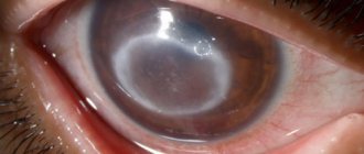
A chalazion (or chalazion) is a chronic inflammation of the eyelid margin around the meibomian gland, resulting from a blockage. Externally, chalazion is very similar to stye; both diseases manifest themselves in the form of a small nodule. Unlike styes, chalazions rarely resolve on their own. It has its own signs, symptoms, stages and treatment regimen. There are several types of chalazion. It can be internal (located inside the eyelid) and external. There is a chalazion of the upper eyelid and a chalazion of the lower eyelid. When we are healthy, our sebaceous glands produce fatty secretions. It moisturizes the mucous membrane of the eye, facilitates the movement of the eyelids during blinking. When the meibomian gland ducts are blocked, a chalazion can develop. Most often, the pathology occurs in people over 30 years of age. Chalazion does not threaten human life. In addition to pain, it most often causes only cosmetic discomfort.
What is a chalazion?
In ophthalmology, a chalazion of the eyelid is a benign tumor-like growth
. It is a capsule closed in the thickness of the mobile eyelid, filled with sebaceous secretion, sometimes pus and dead particles of the epithelium. Upon closer examination, it turns out that a chalazion is an inflamed meibomian gland, connected to the surface by thin ducts that open onto the mucous membranes of the eyelids in contact with the eye. These structures are located in the cartilage of the lower and upper eyelids and are responsible for protecting the cornea. They secrete a secretion that forms a protective film that prevents the evaporation of tears and reduces friction between the eyelid and the surface of the eye.
Under certain conditions, these ducts become blocked and a chalazion begins to develop. First, the secretion it produces accumulates in the gland inside the eyelid, then it stretches its walls, which provokes low-grade inflammation. At the initial stage, the process looks like barley, but is located not along the edge of the eyelid, but at some distance from it. Unlike stye, a chalazion of the eye is not accompanied by pain and looks like a normal flesh-colored lump on the outside and grayish on the side of the mucous membrane in contact with the eye.
Despite the absence of an acute inflammatory process, the chalazion cavity may contain purulent exudate.
Children and adults over 35 years of age are at greater risk of finding out what a chalazion is on the eye. This pathology occurs extremely rarely in adolescents and young adults. When making a diagnosis, the doctor will have to differentiate chalazion from barley, uveitis, keratitis and other inflammatory diseases on the eyelids and outer membranes of the eye.
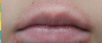
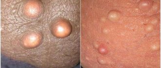
Prosyanka
This is one of the most harmless bumps, the appearance of which on the eyelid does not cause any trouble other than aesthetic discomfort. Millets, also known as milia, come in different sizes. From the smallest - smaller than a poppy seed, to quite large - the size of an average grain of rice. Milia can appear on both the lower and upper eyelids with almost equal frequency. At their core, they are whiteheads localized in the eye area.
Milia can appear in anyone, even in those who have never experienced skin problems. It is better to remove millet from a cosmetologist, because only a specialist can guarantee safety.
To prevent milia, you need to monitor your diet and from time to time make masks to exfoliate dead skin particles that can close pores and clog the ducts of the sebaceous glands.
Causes
The main cause of a chalazion is a blockage of the meibomian gland duct.
followed by an inflammatory process. In most cases, the source of the problem is primary inflammation of the eyelid margin or the gland itself. Chalazion also has secondary causes, that is, not related to eye diseases. Among them, doctors call somatic pathologies:
- the digestive system, in which lipid metabolism is disrupted, and the synthesized fat secretion becomes excessively thick, viscous, and prone to accumulation in the ducts;
- chronic infectious skin diseases - seborrhea and demodicosis, in which parasites and inflammatory processes lead to scarring of the ducts, their gradual narrowing and disrupt the outflow of secretions;
- endocrine diseases, in particular diabetes mellitus, in which immune defense is disrupted and the activity of microorganisms on the surface of the eye and mucous membranes of the eyelids increases;
- deficiency of certain vitamins;
- congenital features associated with increased activity of the sebaceous glands - according to statistics, those with oily skin are more likely to suffer from eye chalazion.
Another common cause of chalazion
is a violation of eye hygiene:
- improper storage and installation of contact lenses;
- contact of dirty hands with eyes;
- using low-quality or expired eye drops;
- using non-sterile tools to create makeup, etc.
Chalazion can also develop against the background of oncology of other organs or the eyelid itself. In the first case, the neoplasm occurs after chemotherapy due to a general decrease in immunity, and in the second it can be a malignant neoplasm, which is mistaken for a chalazion.
Xanthelasma
It is more likely not a lump, but a flat plaque. The problem of xanthelasma is more often encountered by women with hypercholesterolemia, diabetes and a number of other diseases. Xanthelasmas are yellowish in color and rise very slightly above the surface of the skin. They can appear on the eyelids, the skin near the eyes, and on the face. A single lump of xanthelasma is rare; as a rule, they appear in groups and never go away on their own. This is due to the fact that xanthelasma is the result of a lipid metabolism disorder caused by the underlying disease. If such a problem occurs, you must notify your doctor, who will advise what to do.
Stages of development
Regardless of the observed causes of chalazion, it always develops according to the standard pattern. Ophthalmologists distinguish 3 main stages in the formation of a chalazion on the eye:
- Zero stage
, blockage of the duct outlet. There are no external manifestations, since the size of the neoplasm does not exceed 2 mm in diameter. At this stage, sebaceous secretion accumulates inside the gland, inflammation is absent or mild, and there is no pain. Pathology at this stage is discovered accidentally during cleansing of the eyelid, applying makeup and other manipulations. - Stretching of the meibomian gland
with the formation of a noticeable hailstone on the eyelid. The sebaceous secretion thickens, the chalazion increases in size and becomes harder. Due to the pressure created by the neoplasm on surrounding tissues, minor pain and discomfort in the eye may occur when trying to palpate the pea. When an infection occurs, swelling and slight hyperemia of the eyelid over the neoplasm may be observed. The size of the chalazion at this stage does not exceed 3-4 mm. - Encapsulation of the chalazion
, further growth of the hailstone or its spontaneous resorption.
Sometimes, in the absence of therapy and the persistence of the factors that influenced the formation of a tumor in the eye, the growth process of the chalazion is complicated by intense inflammation. At the same time, in addition to fatty secretion, purulent exudate can accumulate in the capsule. It can melt the shell of the chalazion and lead to spontaneous leakage of its contents onto the outer surface of the eyelid or, more often, onto the cornea of the eye. In the first case, a lesion remains at the site of the neoplasm, a fistula is formed - a duct, which periodically opens, and the contents of the gland emerge from it. In the second case, in addition to the formation of a fistula, there is a possibility of infection spreading to the membranes of the eye.
In what cases is it necessary to consult a doctor?
Warning signs indicating the need to visit a doctor are:
- increase in bud density ;
- difficulty blinking and the lump blocking the field of view;
- the size of the seal increases and becomes more than 6-10 millimeters in diameter;
- the patient begins to develop a fever and headaches , but the symptoms cannot be associated with a cold (there is no sore throat and general deterioration in well-being);
- the eyelid begins to swell a lot;
- itching, burning and uncontrollable profuse lacrimation appear;
- Over time, photophobia increases.
Keep in mind! All these signs indicate the accumulation of pus in the lump in critical quantities.
In such cases, a doctor should be consulted to take emergency measures , but even before such dangerous symptoms appear, conservative drug treatment should be started.
Symptoms
With chalazion, the symptoms are not similar to inflammatory diseases of the eyelids, although the appearance of the neoplasm at different stages of development may resemble barley, blepharitis, conjunctivitis and other pathologies. It is distinguished from them by its slow development and the absence of a clinical picture characteristic of acute inflammation.
Chalazion can be distinguished from the mentioned pathologies by the following signs:
- absence of acute inflammation in the tissues surrounding the hard capsule, expressed by redness and swelling;
- no pain when blinking;
- absence of purulent discharge in the lumen of the duct;
- mobility of the hard capsule when palpated.
The main symptoms of this eye disease are
ophthalmologists call the gradually increasing sensation of a foreign object under the eyelid, in the place where the altered meibomian gland is located. Visually, the eye may appear smaller than the other due to the thickening of the eyelid. This feeling appears when the pea reaches a diameter of about 3-5 mm. At the same time, the patient may be concerned about:
- itching of the eye in the place where the pea is located;
- peeling and thinning of the skin over the lesion;
- mild hyperemia, which appears only during periods of increased inflammation of the gland;
- moderate lacrimation.
When the diameter of the neoplasm exceeds 5 mm, the chalazion of the eyelid not only irritates the membranes of the eye, but also puts constant pressure on them. In addition to the already mentioned itching, watery eyes and flaking of the skin, such a large neoplasm can affect visual acuity. It puts pressure on the eyeball, which leads to increased eye pressure and blurred vision. Chalazion can also cause damage to the cornea with subsequent thickening or clouding. All this provokes a significant deterioration in vision.
The most pronounced discomfort is caused by a chalazion of the upper eyelid.
Larger capsules form on it. The degree of discomfort and the participation of the eyelid in moistening the eye are affected - the upper mobile skin fold makes movements with a wider amplitude than the lower one, so any swelling on it is felt better. This type of chalazion is also more noticeable visually - when the upper part of the palpebral fissure is affected, the organs of vision look asymmetrical, and the blinking mechanism is disrupted.
During periods of exacerbation, the chalazion increases in size, tightens the skin and lifts the inner lining of the eyelid, which causes the patient to worry about:
- burning, feeling of sand in the eye due to insufficient hydration of the cornea;
- inflammation, damage to the conjunctiva, which is expressed by redness of the eye, lacrimation, discharge of pus and pain;
- inversion of eyelashes into the eye, resulting in irritation of its membranes, formation of a vascular pattern on the sclera or pinpoint hemorrhages.
Most often, the described symptoms appear in winter, after hypothermia. Chalazion also worsens in the summer after the opening of the beach season, among vacationers in other climatic regions. The statistics are explained by a temporary decrease in immunity and poor eye hygiene when relaxing in open water. In children, chalazions in the eyes often become inflamed in the summer, when children play in sandboxes, dry pools, etc.
Diagnostics
The use of instrumental methods to diagnose chalazion is not required, since even an inexperienced ophthalmologist can distinguish this pathology from other eye diseases. When interviewing the patient, the doctor will find out:
- is there pain when blinking - its absence is interpreted in favor of blockage of the meibomian gland duct;
- is there any sticking of the eyelid with yellow, white or greenish pus in the morning - with a chalazion this phenomenon should not occur;
- how long ago the tumor was first noticed - a period of more than 2 weeks is interpreted in favor of the described pathology.
Next, the doctor examines the eye: palpates the affected eyelid, turns it inside out to examine the inner surface. To determine the causes of chalazion, the ophthalmologist may prescribe additional tests:
- laboratory blood tests (general clinical, biochemistry) to identify endocrine, metabolic, infectious and other disorders;
- bacteriological blood culture for pathogens of systemic infections;
- culture of a smear from the eyelid for staphylococcal infection;
- inoculating a smear from the eyelid for pathogens of gonococci, trichomonas and other pathogens of STIs that can affect the mucous membranes of the eyes;
- stool analysis for eggs of worms, lamblia and other parasites;
- microscopic analysis of epithelial scrapings for demodex and other skin parasites;
- immunological screening.
The doctor may not necessarily prescribe all of the tests listed. It is enough to limit ourselves to the most probable causes of the pathology identified during the collection of individual and family analysis. In most cases, the doctor limits himself to a general clinical blood test and referring the patient to a consultation with a dermatologist and therapist.
Prices
| Name of service (price list incomplete) | Price |
| Appointment (examination, consultation) with an ophthalmologist, primary, therapeutic and diagnostic, outpatient | 1950 rub. |
| Consultation (interpretation) with analyzes from third parties | 2250 rub. |
| Prescription of treatment regimen (for up to 1 month) | 1800 rub. |
| Prescription of treatment regimen (for a period of 1 month) | 2700 rub. |
| Consultation with a candidate of medical sciences | 2500 rub. |
| Biomicroscopy of the anterior segment of the eye | 500 rub. |
| Selection of glasses with plain lenses | 600 rub. |
| Subconjunctival injection (1 eye) | 500 rub. |
| Removal of a foreign body from the conjunctiva, from the eyelids | 800 rub. |
Treatment
According to statistics, treatment of chalazion takes from 2 weeks when diagnosed in the early stages to several weeks if the disease is diagnosed in an advanced form. If you contact an ophthalmologist before the formation of a dense capsule, treatment is carried out using simple means and the tumor is eliminated in the same period as with barley, that is, 2-5 days. However, in practice this happens extremely rarely, because before the gland is encapsulated, the chalazion is practically invisible and does not cause discomfort.
At the initial stages, chalazion treatment is carried out using antiseptics, antibiotics and anti-inflammatory drugs in the form of drops and ointments. They can be single-component or contain additional substances that enhance the effect of the main components.
The most effective are:
- ointments and drops with antibiotics - “Vigamox” (active substance moxifloxacin), “Floxal” (ofloxacin), “Oftaquix” (levofloxacin), “Tsiprolet” (ciprofloxacin);
- combination drugs with antibiotics and hormonal anti-inflammatory substances - “Tobradex” (tobramycin with dexamethasone), “Floxadex” (ciprofloxacin with dexamethasone), “Garazon” (betamethasone with gentamicin).
In most cases, doctors tend to use combination drugs, especially if the chalazion has become chronic or recurrent. Such agents actively suppress pathogenic microflora, reduce inflammation and prevent purulent complications. When prescribed in a timely manner, medications from this group relieve swelling from the walls of the meibomian gland ducts and restore the outflow of secretions.
If the neoplasm is not bothered by unpleasant symptoms and there are no signs of inflammation, ophthalmologists recommend physiotherapy, combining gentle dry heating of a pea with subsequent massage of the eyelid. Warming can be performed in a clinic or private clinic with special equipment. At home, it can be replaced in the following ways:
- place a fabric bag with salt or sand heated in a frying pan on the eyelid for 5-10 minutes;
- iron the cotton fabric with a heated iron and apply it to the eye until it cools, repeat 5-7 times.
Gentle heating helps to soften the thickened secretion, which comes out of the duct during subsequent massage.
Important to remember
that this method cannot be used to treat a chalazion that has arisen against the background of an infection of the eye, as well as one that occurs with obvious inflammation of the meibomian gland. The methods are suitable for eliminating pathology caused by metabolic disorders, chronic gastrointestinal diseases, etc.
After the procedure, you need to rinse the eyelid with warm water using a cotton pad or sterile napkin. After washing, you can treat the eye with any antiseptic. You can also use:
- “Blepharogel” with hyaluronic acid to restore the structure and moisture of the eyelids;
- “Blephaclean” eye hygiene wipes with antiseptics, hyaluronic acid, antioxidants and substances that regulate the activity of the sebaceous glands.
These products can be used not only after a massage, but also during it. The active substances in their composition help soften the sebaceous secretion of the glands and remove it from the ducts of the glands. Despite the enormous benefits for eye health, it is recommended to use them no more than twice a day.
If the chalazion is opened on your own, it is recommended to continue therapy according to the regimen prescribed by the doctor. To prevent infection of the resulting wound, quickly and efficiently cleanse the capsule and tissue regeneration, it is recommended to use:
- drops with antibiotics - “Signitsef”, “Tobrimed”, “Uniflox”;
- ointments with antibiotics - tetracycline, Floxal, erythromycin;
- corticosteroids in the form of ointment - “Hydrocortisone” 0.5%, “Combinil Duo”, “Floxadex”;
- homeopathic ointment "Traumel".
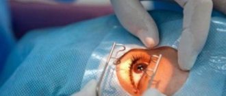
If conservative methods do not lead to improvement or there is a tendency for the tumor to grow, surgical methods are used to treat chalazion. For children, operations are performed under general anesthesia, and for adults, under local anesthesia. There are several methods for removing tumors:
- Classical desquamation of the capsule and its contents.
To carry it out, the doctor makes an incision in the skin of the eyelid and through it, using a curette (a curved sharp instrument), scrapes out the chalazion, then cleans the cavity, treats it with antiseptics and applies sutures. The intervention lasts no more than 15 minutes. The sutures are removed after 5-7 days. - Enscaping the capsule through an incision on the inside of the eyelid
. The doctor everts the eyelid, makes an incision and performs the procedure as in the previous case. There are no stitches with this method. For better healing and prevention of infection, apply antibiotic ointment behind the eyelid and cover the eye with a sterile bandage. - Laser removal of chalazion
. The doctor makes an incision on the outer part of the eyelid and evaporates the capsule with its contents layer by layer. Treatment with antiseptics and sutures are not necessary for this type of intervention - they will be replaced by a scab formed by the action of the laser on the tissue.
After any intervention, slight swelling remains in the eye for a week, and the patient may experience moderate pain.
Removal operation
Neoplasms 5 mm in size, long-existing or prone to recurrence must be removed surgically. The operation is usually performed on an outpatient basis, under local anesthesia, and its duration does not exceed 20–30 minutes. The doctor grabs the area of the eyelid with the chalazion using a fenestrated clamp, then opens the neoplasm and removes it along with the capsule. If there is a fistula tract, it is opened and excised along its entire length. After several stitches are placed, a pressure bandage is applied to the area of the operated eye. For a week after surgery, you will need to instill anti-inflammatory drops or apply ointment.
Prevention
To prevent chalazion, it is enough to maintain visual hygiene:
- thoroughly clean your eyelids after sleep;
- remove makeup before going to bed;
- do not apply cosmetics and creams along the edges of the eyelids and in the eyelash line;
- store contact lenses in sealed, sterile containers with fresh solution;
- pick up and install lenses with clean hands or a sterile instrument;
- do not touch your eyes with dirty hands;
- if you are prone to inflammation of the eyelids, use “Blepharogel” and “Blephaclean” wipes for daily eye care;
- promptly eliminate problems with digestion, metabolism and hormonal imbalances;
- avoid hypothermia.
If you find a small hailstone on your eyelid, you should immediately go to an ophthalmologist with the problem in order to have time to deal with it using conservative methods.
Preventive measures
Prevention includes following the recommendations:
- Strengthening immune forces
. This should always be done, not only when there is a threat of hordeolum or in the off-season. It is recommended to take vitamins, give preference to fresh vegetables and fruits, move more, be in the fresh air, and follow a daily routine. - Timely elimination of ophthalmological disorders
. If you are prone to eye diseases, you should regularly visit an ophthalmologist. Self-diagnosis and self-medication usually contribute to the transition of pathology to a chronic form. - Compliance with hygiene standards
. You need to have your own towel, wash your hands with soap, change bed linen every 1–2 weeks, and not use other people’s cosmetics. - Developing the habit of not touching your eyes
. This should not be done even in a normal state, when a person is not bothered by any problems. This rule also applies to the face - you should not touch it during the day with unwashed hands.
Simple preventive measures will help avoid inflammation and keep your eyes healthy.
Consequences and complications
With timely diagnosis and treatment, chalazion has no consequences. If you ignore a hailstone for a long time and allow it to grow to 5 millimeters or more, there is a risk of complications:
- transfer of infection to the conjunctiva, cornea;
- chronic compression of the eyeball and irreversible decrease in visual acuity;
- formation of ulcers on the cornea;
- development of phlegmon of the century;
- fistula formation.
Treating these pathologies is tens of times more difficult and expensive than eliminating a small chalazion. But even with a successful outcome, scars will remain and the shape of the palpebral fissure will change. Therefore, you should not hope that the tumor will resolve on its own. It is better to enlist the help of an ophthalmologist and undergo a course of treatment.
Useful video
From this video you will learn how to distinguish a chalazion and a stye on the eye:
Lumps on the eyes usually appear gradually , the process can last several days, weeks and even months depending on the original cause.
But the person himself needs to pay attention to such changes.
And at the first obvious signs of the development of such formations, it is better to play it safe and undergo an examination in order to identify possible pathological seals in time.