General information
Allergic diseases have been occupying a leading position in the structure of general morbidity among the population for a long period. Among allergic pathologies, a special niche belongs to allergic dermatoses . According to the literature, the prevalence of allergic dermatitis in the human population varies between 15-25%, with young people and children more often affected, while in older people, due to age-related involution of the immune system, allergic dermatoses develop relatively rarely. Allergic dermatoses are represented by several types. The most common ones include:
- Allergic contact dermatitis develops when the mucous membrane/skin is exposed directly to an allergen. It develops predominantly on the skin in the area of contact with the allergen (on the face or on the arms or legs), but can extend beyond the site of action of the external allergen. Disseminated/generalized rashes may develop much less frequently.
- Toxic-allergic dermatitis (allergens enter the body through the digestive tract, respiratory tract or by injection through the blood).
- Atopic dermatitis (a chronic relapsing disease caused by the genetic predisposition of the human body to a certain type of allergen).
The ICD-10 code for allergic dermatitis is determined by the type of dermatitis: L23 Allergic contact dermatitis; L20 Atopic dermatitis; L27 Toxic-allergic dermatitis. Due to the specific etiology, pathogenesis, clinical picture and treatment of each type of allergic dermatitis, it is not possible to consider them in the scope of one article, so we will consider only allergic contact dermatitis (ACD), which in most cases is a manifestation of a cell-mediated delayed (late) allergic reaction. type (type IV hypersensitivity reaction), which occurs in response to contact with a specific skin allergen. In fact, ACD is the result of sensitization (hypersensitivity) of the body's immune system to one/several specific allergens, which leads to the occurrence (relapse) of an inflammatory reaction on the skin.
The number of visits to dermatologists by patients with signs of contact allergic dermatitis is at least 10% of all visits to a dermatologist. Moreover, in 4-5% they are due to the influence of professional factors. Contact allergic dermatitis is more often registered in women, which is due to their more frequent contact with skin allergens (jewelry, detergents/cosmetics, etc.). The development of allergic dermatitis can occur as a reaction to exposure to any substance. In this case, the leading importance is not the nature of the stimulus, but the individual’s individual sensitivity to it. The concentration of the irritant, the area of its impact and the route of penetration into the body are not decisive.
Publications in the media
Contact dermatitis is an acute or chronic inflammatory skin disease caused by the irritating or sensitizing effect of exogenous factors. Incidence: 669.2 per 100,000 population in 2001.
Classification • Primary irritant dermatitis (simple contact dermatitis) • Allergic contact dermatitis (ACD) • Phototoxic dermatitis (see Photodermatitis).
Etiology and pathogenesis • Primary irritant dermatitis is associated with skin damage caused by exposure to irritating substances: chemicals (acids, alkalis, phenol), some plants (euphorbia, nettle, ash, caustic buttercup, ivy, hogweed, poison sumac), physical (UV -rays, high and low temperatures, etc.), mechanical (friction, prolonged pressure) irritants cause an inflammatory reaction in any person • ACD is caused by sensitization and the development of a type IV allergic reaction. The most common causes of ACD: salts of chromium, nickel, cobalt (jewelry, zippers, hooks, watches and bracelets); dyes (ursol, n-phenylenediamine derivatives, etc.); plants (sumac, oak, primrose); latex and various additives used in rubber production; cosmetical tools; LS • Phototoxic contact dermatitis occurs under the influence of the UV part of the solar radiation spectrum on the skin after exposure to certain substances.
Risk factors • Occupational: contact with occupational allergens • Contact with household chemicals.
Clinical picture
• A pathognomonic sign is a sharply demarcated edge of the lesion.
• The process primarily involves areas of skin with a thin epidermis (eyelids, genitals, etc.).
• The skin of the palms and soles is most resistant to irritation; The skin of deep folds is not affected.
• Forms of contact dermatitis •• Simple contact dermatitis - erythematous, vesiculo-bullous, necrotic-ulcerative •• ACD ••• Acute form: papules, blisters, blisters with surrounding erythema, oozing, itching. Initially, rashes appear only at the site of contact with an irritating substance or allergen; subsequently they can spread ••• Chronic form: thickening with lichenification, erythema, peeling, and in some cases erosion.
Research methods • If ACD is suspected, a skin patch test is performed with a standard set of contact allergens attached to a patch tape that fixes them on the skin for 48–72 hours. The reaction is assessed 20 minutes after removal of the allergen • Identification of a possible photosensitizer.
Differential diagnosis • Infections caused by HSV • Bullous pemphigoid • Seborrheic dermatitis • Atopic dermatitis.
TREATMENT
Management tactics • The influence of a possible etiological factor should be eliminated • Diet excluding spicy foods, alcoholic beverages; limiting table salt and carbohydrates.
Drug therapy
• Locally •• Cold disinfectant lotions with 2% resorcinol solution, 3% boric acid solution, Burov's fluid (1:40 dilution) •• HA ointments with high activity, for example, fluacinolone acetonide (0.025% ointment) 3-4 times a day, preferably under a compress. It is recommended to apply HA creams with lower activity to the skin of the face and skin folds. •• For secondary infection, antibiotics (for example, gramicidin, gentamicin, erythromycin).
• Systemically •• GK (only in severe forms with a large area of damage), usually prednisolone 0.5-1 mg/kg/day with gradual withdrawal over 10-14 days •• Antihistamines - hydroxyzine 25-50 mg 4 r/day or diphenhydramine 25–50 mg 4 times/day •• If a secondary infection occurs, antibiotics: erythromycin 250 mg 4 times/day.
Complications • Attachment of pyogenic, yeast infection • Malignancy in radiation dermatitis (radiation cancer) • Transformation of allergic dermatitis into eczema.
The prognosis is favorable.
Prevention. They recommend compliance with hygiene rules, rational employment with the exclusion of industrial irritants and allergens, compliance with occupational safety and health regulations, individual prevention (working clothes, protective gloves, anti-aerosol respirators, protective neutralizing creams, surfactants, etc.), elimination of household chemicals allergens.
Reduction. ACD - allergic contact dermatitis
ICD-10 • L23 Allergic contact dermatitis • L24 Simple irritant contact dermatitis • L25 Contact dermatitis, unspecified
Application. Enteropathic dermatitis is a disorder (including hereditary absence of zinc-binding factor, 201100, r) of zinc absorption (manifests with the beginning of complementary feeding of the child), characterized by blistering rashes around natural openings (eyes, nose, mouth, anus), as well as in the area of the elbows, knees, inguinal-femoral folds, on the thighs, buttocks, where scaly plaques subsequently form; accompanied by hair loss, onychopathies, photophobia and dysfunction of the gastrointestinal tract (diarrhea, steatorrhea, profuse fetid stool). The disease is often complicated by fungal and purulent infections, stomatitis, blepharitis, malnutrition, mental disorders, and developmental delays. Causes: zinc deficiency, impaired zinc absorption, hepatitis B. Treatment: zinc sulfate 30–150 mg/day orally for 3 months. The prognosis with timely initiation of treatment is favorable. Synonyms: Danbolt-Kloss syndrome, Brandt syndrome. ICD-10. E83.2 Zinc metabolism disorders.
Pathogenesis
The pathogenesis of contact dermatitis is a delayed-type allergic hypersensitivity reaction that develops after contact of the allergen with the skin after 15-48 hours. After the allergen gets on the skin, it binds to tissue proteins to form compounds - antigen , which can cause an allergic reaction. Next, the antigen in the membrane molecules of T-lymphocytes is absorbed by Langerhans cells, producing interleukins and interferon gamma , which enhance the immune response and inflammatory reaction.
Activated T lymphocytes migrate through the lymphatic vessels to regional lymph nodes, where they undergo antigen-dependent proliferation and differentiation. T-lymphocytes that have undergone “specialization” participate in the immune response, and the rest are memory cells, which determine a rapid and pronounced response in cases of repeated contact with the allergen. The accumulation of T-lymphocytes that recognize the allergen occurs over 10-15 days, after which T-lymphocytes leave the blood and populate the peripheral organs of the immune system.
Activation of memory cells, rapid accumulation of macrophages and lymphocytes occurs upon repeated contact with the allergen. In the dermis, as a result of the development of an allergic reaction, a lymphoid-macrophage infiltrate is formed with pronounced immune damage to the skin, mainly at the sites of penetration/localization of the allergen and perivascularly, where helper-inductor T-lymphocytes . Under the influence of cytokines, the death of cellular elements of the skin occurs and its structural and functional usefulness is disrupted, skin necrosis . Since contact of the allergen occurs with a limited area of the skin, monosensitization of the body initially develops, however, in the future, the possibility of developing polyvalent sensitization with the risk of transition from allergic dermatitis to eczema cannot be ruled out. Relief of an allergic reaction occurs as the allergen is eliminated from the body. Below is a schematic diagram of the pathogenesis of an allergic reaction.
Diagnosis of allergic contact dermatitis
Allergic contact dermatitis (ACD) (according to ICD classification L23) is a skin disease caused by sensitization to biological or chemical allergens that occurs when the skin comes into contact with low molecular weight components [1].
ACD is characterized by local inflammation of the skin with typical hyperemia, edema, and desquamation. In severe cases, symptoms characteristic of true eczema or bulla develop. The development of a skin process is usually detected at sites of contact with the causative allergen [1, 2].
There are two types of contact dermatitis:
- non-allergic contact (irritant or simple) dermatitis (NCD);
- allergic contact dermatitis (ACD).
There is no immune response at the heart of NCD. NCD occurs when the skin comes into direct contact with a strong chemical. Inflammatory changes in the skin are not associated with the development of an allergic reaction. The causes of NCD are the irritating effects of chemical and biological agents. NCD occurs almost immediately after the first contact with the stimulus. NCD is provoked by such factors as: acids, alkalis, phenol, detergents, poisonous secretions of plants and animals, cosmetic additives, gasoline, cement, paints. With accompanying physical factors (friction and pressure of clothing, insolation), NCD can become severe. The skin of the hands, face, and perineum is most often affected by NCD.
Let us give the main examples of NKD.
- “Diaper dermatitis” is a skin reaction to the irritating effects of urine and feces due to the greenhouse effect that occurs under diapers and diapers.
- Contact dermatitis that develops in humans when exposed to low concentrations of chlorine. It can be diagnosed among medical personnel working with disinfection material and in operating rooms; in patients visiting the pool.
- Dermatitis caused by contact with the secretions of a poisonous jellyfish in the sea.
- Contact dermatitis caused by exposure to the juice of Sosnowski's hogweed, "poison ivy", which, when exposed to sunlight, has an almost burn effect.
- Occupational dermatitis of tilers in contact with adhesive mixtures.
ACD is always based on an allergic reaction. Most often, an immune disorder is characterized by the development of a delayed-type allergic reaction, when an antigen (allergen) that gets on the skin is captured by antigen-presenting cells of the epidermis and, partially transformed, penetrates the lymph nodes, where cells of the immune system - T-lymphocytes - meet this foreign agent. Sensitized lymphocytes infiltrate areas of skin contact with the allergen, causing a whole cascade of inflammatory reactions. Unlike IgE-mediated reactions, which are clinically easily recognized because they occur extremely quickly after contact of the allergen with the body (from several minutes to an hour), delayed-type hypersensitivity reactions develop no earlier than 48 hours later.
Identification of the causative sensitizing factor only by clinical manifestations due to the long period of development of such reactivity is often not possible. In addition, the difficulty of diagnosis lies in the fact that it is often difficult to trace the connection with the trigger effect of any chemical reagent, since the aftereffect is observed. That is, contact with the allergen has already been eliminated, and the clinical picture of dermatitis is observed after a day, or even two. In extreme cases, the reaction may develop much later (more than 7 days) after contact with the allergen. Allergic dermatitis occurs only in sensitized people, and the severity of clinical manifestations depends in each case only on the degree of sensitization, i.e., on the level of hypersensitivity of the body and, especially, on the hereditary predisposition to allergic reactions [3, 4].
ACD is characterized by burning, itching of the skin and the appearance of redness and swelling upon repeated contact with external irritants. If the contact was short-term, the disease lasts several days, with long-term or frequent contact with allergens - many months or years. In the acute form of the disease, burning, itching and swelling of the skin, redness (erythema), nodules (papules), blisters (vesicles), erosions (defects of the epidermis), crusts, and peeling appear. In the chronic process, the skin is covered with nodules, may peel off, and is characterized by increased skin pattern (lichenization), scratching (excoriation), subject to increased sensitivity (sensitization) to allergens.
V. A. Ado made a great contribution to the study of pathogenetic mechanisms of the development of chemical allergies in our country. He was the first to reproduce chemical allergies in experimental animals. To do this, the animals were previously sensitized with a conjugate consisting of a high-molecular-weight globulin combined with a low-molecular-weight chemical substance. When a chemical allergen was applied to the skin of a sensitized animal, dermatitis developed. Thus, the sensitizing activity of many low-molecular compounds that provoke the development of, for example, occupational dermatoses has been proven [5].
When collecting anamnesis, special attention is paid to the connection between the onset or exacerbation of the disease with contact with chemical allergens. As a rule, patients do not find a causal relationship between the disease and contact with the allergen. Most often, the supposed trigger of the disease, in their opinion, is food, nervous stress or an infectious disease. Even more often, given the subacute onset, patients find it difficult to answer the question of what they attribute the onset to. At the same time, all patients note an exacerbation of the disease upon contact with household chemicals. This is especially common among women who do housework without using latex gloves. Most report improvement in their skin while on vacation when they avoid contact with detergents for two to three weeks. Most often, with ACD, lesions are observed on the skin of the palms and backs of the hands, face, neck, abdomen, legs and feet.
Many patients suffering from allergic contact dermatitis have contact with occupational hazards. The most susceptible to the development of this pathology are hairdressers, builders, photographers, chemical laboratory workers, car service and cement plant workers, vivarium workers, and medical personnel working in operating rooms. They associate the exacerbation of the disease with working with chemical reagents, contact with latex, paints and varnishes, lubricating oils, and cosmetics. The most significant and most often causing reactions are recognized as: metal ions (nickel, chromium, cobalt), ointment components (lanolin, local anesthetics, fragrances, parabens, Peruvian balsam), paints, rubber, preservatives, formaldehydes [4, 5].
Diagnostics to identify the causative chemical agent that causes ACD is carried out using a set of application tests of the so-called European chemical panel (Allertest). We tested 140 patients diagnosed with allergic contact dermatitis. The results of this testing are presented.
Positive samples were detected in 124 patients out of 140 examined. Most patients had more than one positive test. The intensity of allergic reactions varied (from weakly positive to strongly positive reactions).
Most often, in 40.7% of patients, a reaction to metal ions, especially nickel sulfate, was recorded. Moreover, the average size of the papule and hyperemia, estimated in diameter as 8.3 ± 3. mm/15.3 ± 6.6 mm, was the largest compared to reactions to other ingredients.
When interviewing these patients, only 34 out of 57 noted a reaction to metal objects (buttons, jewelry). They did not limit their contact with metal objects. Women often recalled that for some reason they were unable to wear earrings earlier due to inflammation of the earlobe. The localization of dermatitis was characteristic: the skin of the abdomen at the level of the navel (the place of friction of a buckle, button or zipper); skin of the 2nd and 3rd phalanges of the right hand (point of contact with cutlery), skin of the palms, earlobes, neck (place of friction of jewelry). With chronic contact with metals, the disease was characterized by symptoms of true eczema. All patients reacting to the allergen nickel sulfate were given recommendations: eliminate contact with metal objects to a minimum, eliminate nickel-containing utensils, spoons and forks from everyday use, exclude foods from cans from the diet, coat door handles and bathroom faucets with polyurethane varnish, keys, scissors and knives. Patients' attention was focused on the fact that nickel salts have the greatest harmful effect when in contact with moisturized skin, even if the moisture is caused by increased sweating [5].
Eight patients had a reaction to lanolin. These were weak positive samples, not exceeding 5 mm in diameter. However, it turned out that it was these patients who noted that after using certain ointment forms or creams, the condition of the skin on their hands sharply worsened a day later. These patients were advised to carefully familiarize themselves with the basis of all cosmetic and medicinal products in order to eliminate the dominant component - lanolin, refuse to use cosmetics, wear woolen mittens and gloves, and use rectal and vaginal suppositories and gels. In the case of lanolin content in some types of cosmetics, when patients indicated that it was well tolerated, it was recommended to use only these proven products, taking into account the fact that not every type of lanolin causes allergic reactions.
In 20–27% of patients, positive reactions were detected to the components contained in many cosmetics (fragrance mixture, quaternium-15, parabens). Skin lesions were most often observed in the face and back of the hands. One patient suffered from an allergic reaction to shower gels, which caused burning and itching all over her body. Four patients had a reaction to the eye cream. After giving up cosmetics containing a reactive component, the condition quickly improved [6].
We observed one of the most severe cases of contact dermatitis in a 14-year-old girl after receiving a tattoo, which she received at a resort in Bulgaria. Pain, swelling, and severe itching appeared locally at the tattoo site after 12 hours. When examined on the skin of the right shoulder over the pattern applied with paraphenylenediamine paint, pronounced hyperemia, severe pain when touched, and swelling, accompanied by severe itching, were observed. The skin areas were covered with pustular elements with serous discharge and crusts.
After treating the skin with topical disinfectants and removing the tattoo whenever possible, the affected area was treated with an aerosol preparation containing a topical corticosteroid and an antibiotic. A course of treatment with a third generation antihistamine was prescribed systemically. The patient was recommended to undergo dry cold applications locally during the first two days. After a week, the swelling subsided, and crusts and peeling of the skin remained at the places where the tattoo was applied. And after another week, the process was completely stopped, only pigmentation remained in the area of the drawing lines. A month later, the patient underwent a 72-hour patch test using a standard set of European panel of chemical allergens. But after 24 hours the patient complained of itching under one of the application chambers. After removing the test, a blister with serous exudate developed at the site of application with paraphenylenediamine.
In another patient with a reaction to paraphenylenediamine, ACD was observed on the scalp, with the development of angioedema after using hair dye. Reactions to the component of dyes and photographic reagents (paraphenylenediamine), detected in 17.8% of tested patients, were more pronounced and averaged 9.3 ± 5.6 mm for papule and 16.5 ± 6.2 mm for hyperemia. Some patients regularly worked with photoreagents in a hairdressing salon. Recommendations were given to eliminate contact with paints that contain paraphenylenediamine, mascara, photo reagents, and printer ink [7].
A special place among the causes of ACD is occupied by an allergic reaction to rubber and its components. Almost all patients interviewed believed that the only method of preventing contact with harmful reagents was the use of latex gloves. Almost every third patient had a reaction to some rubber component. First of all, a thorough survey was carried out about possible contact with rubber. Occupational ACD included diseases among medical personnel using rubber gloves, film editing mechanics, and construction workers who have contact with rubber glue. ACD was detected in the majority of patients with hand eczema who used rubber gloves for housework for protection. Two men and one woman had reactions to condoms. Two patients had skin reactions to rubber drainage used after surgery [8, 9, 10].
To protect the hands of all patients with a reaction to rubber and its components, it was recommended to use polyvinyl chloride gloves or a cloth glove with a latex one on top. It was suggested to choose powder-free gloves. Before using the gloves, it was recommended to lubricate the skin of the hands with a cream containing protective silicone, after which they should wash their hands with soap and again treat them with an emollient cream. The need to avoid contact with clothing containing latex, rubber shoes, condoms, rubber protecting the steering wheel and levers of a car, tennis rackets, etc. was discussed with patients.
Discussion
ACD is a chronic skin disease caused by sensitization to chemical allergens that occurs when the skin comes into contact with low molecular weight components. These molecules conjugate with large proteins, forming ligands and chelatons that are recognized by T lymphocytes as foreign antigens. It is well known that the clinical manifestations of ACD are polymorphic and can manifest as erythema, papules, urticaria and bullae. Timely diagnosis and elimination of the causative chemical agent can quickly relieve symptoms and stop the transformation of dermatitis into a chronic form.
Literature
- Friedmann PS ABC of allergies: Allergy and the skin. II–Contact and atopic eczema // BMJ. 1998, 18; 316 (7139): 1226.
- Goldberg AM, Maibach HI Dermal toxicity: alternative methods for risk assessment // Environ Health Perspect, 1998; 106(Suppl 2): 493–496.
- Dearman RJ, Moussavi A, Kemeny DM et al. Contribution of CD4+ and CD8+ T lymphocyte subsets to the cytokine secretion patterns induced in mice during sensitization to contact and respiratory chemical allergens // Immunology. 1996; 89(4):502–510.
- Ado A. D. Private allergology. M. 1976. 512 p.
- Ado V. A. Allergy. M.: Znanie, 1984. 160 p.
- Groot AC Labeling cosmetics with their ingredients // BMJ. 1990; 23; 300 (6740): 1636–1638.
- Brancaccio RR, Brown LH, Chang YT et al. Identification and quantification of para-phenylenediamine in a temporary black henna tattoo // Am J Contact Dermat, 2002; 13:15–18.
- Binkley HM, Schroyer T., Catalfano J. Latex Allergies: A Review of Recognition, Evaluation, Management, Prevention, Education, and Alternative Product Use // J Athl Train. 2003; 38 (2): 133–140.
- Worth A., Arshad SH, Sheikh A. 10-Minute Consultation: Occupational dermatitis in a hairdresser // BMJ, 2007; 335:399–400.
- White IR Occupational Dermatitis//BMJ, 1996; 313:487–489.
M. A. Mokronosova , Doctor of Medical Sciences, Professor of the I. I. Mechnikov Research Institute of Vaccines and Serums of the Russian Academy of Medical Sciences , Moscow
Classification
The classification is based on the clinical symptoms (course) of the skin process, according to which the following are distinguished:
- An acute course, manifested by pronounced bright red hyperemia with predominantly exudative morphological elements (spots, papules, blisters, erosions, weeping). Dermographism (local change in skin color due to mechanical irritation) is persistent, red.
- Subacute course. Hyperemia is less pronounced, pinkish-red in color. In addition to exudative elements, scales, crusts, and infiltration may be present on the skin, mainly at the base of the morphological elements. There is no weeping. Dermographism is not persistent, red.
- Chronic course. Hyperemia of a bluish-reddish color. Exudative elements are practically absent, in places there are scales, crusts, and lichenification. There is no weeping. Dermographism is mixed - red with a transition to white.
Causes
As already indicated, the cause of the disease is sensitization of the body’s immune system to an allergen/several specific allergens, causing the occurrence/exacerbation of an inflammatory reaction on the skin. Allergens can be a wide range of chemicals that a person comes into contact with at home or at work. Substances most commonly associated with allergic contact dermatitis include:
- Metal ions (nickel, chromium, aluminum, cobalt), which are widely used in the manufacture of dishes, coins, jewelry, etc.
- Rubber products (latex) – used for the production of toys, pacifiers, rubber gloves, condoms.
- Perfumes/decorative cosmetics, skin care cosmetics.
- Medicines for topical use containing hormones, antibiotics , herbal supplements.
- Household chemicals (powders, detergents for washing dishes, caring for furniture, etc.).
- Synthetic materials for making clothing.
- Occupational allergens are various chemicals that come into contact with during the production process (paints, inks, formaldehyde and phenol-formaldehyde resins, epoxy compounds, pigments, pesticides, chromium compounds, nickel compounds, platinum salts, etc.).
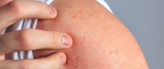
Even a flea bite (insect allergy) can provoke the development of an allergic reaction. As is known, flea allergic dermatitis often develops in animals (dogs, cats, small rodents) when fleas appear and actively reproduce. Although humans are not a permanent host for fleas, fleas from animals can nevertheless jump onto a person and bite through the skin, releasing saliva into the wound. If a person has hypersensitivity to the enzymes in flea saliva, an acute reaction develops - the bite sites become red, swollen, itchy, and when scratching them, a secondary infection may occur (Fig. below).
The development of allergic dermatitis is promoted by:
- Genetic predisposition of the body to allergic reactions.
- Neuropsychiatric disorders.
- Hatology from the gastrointestinal tract, including dysbiosis .
- Chronic skin diseases.
- Decreased humoral/cellular immunity .
- The presence of foci of chronic infection in the body ( caries , tonsillitis , adnexitis , etc.).
- Increased sweating.
- Occupational sensitization.
Also, the development of allergic contact is facilitated by the thinning of the stratum corneum of the skin, i.e., when it becomes thinner, dermatitis develops faster.
Diagnostics
Often, with a thorough interview of the patient, it is possible to determine which product or chemical agent caused the allergic reaction, however, to make a final diagnosis, an allergy test should be performed.
There is a standard Allertest that allows you to identify hypersensitivity to certain substances. It consists of 2 plates, on the surface of which 24 reagents are applied. After preliminary wetting with water, the system is glued to the skin of the forearm or shoulder. The result is assessed after 72 hours.
After removing the plates, the patient is examined for the following manifestations:
- erythema (+);
- erythema. papule (++);
- erythema, papule, vesicle (+++);
- erythema, papule, vesicle, edema (+++).
To verify the results obtained, the patient should also be examined one day after the tests. The reaction must be saved. Also, a similar test can be carried out without using the above system, but by applying the suspected allergen to the skin and fixing it with an adhesive plaster.
The disadvantages of this method are that the body is sensitized to the test substances, and only the presence of a reaction to the tested agents is visualized, which may not be the cause of the primary occurrence of allergic dermatitis.
You might be interested! Skin dermatitis: types, symptoms, photos and treatment methods
Symptoms of allergic dermatitis in adults
Allergic contact dermatitis in adults manifests itself primarily in areas of the skin exposed to the allergen, but clinical manifestations can significantly extend beyond the areas of exposure to allergenic agents. The main types of allergic rashes are represented by erythematous, papular or vesicular elements, which can be present on the skin of any part of the body (face, arms, legs, torso).
As a rule, the symptoms of allergic dermatitis develop against the background of erythema and are accompanied by burning, itching, and a feeling of heat. In this case, the allergic rash has a mildly expressed polymorphism of rashes in the form of vesicles, papules, erosions, scales and crusts. Symptoms of allergic dermatitis after cessation of contact with the allergen quickly and completely regress, however, in the event of repeated contact with the allergen, rapidly developing relapses of allergic contact dermatitis are observed.
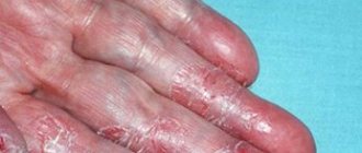
What does an allergic rash look like in adults with allergic contact dermatitis? The figures below show characteristic symptoms in adults with allergic contact dermatitis.
Contact allergic dermatitis on the face
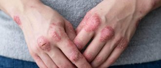
Contact allergic dermatitis on the hands
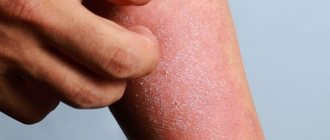
Allergic contact dermatitis on the legs
The severity of symptoms of contact dermatitis depends directly on the chemical activity of the allergen and the duration of contact with it. Chronic changes in the skin evolve sequentially (from transient erythema to vesicles or severe edema with blisters/ulcers and their combination). Often, rashes are characterized by a certain localization or grouping, which indicates the specificity of the effect of the antigen.
For example, a linear stripe indicates exposure to an irritant or exogenous allergen, and a ring-shaped stripe of erythema (under the belt/watch band) indicates the location of the allergen. During aerogenic contact, exposed skin areas are predominantly affected, for example, by perfumed aerosols.
With general exposure to the substance, rashes can be widespread throughout the entire skin. Typically, the rash manifests itself 15-48 hours after exposure to the allergen. In cases of chronic allergenic effects on the body in the presence of functional changes in the activity of the central nervous system, immunological and endocrine systems, as well as in cases of inadequate therapy, ACD can transform into eczema and be accompanied by the development of polyvalent sensitization.
What is allergic dermatitis?
Allergic dermatitis is a skin disease that manifests itself after contact of a person’s skin with an allergen of various natures. In this case, the body’s reaction manifests itself not only locally (in the area of contact), but generally (throughout the body).
ICD-10 code
According to ICD-10 (International Classification of Diseases), the class of allergic contact dermatitis is designated by code L23.
This class includes:
- allergic contact dermatitis caused by metals (L0);
- allergic contact dermatitis caused by adhesives (L1);
- allergic contact dermatitis caused by cosmetics (L2);
- drug-induced allergic contact dermatitis (L3);
- allergic contact dermatitis caused by dyes (L4);
- allergic contact dermatitis caused by other chemicals (L5);
- allergic contact dermatitis caused by food (L6);
- allergic contact dermatitis caused by plants other than food (L7);
- allergic contact dermatitis caused by other substances (L8);
- allergic contact dermatitis, cause not specified (L9).
Tests and diagnostics
The diagnosis of allergic contact dermatitis is made on the basis of the clinical picture, medical history, physical examination and the results of skin patch tests. Of particular importance is the medical history (in dermatovenereology), according to which it is necessary to carefully study the history of the development of the disease and especially the factors contributing to the disease. To identify a specific allergen, skin patch tests with allergens (standard test kits) are used. Differential diagnosis is made with atopic and seborrheic dermatitis , simple contact dermatitis , herpetic skin lesions, psoriasis , eczema .
Allergic dermatitis in children
As practice shows, in most cases children develop atopic dermatitis , which is due to genetic factors, the child’s living conditions and the individual characteristics of the skin structure. Research results indicate an immune mechanism for the development of atopic dermatitis. According to Komarovsky, atopic dermatitis first appears in children under 2 years of age, often in infants.
At the same time, adequate atopic dermatitis in many cases goes away without a trace by 3-5 years, but in the absence of therapy it can progress significantly and clinically manifest itself throughout adult life. What does atopic dermatitis look like in children? The main manifestations are the presence of atopic allergy , manifested by a red rash with clear or blurred contours, itching, eczema and the presence of altered vascular reactivity. At the same time, different localization of the lesion is possible on the skin of the face, limbs, torso, neck, but the most common atopic dermatitis in children is on the face. The morphology of the rash elements varies significantly depending on the form of the inflammatory process (acute, subacute or chronic). Below is a photo of allergic dermatitis in children.
How to cure allergic dermatitis in children? Treatment is complex, including elimination of trigger factors, an elimination diet, external medications and systemic therapy (Fig. below).
What does allergic dermatitis look like?
Allergic dermatitis manifests itself in the form of a polymorphic rash, in which various morphological elements are distinguishable:
- roseolous-erythematous rashes;
- papules;
- vesicles;
- areas of skin with severe swelling.
In this case, the true polymorphism of the rash is determined - the simultaneous appearance of various components of the rash.
Photo
The photograph shows a typical clinical picture of allergic dermatitis. The patient has both ordinary pink-red spots and blisters filled with clear serous fluid in one area of the skin. On the site you can also see photos of allergic rashes in children.
Diet for allergic dermatitis
Hypoallergenic diet
- Efficacy: therapeutic effect after 21-40 days
- Timing: constantly
- Cost of products: 1300-1400 rubles. in Week
The duration of the elimination diet is at least 6-8 months. It is dietary nutrition that can reduce the general state of hyperreactivity. In this case, the diet menu should correspond to the age/gender needs for basic nutrients and calorie content. The nutritional menu for adults should exclude to the maximum all foods that have high allergenic activity (coffee, chocolate, cow's milk, strawberries, cheeses, etc.). Also, nutrition provides for the exclusion/limitation of products containing food additives (antioxidants, flavorings, preservatives, dyes).
Prevention
Prevention of ACD is based on eliminating the provoking factor, which is achieved by removing the allergen-significant factor from everyday life through the use of personal protective equipment for mucous membranes and skin (wearing gloves, special protective clothing, protective creams).
The patient must know his individual allergens to which he develops a reaction. For example, patients with a nickel allergy should not use nickel-plated utensils, wear stainless steel jewelry, or allow fasteners/rivets on jeans or underwear to come into contact with the skin. If you react to specific perfumes and cosmetics, they should be excluded from use. If you react to latex, use vinyl gloves.
Treatment of allergic dermatitis
We previously discussed the treatment of perioral dermatitis.
Diet
A necessary stage of treatment for allergic dermatitis is following a diet that eliminates contact with the allergen.
There are 2 groups of diets:
- Elimination. This group of diets is prescribed for food allergies, while the food product that causes the allergic reaction, products made on its basis, as well as substances that can cause cross-allergy are excluded from the diet, for example, people with an allergy to avocado have a cross-allergy to latex products.
- Basic. The basic group of diets is used for dermatitis of non-food nature. Foods that can cause antigenic stimulation of the body are excluded from the diet.
Medications
General treatment consists of the following types of medications:
- H-1 blockers - histamine receptors (loratadine, suprastin, Zyrtec, Cetrin). This group of drugs is used for directly developed manifestations of allergic dermatitis. The presented medications (antihistamines) block the activation of receptors by histamine (a mediator of inflammation), as a result of which the manifestations of allergies are stopped.
- Cell membrane stabilizers (ketotifen). Membrane stabilizers are used to prevent relapse of the disease upon repeated contact with the allergen.
- Glucocorticoids (prednisolone, dexamethasone). Glucocorticoids are intended for the treatment of dermatitis complicated by a secondary infection.
Ointments
Local treatment involves applying medicinal substances to the surface of the affected skin. Antihistamine ointments, for example Bepanten and Fenistil, can be applied to the affected areas.
For allergic dermatitis, 2 categories of ointments are used:
- Non-hormonal. These ointments are used most often. They have anti-inflammatory, wound-healing effects, reduce itching, and relieve swelling. This group includes:
- eplan,
- panthenol,
bepanten,- exoderil,
- histan,
- elidel,
- protopic,
- losterine,
- thymogen,
- naftaderm,
- we see,
- Zinocab and many other analogues.
- Hormonal. Ointments based on hormonal drugs are used only if other treatment methods are ineffective, as well as when there is a threat of allergic dermatitis turning into eczema. They have an immunosuppressive effect, inhibit the immune system, as a result of which their use may be accompanied by side effects (hypopigmentation, skin atrophy). Thus, they should be used only under the supervision of a doctor - a dermatologist or allergist. Representatives:
- celestoderm,
- advantan,
- flucinar,
- fucicort,
- Akriderm ointment and others.
Folk remedies
Most folk remedies for the treatment of this type of dermatitis are aimed at eliminating inflammation on the skin, itching, and also to stimulate wound healing.
Traditional recipes:
- Compresses and dressings based on herbal decoctions. For this purpose, decoctions of string, calendula, burdock, and lemon balm are used.
- Baths with decoctions of chamomile, nettle, valerian.
- Tea with infusion of cornflower, chamomile, periwinkle.
- Celandine applications.
- Using tar soap, which has an anti-inflammatory and antiseptic effect. It also enhances reparative processes in the skin. However, a contraindication to its use is the presence of weeping erosions on the skin.
- Sea buckthorn and tea tree oils can be used both as applications and in the form of ointments, when mixed with baby cream.
List of sources
- Ivanov, O. L. Allergic contact dermatitis and associated allergic dermatoses: modern ideas about etiology, pathogenesis and diagnosis / O. L. Ivanov, E. S. Fedenko // Ros. magazine leather and venereal diseases. 2010. -No. 4. - P. 47-51.
- Korsunskaya, I. M. Therapy of contact dermatitis in adults and children / I. M. Korsunskaya, O. B. Tamrazova, T. A. Shashkova // Vestn. dermatology and venereology. 2006. - No. 4. - P. 46-46.
- Yu. Ilyina, N. I. Allergic skin diseases in clinical practice / N. I. Ilyina, E. S. Fedenko // Ros. allergol. magazine 2005. -№3.-S. 55-67.
- Luss L.V., Erokhina S.M., Uspenskaya K.S. New possibilities for diagnosing allergic contact dermatitis // Russian Journal of Allergology. 2008. No. 2. P. 28-35.
- Immunological mechanisms of development of allergic dermatoses / R.T. Kazanbaev, V.I. Prokhorenkov, T.A. Yakovleva, E.Yu. Vasilyeva // Siberian Medical Review. - 2013. - No. 4. — P.9-13.