18.01.2019
Introduction from D.S.: the article may seem a bit dry and boring if your work is not related to dermato-oncology. If this is the case, you can only read my explanations, they will be highlighted in gray. I have also highlighted the most important passages in the original text bold italics . Reading these fragments will be enough to form an opinion about the problem of spots on the genitals.
Alexandra M. Haugh, BA, Emily A. Merkel, BA, Bin Zhang, MS, Jeffrey A. Bubley, BA, Anna Elisa Verzi, MD, Christina Y. Lee, BA, and Pedram Gerami, MD
A clinical, histologic, and follow-up study of genital melanosis in men and women
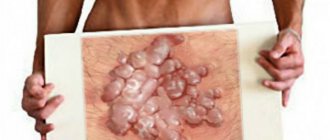
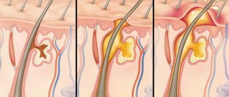
Description of the problem: Clinically, genital melanosis is difficult to distinguish from melanoma. There is insufficient data on the risk of developing melanoma due to genital melanosis.
Objectives of the study: To analyze and characterize lesions that are clinically defined as genital melanosis, and to assess the risks of developing genital melanoma and melanoma at other body sites using a retrospective cohort study.
Methods: observation of 41 patients with genital melanosis, as well as analysis of the results of histological examination of the formations.
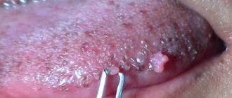
Results: Genital melanosis may be difficult to distinguish clinically from genital melanoma. However, the first condition develops at an earlier age than the second. Most lesions identified as genital melanosis stabilize or regress. 5 of 41 patients had a history of melanoma. Only one of them had genital melanoma. Most of the formations were characterized by melanocytes with suprabasal migration.
Limitations: The studies were conducted on a small number of patients for a limited time.
Conclusion: patients with genital melanosis, especially with atypia according to histological examination, require regular monitoring. This is necessary to detect the development of melanoma in time.
Genital melanosis is rare. This condition is observed in 0.01% of patients who consult a dermatologist [1, 2]. There is insufficient scientific data on this issue. Manifestations of genital melanosis are atypical. They are characterized by large areas of mucosal pigmentation that are clinically indistinguishable from melanoma. Almost nothing is known about the role of genital melanosis in the development of genital melanoma and melanoma of other skin and mucous membranes.
During the study, researchers observed 41 patients with genital melanosis. They also studied the results of histological studies. Finally, the researchers analyzed the history and incidence of melanoma in patients with genital melanosis. The data obtained should help clinicians choose the right diagnostic and treatment tactics when treating patients with genital melanosis.
Methods
The study involved 41 patients with genital melanosis. The criterion for participation was the presence of a mass larger than 1 cm or multiple associated lesions whose total size exceeded 1 cm.
The patients were divided into two groups according to the size of the formation. The first included participants with formations larger than 1.5 cm, the second included participants with formations less than 1.5 cm.
Biopsies were performed in 35 of the 41 patients. During the histological examination, pathologists assessed, among other things, the following data:
- The number of melanocytes per square millimeter of the epidermis.
- Presence of nuclear atypia.
- Location of melanocytes in the epidermis.
Five patients were biopsied multiple times. Samples with the highest number of melanocytes per square millimeter of epidermis were used for overall statistical analysis.
Treatment
Treatment methods are selected depending on the stage of degenerative changes, the risk of malignant tissue degeneration, the woman’s age and concomitant diseases. Kraurosis is an intractable disease, but modern methods of conservative therapy can prevent further progression of the disease. Attention is paid not only to eliminating the root cause of kraurosis, but also to correcting external defects.
Treatment methods:
- Prescribing antibiotics, antifungal or antiviral drugs. This treatment is prescribed for confirmed infection.
- Topical use of corticosteroids and antihistamines. Ointments and gels based on these medications alleviate the inflammatory process and improve blood supply to tissues.
- Local agents based on estrogen and progesterone. Due to disruption of endocrine functions, the patient’s body may have a deficiency of these hormones.
- Removal of papillomas using laser or radio wave coagulation. Cryosurgery may also be prescribed.
- Physiotherapy. Warming tissues using high-frequency current improves skin regeneration.
- Block of the pudendal nerve using novocaine injection.
- Surgical treatment using ablation and exposure to cold (liquid nitrogen). This method of correction is prescribed when conservative therapy is ineffective.
The gynecologist must prescribe a therapeutic diet and vitamin supplements to the patient. If anxiety is expressed, a consultation with a psychotherapist is carried out. Sedatives improve stress resistance.
results
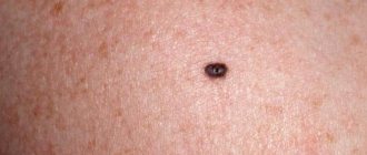
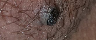
Detailed clinical examination results were available for 38 of the 41 study participants. In 17 cases, the genital lesions were irregular in shape and color. In 22 cases, the lesions had at least two colors, including several shades of brown and black, zones of white or gray regression, and mixed zones of hyper- and hypopigmentation.
The size of 12 lesions exceeded 1.5 cm. 22 patients had multiple lesions, and 19 patients had only one.
The researchers found no significant correlations between clinical manifestations and histological results.
The researchers also checked for a history of human papillomavirus (HPV) infections and inflammatory dermatoses. HPV and inflammatory dermatoses are considered risk factors for the development of genital melanosis. During the analysis, no connection was found between HPV infection and inflammatory dermatoses with clinical or histological manifestations of genital melanosis.
The history of melanoma in close relatives and the person himself was analyzed in 32 and 34 patients, respectively. 7 out of 32 had melanoma in relatives, and 5 out of 34 had melanoma in their personal medical history. Only one patient was diagnosed with genital melanosis and genital melanoma simultaneously. Those. on the genitals of one person in different places (!) both genital melanosis and melanoma were present (approx. D.S.)
No correlation was found between family history of melanoma and clinical manifestations of genital melanosis.
In patients with melanoma, a tendency towards an increase in the number of suprabasally located melanocytes was revealed. The average number of melanocytes per square millimeter was 53.6. In patients without melanoma, this figure was 35.5.
Of the three patients with nuclear atypia in melanocytes, two had a history of melanoma.
Follow-up history is known for 30 of the 41 patients. None of the 30 participants developed genital melanoma. In one patient, genital melanosis developed 10 years after identification and successful removal of genital melanoma.
Detailed descriptions of clinical examination findings are available for 23 patients. Nine of the 23 study participants had unchanged genital lesions over 33 months. Ten of the 23 patients had no evidence of residual lesions 56 months after diagnosis of genital melanosis. Only 2 out of 23 people had relapses after excision of the formations. No correlation was found between the recurrence rate and any histological or clinical features.
What color of the labia is considered normal?
The concept of normal for the color of the labia is very relative. For example, among Europeans, Americans and Scandinavians, red labia prevail, with varying degrees of saturation, from pale to bright pink or brick red. Asians and women from the African region most often have brown labia, ranging from the lightest beige to dark, almost chocolate shades. In most cases, pigmentation is inherited and appears from generation to generation with minor changes.
In some cases, the color of the labia may be heterogeneous:
- different colors of the right and left labia;
- there is marbling (two or more colors mixed in the form of small specks);
- There is uneven coloring with a predominance of one color.
Such “spotting” is also considered normal, especially if the girl was born to parents from different racial groups. We can talk about pathology or deviation from the norm only if pigmentation changes throughout life.
The discussion of the results
During the study, doctors studied data from clinical observation and histological analysis of 41 patients with genital melanosis. In 22 patients, the formations had several colors; in 11 patients they were more than 1.5 cm.
Researchers considered the average age of onset of this condition to be one of the clinical signs of benign genital melanosis. Among the study participants, it was 41 years. Genital melanoma develops on average in the sixth decade of life.
The researchers concluded that it is necessary to avoid unnecessary surgical intervention for genital melanosis. Clinicians consider it necessary to excise and send for histological examination all formations that seem suspicious to them. In the case of genital melanosis, this approach is not necessary. Most of the lesions examined during the study with clinical atypia, such as multiple colors and larger than 1.5 cm in size, were found to be benign.
Excisional biopsy in the genital area not only causes discomfort for the patient. The operation can harm sexual health and disrupt urinary function. The cosmetic result of the operation may be unfavorable.
Researchers have found that in most cases of genital melanosis, it is sufficient to actively monitor patients. Only one of the 41 study participants had both genital melanosis and genital melanoma. In this patient, genital melanosis was discovered almost 11 years after the diagnosis of vulvar melanoma. The formation was excised, and according to the results of histological examination it was recognized as benign.
During 30.5 months of follow-up, none of the study participants developed genital melanoma due to genital melanosis. In 19 patients who came for examination, the formations stabilized, decreased or disappeared. One patient was diagnosed with recurrence of genital melanosis after excision of the lesion.
Based on these data, the researchers concluded that the risk of genital melanosis transforming into melanoma is so small that it is difficult to measure.
The incidence of genital melanosis in the population is unknown. According to studies [1, 2], this condition occurs in 0.011% of patients who come to see a dermatologist. The incidence of genital melanoma is 0.19 cases per 1 million men and 1.8 cases per 1 million women. [4]
The study found that 15% of the experiment participants had a history of melanoma. In only one out of five cases the tumor was found in the genital area. The average age of patients with melanoma was 39 years.
Scientists have confirmed an increased incidence of genital melanosis in patients with a history of melanoma. Such patients have a suprabasal distribution of melanocytes, as well as an increased number of melanocytes in the epidermis.
Genital melanosis with clinical signs of atypia is recognized as an indication for active monitoring of patients, including examination of the whole body with a dermatoscope. However, according to the study, genital melanosis cannot be considered a precursor to genital melanoma. Therefore, in most cases, in patients with genital melanosis, radical surgery with excision of formations is not indicated.
The materials are intended only for readers over 18 years of age.
The labia minora look different for everyone. There are no standards for beauty or non-beauty in this area. As they say: “There are no comrades according to taste!” Why do some women decide about the need for surgery to change their shape and size?
The main reason why my patients come to me is a general dissatisfaction with the appearance of the labia minora. Feminists argue that men impose these beauty standards on women. Judging by my patients, this is not true; most often this issue is not discussed with sexual partners at all. At the same time, there are a lot of fans of the labia majora and elongated labia minora, so I always recommend talking about this with a man, precisely the one whose opinion is important to you. It must be remembered that it is extremely difficult to enlarge what has been cut off.
Why should you read this?
First of all, this article is intended for women who decide on an operation to remove excess tissue of the labia minora simply based on advertising and imposed information that this is correct. Unfortunately, there is very little information on blogs about the fact that a large labia minora is normal and beautiful for many people. At the same time, there is absolutely no need to feel insecure, shy, it is better to stop and just listen to yourself, understand what exactly you need, and maybe, in a second place, find out what your husband wants, namely a husband, and not a temporary sexual partner . If you are satisfied with everything, then move on with your life, but if not, then before going for an operation, in my opinion, it is advisable to find out the opinion of your loved one.
This article should also be of interest to those who are absolutely convinced of the correctness of the decision made, because the photographs presented, taken in the postoperative period, will show, in my opinion, the most positive and close to ideal result of labiaplasty.
What this area should look like before and after labiaplasty
I’ll just present the anatomy, that is, what should be present and what should not be. The size of the labia minora should ensure the closure of the entrance to the vagina in a non-excited state, and how many centimeters they are is individual. The clitoris should be covered with folds, again at rest; during sex it opens slightly and can be visible. Labiaplasty should also ensure the preservation of these structural features; the labia should not be excessively removed and the clitoris should not be opened, otherwise it is already called “female circumcision” and is an extremely dangerous and harmful procedure.
The folds of the clitoral hood are pulled back with your fingers, and it is exposed; this normally happens only during sex or sexual arousal.
Beautiful labia minora before and after surgery
Clinical case: labiaplasty was performed using the marginal wedge resection technique.
A rather complex clinical case from the point of view of surgical intervention, doubling of folds in the area of the clitoral hood. If you look from top to bottom, you can see three levels, similar to Christmas tree branches, the last of which is the labia minora themselves. A marginal wedge resection and correction of the clitoral hood were performed.
The clinical case is pigmentation and, according to the woman, excessively enlarged labia minora. After giving birth, I was worried about the “opening of the vagina.” A marginal wedge labiaplasty with vaginoplasty and restoration of the pelvic floor muscles was performed. If the scope of the operation was limited only to resection of the labia minora, then the situation with the “gaping” of the vagina would only worsen immediately after the intervention.
So, if these photographs, for example, before the operation seem beautiful to you in different positions or, on the contrary, you like the look of the labia after the intervention more, then this indicates that only you have the right to choose that same aesthetic standard. I repeat, the main thing is that the rules for maintaining anatomical and physiological characteristics are followed. Good luck and successful choice!
SUMMARY FROM D.S.:
This study shows that age spots on the genitals are, in the vast majority of cases, NOT DANGEROUS. However, researchers note a possible increased risk of developing skin melanoma (not just the genitals, but also other areas) in people with genital melanosis. This means that if you have similar pigment spots, they must be examined annually with a dermatoscope by an oncologist or dermatologist, just like ALL other pigmented skin formations.
Why do the labia change color?
Changes in the color of the genitals can be caused by factors independent of genetics. The most common of them:
- Hormonal fluctuations - puberty or menopause. Young girls sometimes develop labia blue or a paler shade than before. In women, during the period of extinction of sexual activity, on the contrary, an increase in pigmentation or redness of the labia is observed. A similar effect occurs when taking hormonal contraceptives.
- Injuries or rubbing of the perineum lead to increased pigmentation with a predominance of blue and red shades. Often after rough sexual intercourse, purple labia are observed. Such conditions are accompanied by swelling and pain.
- Increased pigmentation of the external genitalia may indicate pregnancy, intoxication of the body or oncology. Experts note that black labia in European women are observed with cancerous tumors of the pelvic organs, and with Cushing and Addison syndrome, in addition to the genitals, other parts of the body (armpits, knees, lower abdomen) also darken.
All of these conditions require consultation with a gynecologist and other specialists. If there is no severe pathology behind the change in pigmentation, a woman can go to a beauty salon, where a procedure for whitening or lightening the labia will be performed. It will help restore an aesthetic appearance to the genitals.
The article was checked for compliance with medical standards by a leading specialist at the Polyclinic+1 clinic, a dermatovenerologist, urologist, and mycologist.
Malashenko Vladimir Alexandrovich