Benign skin tumors can have different shapes, structures, colors, sizes, and be localized in different parts of the body. Depending on whether a pimple or mole has formed on the skin, treatment is determined. If atypical neoplasms appear, you should consult a specialist doctor for diagnosis and solution to the issue of removing skin elements. If you try to eliminate the problem yourself, you can injure the skin and provoke negative consequences - growth and malignancy of the tumor.
Seborrheic warts - causes of formation
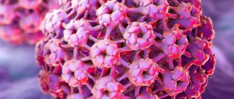
The causes of seborrheic warts are unknown. But it is reliably clear that they are not related to the HPV papillomavirus, although their name indicates this. Thus, a seborrheic wart has nothing in common with such epidermal lesions as true condylomas, papillomas and warts.
However, there are some factors that predispose you to developing seborrheic warts:
- Age
. Warts most often appear in people over 35 years of age. Many people have more than a dozen of them; - Genetic predisposition
. The presence of seborrheic warts or other epidermal nevi in immediate family members; - Conditions associated with actinic keratosis
or impaired sebum production.
Moles, neoplasms, ultraviolet radiation and HIV: an interview with a dermatologist
How can you spot a suspicious mole yourself, distinguish it from a normal one, and manage to remove it before it’s too late? Do you need to examine moles yourself at home? Is it true that sunbathing is strictly forbidden, or is it still possible a little? How are skin diseases related to HIV? Dermatologist-oncologist at the Rassvet clinic, Tatyana Kuzmina, answers these and other questions.
— How often should I undergo a preventive examination with a dermatologist if nothing bothers me?
— It depends on the results of the initial examination by a dermatologist. I examine all formations on the skin, and if there are suspicious ones, I highlight them, measure them and draw up a map of nevi (moles). If there are formations that require dynamic monitoring, then we invite patients to an appointment once a year. Also, once every six months or a year, you need to come for an examination to a dermatologist for those who have many formations (in this case, only a nevus is considered a formation) on the skin, more than fifty to one hundred. Such people are classified as patients with dysplastic nevus syndrome: they develop new moles all the time and are more likely to change towards dysplasia (a condition that is a precursor to melanoma).
We also invite adults over forty for examination once a year - if at this age a new nevus appears on the skin, then there is a higher risk that it will turn out to be something malignant. If a person is under forty and at the initial appointment there are no formations that would cause suspicion, and there is no tendency that something suspicious may appear in the future, then the appointment will only need to come once every three years. But this is provided that the person at home independently observes nevi and conducts self-examination at least once every six months.
— What should you pay attention to at home and how to conduct independent examinations between visits to the doctor?
— At home, from time to time you will need to check the parameters of the tumor map. If you see any dynamics: growth, changes in color, coloring, then you need to come to the appointment earlier. For example, if a mole is actively growing (by more than two millimeters per year), this should alert you. Suspicious formations also include nevi larger than a centimeter, asymmetrical, with uneven coloring and borders. If such formation begins to change, it is better to come and show it to the doctor.
But we also need to keep an eye on new formations. Until the age of forty, a person may develop new nevi, this is normal, but if they are black, then one should be wary. When one appears, regardless of its size, you need to consult a doctor. If you are not sure whether the lesion is black or not, take a black object, such as a phone, apply it to the nevus and compare. If the lump is dark brown in color, it is most likely benign.
Tatyana Kuzmina, dermatologist-oncologist at the Rassvet clinic.
— What does a doctor do if, during an examination, he reveals something suspicious?
— If a nevus seems suspicious, we always suspect a malignant pigment formation, melanoma in situ, and suggest removing it. Depending on the visual assessment, we determine what it could be: a benign nevus that has just begun to change, or an already changed formation.
In the first case, we remove the nevus with a certain indentation, in the second - a more serious intervention; additional examinations need to be carried out to find out whether the process is only in the skin or has already spread beyond its borders. If this formation turns out to be melanoma in situ or severe dysplasia, then everything ends with surgical removal, and the materials after the operation are sent for histology. If the histology results indicate stage 1 or 2, then this is a good prognosis, and in the future you can count on a complete cure. In the third or fourth stage, treatment must be continued, but final recovery is unlikely.
Therefore, it is very important to visit a doctor at the first signs of changes in the skin, when color or size has just begun to progress. Melanoma skin cancer develops quickly, a small dark spot of two millimeters may appear on the skin, this will already be melanoma, and active measures will need to be taken to remove it. No need to wait. If the patient manages to see a doctor within six months, then, as a rule, the formation does not have time to move to the stage when we cannot help him and additional treatment in addition to surgery will be required.
— How can you reduce your risk of getting skin cancer? Don't sunbathe at all? Sunbathing only with photoprotection?
— Melanoma is triggered by sunburn. The more active the sun, the easier it is to get a sunburn. The activity of the sun increases as one approaches the equator - in countries close to it, for example in Thailand, where people now often travel for several months, ultraviolet radiation is of higher intensity (the activity index on a scale from 0 to 10 reaches its maximum values, the sun's rays damage the genetic apparatus of cells more strongly skin). Such radiation can provoke melanoma without visible sunburn.
No protective equipment can completely resist radiation, but with photoprotection and photoprotective clothing, you can avoid skin damage and reduce the risk of developing melanoma. However, even with protective equipment, ultraviolet radiation will accumulate - we call it chronic ultraviolet radiation, which increases the risk of developing non-pigmented skin cancers, in particular basal cell carcinoma. This is a type of tumor that develops from non-pigmented skin cells of the epidermis (keratinocytes) rather than from pigment cells (melanocytes).
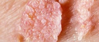
What does a seborrheic wart look like?
Seborrheic papillomas are characteristic mushroom-shaped skin rashes. However, such a wart can be confused with other skin lesions, such as actinic keratosis or some malignancies, mainly cutaneous melanoma. It also happens the other way around, when skin cancer is mistaken for a harmless seborrheic formation.
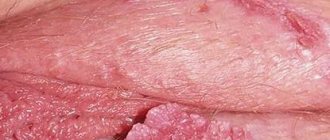
Keratinized pigmented lesion
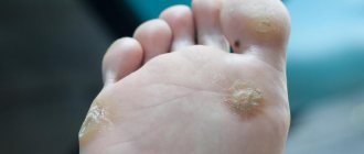
Multiple seborrheic keratomas
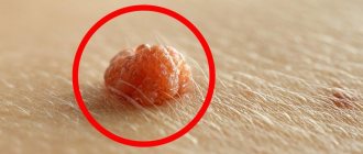
Development of seborrheic papilloma
Therefore, diagnosis and treatment of any neoplasms should be carried out strictly by an experienced dermatologist. It is strictly prohibited to independently diagnose yourself, much less remove tumors at home.
Here are the features that distinguish a seborrheic wart from other skin lesions:
- Seborrheic papilloma is well demarcated from healthy skin. It gives the impression of being superimposed on the surface.
- The size of the formation ranges from a few millimeters to several centimeters.
- On the surface of the nipple there are numerous depressions containing an accumulated mass of callus epidermal cells and sebum.
- Depending on the stage of development, the color of the nipple varies from a color similar to healthy skin to dark black, which is not a sign of melanoma.
Seborrheic warts are not accompanied by symptoms such as pain, itching, burning. The exception is mechanical irritation, for example, when changing clothes.
Appearance mechanism
You can confuse the inflammatory element (acne, blackhead, pimple) with red-type moles (angiomas, red, pink vascular nevi).
A mole similar to a pimple and pimples in the form of moles appear under the influence of external, internal etiological provocateurs. The formation of inflammation of the skin and vascular elements occurs under the influence of factors:
- Physical, chemical injuries, burns. Hyperproduction of melanocyte cells is provoked, an increase in pigmentation in the lesion, vascular formations are involved, the mole is similar in appearance to acne.
- Infectious lesions of the skin surface in the absence of personal hygiene provoke the development of nodular acne, which is similar in appearance to angiomas.
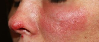
- Due to hormonal imbalances, age spots and hormonal rashes may appear. The difference is that moles are pointlike and isolated, while inflammatory elements are distributed throughout the body in large numbers. Often a hormonal rash appears on the face, in the nose area.
Pimple
A pimple is an inflammatory element that appears as a result of a malfunction of the sebaceous gland or the addition of a secondary infection (white abscess). The appearance of a growth is dangerous; pus can break into the melanocytes and provoke atypical malignant growth of cellular structures. The basis for the appearance of acne is internal blockage of the sebaceous gland, external contamination of the pores, which disrupts the normal process of renewal of the epidermis by removing subcutaneous fat and desquamation of the epithelium. In case of injury to a mole (tearing off, puncture, scratch, abrasion) or an insect bite (mosquito, bedbug), a pimple may form on top of it, if treatment with antiseptics was not carried out in a timely manner, a bacterial infection was introduced, an irritation reaction to the saliva of the insect occurs, inflammation from damage to the surface layer of the skin or transmission of infection through a bite.
One of the main factors in the appearance is exposure to ultraviolet radiation. Sun rays lead to a decrease in local immunity, disruption of skin metabolism, and increase the risk of inflammatory elements.
Often after sunburn, you can observe white growths - milia, which arise as a result of complete blockage of the sebaceous gland. You can observe excessive formation of acne due to hormonal imbalances in the body during puberty, which is due to hormonal instability and increased sebum production. The ducts do not have time to eliminate subcutaneous fat, the problem of incomplete exfoliation of the epidermis arises, and new inflammatory elements appear.
The basis for disorders of lipid metabolism and general metabolism, which leads to rapid secretion of sebum, is a disrupted diet, addiction to fatty, spicy foods, and lack of sufficient amounts of useful macroelements, vitamins, and minerals.
Moles
Dark pigment spots form on the skin when the production of melanin pigment in melanocyte cells increases. The process of acne appearing on moles depends on factors:
- damage to the skin (sunburn, chemical burn, mechanical damage) can stimulate epithelization, hyperproduction of melanocytes at the wound site;
- hereditary factor in the appearance of formations - provocation of migration, the formation of a large number of melanoblasts at the stage of development of the dermis;
- under the influence of solar insolation or artificial ultraviolet irradiation, there is a change in the production of melanin, which can provoke hyperpigmentation and the appearance of age spots - freckles;
- the appearance of pigment spots can be caused by skin infections, inflammation of the epidermal layer;
- in old age, hyperpigmentation of the skin can occur against the background of thinning of the lipid layer, accumulation of melanocytes, and vascular disorders of the surface capillaries (hemangiomas);
- acne or mole, it can be difficult to determine if there is a red formation - benign vascular tumors that arise when microcirculation processes are disrupted, increased viscosity of the circulatory system, or cardiovascular diseases.
The more active the production, the blacker the formation will be.
Important
Seborrheic warts are benign skin lesions. This means that they cannot turn into cancer cells and be dangerous to health. Seborrheic warts are only an aesthetic problem. Especially when they are on the skin of the face.
The exception is when such a neoplasm is located in a place of constant friction or other traumatic factor. In this case, skin infection may occur. This can result in serious inflammation and even blood poisoning.
Mole removal methods
Surgical removal of moles is the safest option against melanoma-type malignancies, and the surgically removed tissue is submitted for pathological analysis to definitively confirm the diagnosis and ensure that the skin edges are free of malignant cells.
For some types of moles, other removal options are possible, such as dermabrasion, excision of the superficial layers of the skin, or laser ablation.
Doing mole removal yourself using alternative methods is not recommended or medically approved.
People prone to the appearance of moles (usually people with fair skin, blue eyes, blond hair) should undergo regular preventive examinations by a dermatologist, preferably once a year. It is also recommended to avoid prolonged sun exposure and use sunscreen, from sunscreen to hats or light shirts that cover most of the body.
Seborrheic papilloma and cryotherapy
One of the methods for removing seborrheic warts is cryotherapy, that is, freezing the skin lesions with liquid nitrogen. The wart dies off when exposed to very low temperatures.
Cryotherapy
After treatment with liquid nitrogen, literally a few days after the procedure, the color of the wart changes to black, and the skin lesion takes the form of a scab (crust), which disappears from the surface after a month.
Unfortunately, the procedure carries the risk of leaving an unsightly scar, so cryotherapy is not suitable for removing facial warts.
Why a mole can look like a pimple
Moles can look like a pimple for the following reasons:
- due to the similarity of shape and color, small nodular size, the light pink element is difficult to distinguish from a pimple;
- characteristic localization - the sudden appearance of a vascular hemangioma on the face can easily be mistaken for a pimple;
- after hypothermia or overheating in the sun, a primary form of light pink nevus with minimal pigmentation may appear;
- often vascular nevi can disappear on their own or reappear in the same place under the influence of unfavorable internal or external factors;
- one of the reasons for the appearance of a red nevus is a disruption of the digestive tract; the appearance of a new element is provoked after eating junk food, which a person often mistakes for an allergic rash.
Do not rush to remove the growth immediately, wait to make sure the diagnosis is accurate.
Seborrheic wart and IPL laser
Another method for removing seborrheic warts is the use of an IPL laser, which completely burns away the skin lesions, leaving a small concave socket.
IPL laser treatment
Within a few days after the procedure, a scab will form, which will fall off after 2-3 weeks. After treatment, a scar may also remain, but, unlike the previous option, it is almost invisible. Therefore, warts on the face are removed with a laser. A similar effect is also achieved by the radio knife, another modern tool designed to remove moles and warts of almost any type.
Types of moles
First of all, all moles are divided into epidermal and dermal.
Let us first consider the epidermal varieties of nevi. Such moles on the surface layer of the epidermis are, in turn, divided into specific and nonspecific.
Nonspecific moles are divided into:
- A borderline nevus is a mole with which a person can be born, or such a spot can appear at any age. Most often, such moles occur on the arms (palms), legs (feet) and even on the genitals. Visually they look like a spot or papule. The color of such a spot is uniform: it can vary from different shades of brown to black. The size of such spots usually does not exceed 1-2 cm.
- Intradermal nevus is the most common type of mole. Visually, they resemble a hemisphere, which seems to rise slightly above the surface of the skin. Such moles can be located on any part of the skin; the diameter usually does not exceed 1 cm. The color of such a nevus is also always uniform, either different shades of brown or black. Such moles bleed at the slightest attempt to injure them and may decrease in size.
- Complex nevus is a transitional form of congenital nevus, which visually looks like a papule, or a group of papules. Complex moles do not have a specific location.
Specific nevi are:
- Epithelioid nevus is a benign formation, most often they are single, but multiple formations of this type are not less common. Most often they appear on the legs and face. These moles reach 1-2 cm in size, are easily injured, and can disappear spontaneously.
- A mole of balloon-forming cells. This is the rarest type of nevus. Visually presented in the form of a spot, or papule. The main characteristic feature of such moles is their color. This is a brown mole with a yellow rim around the edges.
- Setton's mole is a nevus surrounded by discolored skin. It can be either single or multiple. Most often, such moles appear in people with vitiligo or other autoimmune pathologies in the body. They may disappear spontaneously on their own.
Dermal varieties of moles are:
- The Mongolian spot is a mole of quite impressive size (6-10 cm) in newborns. Localization is in the area of the sacrum, or buttocks, or thighs.
- Nevus of Ota is a type of skin defect, single or multiple, dark blue in color. They are located mainly on the face, in the area of the eyes, nose, and cheeks. They also occur on the mucous membranes of the nose, mouth and even eyes.
- Ito’s nevus is visually the same as the previous type, only the location of such moles is the neck, shoulder blades, and supraclavicular region.
- A blue nevus is a dense and smooth nodule that lacks hair growth. The sizes vary from 1 to 3 cm. The color can be gray or black.
In addition to dermal and epidermal moles, there are mixed types of moles. They, in turn, are divided into combined (which combine the characteristics of several nevi) and congenital. Congenital nevi can be either clear or blurred. They are similar to acquired ones and their main feature is their rather large size: they can be 1 cm, or they can occupy the entire surface of a limb, neck, or torso. A distinctive feature of congenital moles is their inability to disappear spontaneously.
The most dangerous are nevi of melanocytic origin. These are moles that most often transform into a malignant tumor.
Such moles are called dysplastic (atypical) nevi. This kind of moles are not congenital and are exclusively acquired. This is a single or multiple spot, which can be either round or oval of irregular shape. A distinctive feature of such a mole is its uneven edges with an accent in the center in the form of a papule.
The color of such a nevus is also heterogeneous: it can be a variety of brown, red, pink and even red colors. The size of such a nevus is also quite impressive. Such formation is rarely less than 6 mm. Such moles can be located on any skin surface of the body, less often they appear in the face area.
Seborrheic wart - surgical treatment
Removal of skin lesions with a surgical scalpel involves removal of the wart at the epidermal level. This method is suitable for large lesions on the body.
Surgery
It is important to understand that the choice of method for removing seborrheic papilloma should be made by a dermatologist. Some tumors may raise doubts about their origin, so it is important to preserve the removed tissue for analysis. In this case, the doctor will choose a surgical method, since the laser and liquid nitrogen completely remove the affected tissue, so there will be nothing to take for analysis.
ONLINE REGISTRATION at the DIANA clinic
You can sign up by calling the toll-free phone number 8-800-707-15-60 or filling out the contact form. In this case, we will contact you ourselves.
Methods for diagnosing such a mole
To accurately determine whether a mole similar to a pimple has emerged or an inflammatory skin element has appeared, you need to consult a specialist doctor who will conduct a thorough examination and prescribe research methods to accurately determine the origin of the neoplasm.
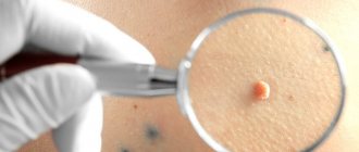
If there are no problematic issues, the doctor may limit himself to examination using a dermatoscope (an optical apparatus with a magnifying glass and a fluorescent lamp to illuminate the surface of the skin); no tests will be needed. To accurately determine the nature of the pigment formation, the doctor can take a biopsy for histological analysis and determine the tissue composition of the formation.
Independent attempts to determine whether a mole or acne may be inaccurate; if you try to remove the formation, negative consequences may occur; it is worth seeking qualified medical help.