A choroidal nevus is a benign tumor of the choroid. Such tumors are detected in almost 10% of people with white skin color and are practically not found among dark-skinned people.
Choroidal nevus is a pigmented formation of a cluster of melanocytes (pigment cells). As a rule, nevi are formed from birth, however, their pigmentation appears later, in the prepubertal period. Therefore, they are found, in most cases, in adults. In women and men, nevi occur with the same frequency.
The usual location for choroidal nevus is the posterior fundus region, behind the equator. However, sometimes it is found in the preequatorial zone and even directly in the equator zone.
At the early stages of their development, such nevi are localized on the superficial layers of the choroid, with age, spreading into its deeper tissues.
Classification of nevi
In ophthalmology, it is customary to divide nevi into stationary and progressive. In addition, among choroidal nevi, typical, atypical and suspicious are distinguished.
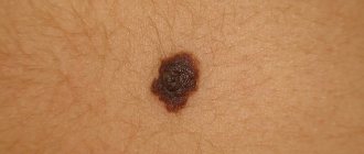
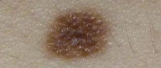
The appearance of a stationary choroidal nevus (typical) is defined as a flat or slightly protruding formation on the fundus, oval or slightly round in shape, gray-green or gray in color. Its size does not exceed 1 - 6 mm in diameter, the boundaries are clear or slightly blurry. Such nevi are characterized by uniform color, lack of growth and changes in the retina. Sometimes drusen (accumulations of particles of cellular metabolic products) are found on its surface. With a stationary choroidal nevus, vision is not impaired.
A distinctive feature of a progressive choroidal nevus is an increase in volume. Its shape may undergo changes, the uniformity of color is disrupted, and the boundaries are blurred. Dystrophic changes occur in the adjacent areas of the retina, and the choroidal vessels may be compressed. In some cases, serous retinal detachment is sometimes observed. Vision often decreases, with spots appearing before the eyes and images are distorted. Progressive nevi are considered to be at high risk of malignancy. At the same time, a nevus is considered progressive when changes in its dynamics are detected. The detection of a primary choroidal nevus in a patient with the above-described signs gives reason to consider it “suspicious”.
Atypical are called nevi that have no color (pigmentless), as well as “halo-nevi” surrounded by a zone of paler choroid (a sign of atrophy). Histologically, they consist of cells with degenerative phenomena, which is considered one of the signs of malignant growth.
Types of eye melanoma
A classification of choroidal melanoma based on morphological characteristics has been developed. Depending on the cellular structure, the following types of this tumor are distinguished:
- Spindle cell.
- Epithelioid.
- Mixed (mixed melanoma).
- Fascicular.
- Necrotic.
This classification has certain disadvantages, since necrotizing choroidal melanoma is determined clinically, but it is impossible to determine its cellular identity due to extensive necrosis. Fusiform and fascicular types have a similar prognosis. In this regard, it is currently customary to distinguish only 2 types morphologically: spindle cell and epithelioid. The mixed form occupies an intermediate position. Its prognosis depends on the predominance of certain cells. Epithelioid cell melanoma of the choroid is considered to have the least favorable prognosis.
Diagnostics
Detection of nevi, as a rule, occurs accidentally during ophthalmoscopy of the fundus. For a final diagnosis, their dynamic observation with regular examinations of the fundus, including examinations with color filters, is necessary. Thus, a choroidal nevus becomes clearly visible in red light, and when examined in green light, the nevus “disappears.” At the same time, only changes in the layers of the retina, which are characteristic of a progressive nevus, remain visible. Ultrasound examination, in some cases, can sometimes reveal the protrusion of the formation. With fluorescein angiography (examination of the fundus vessels using a contrast agent - fluorescein), the presence of stationary nevi is indicated by the absence of changes in the vessels surrounding the choroid. With a progressive nevus, fluorescein angiography detects the leakage of fluorescein through the walls of the vessels adjacent to the nevus.
Stages of development of choroidal melanoma
According to the international classification, there are 4 stages of development of this tumor. Criteria for the prevalence of the tumor process:
- T1 - melanoma size 10 mm or less, thickness - 2.5 mm or less.
- T2 - the size of the neoplasm is 10–16 mm, the greatest thickness is 2.5–10 mm.
- T3 - measuring 16 mm and/or thickness more than 10 mm without spreading beyond the eyeball.
- T4 - the largest tumor size is 16 mm and/or thickness more than 10 mm with extension beyond the eyeball.
There are also 4 clinical stages of choroidal melanoma. Each of them is characterized by certain symptoms of the disease:
- The first, so-called “quiet eye” stage, is characterized by the absence of significant clinical manifestations and complaints. Retinal clouding may be present, and visual field defects may also be detected.
- The second stage is characterized by the appearance of pain in the eyes, inflammation, redness of the eyeball, and swelling of the eyelids.
- At the third stage, choroidal melanoma extends beyond the boundaries of the eyeball, exophthalmos is formed, and the sclera loses its integrity.
- The fourth stage is accompanied by generalization of the process. The patient's general condition is deteriorating. Patients complain of severe pain, body weight decreases, and intoxication increases. Metastases of melanoma appear in internal organs: liver, lungs, bones. Damage to one or another organ provokes the appearance of corresponding symptoms. A further decrease in visual acuity, a feeling of veil or fog before the eyes may be detected. These manifestations are caused by bleeding into the vitreous body and clouding of the lens.
Symptoms of the second and third stages of choroidal melanoma are pronounced when the tumor is located in the central or paracentral part of the fundus. Peripheral localization of the tumor is characterized by a long absence of subjective sensations. In this case, melanoma is detected either by chance or at the stage of tumor disintegration and its secondary manifestations.
Treatment of nevi
For a typical stationary choroidal nevus, special treatment or long-term observation is not required, since the risk of malignant transformation of its cells is extremely low. However, these entities are subject to mandatory documentary registration.
For suspicious and atypical nevi, mandatory photographic recording of the fundus picture, repeated ultrasound examinations and frequent re-examinations (several times annually) are required. Establishing a diagnosis of a progressive nevus makes it necessary to choose immediate treatment, since a progressive nevus is a potentially malignant tumor. For the treatment of such nevi, photo and laser coagulation are recommended. With timely treatment, the prognosis is favorable.
At what stage the malignancy of the nevus begins is impossible to predict. Therefore, the key to the patient’s health is to carefully follow the doctor’s recommendations, regular follow-up visits to a specialist, etc. treatment started as early as possible, if indicated.
In the ophthalmology department, you can undergo a full examination using the latest equipment if you suspect a nevus of the choroid and further treatment (if necessary) from leading retina specialists in Moscow.
To find out the cost of a particular procedure or make an appointment at our clinic, you can call in Moscow 8(499)322-36-36 or number 8(800)777-38-81 (toll-free from mobile phones and for regions of the Russian Federation) daily from 9:00 to 21:00. You can also use the online registration form.
Etiological factors
Most cases of choroidal melanoma are sporadic, that is, caused by one or another mutation of the melanocytic precursor cell, which can give rise to a pathological tumor clone. In addition, there is an assumption about the hereditary cause of this disease. The influence of such a typical provoking factor for skin melanoma as increased insolation for this tumor is also not excluded.
Elderly people are at risk (the average age of tumor manifestation is 60 years). Men get sick a little more often. Those with fair skin and hair, nevi and freckles are prone to developing choroidal melanoma.
A mole under the eye or on the lower eyelid - what does it mean?
A mole under the eye means that its owner has a soft and pleasant character, calmness and balance. He is pleasant to talk to, a good friend and a good colleague. The positive energy of such a person makes him a welcome guest. He is distinguished by optimism and the ability to listen; communicating with such a comrade is always interesting.
Such individuals love animals and children. They are able to take care of even a casual acquaintance who gets lost in a strange city, loses his wallet, or gets into other troubles. They do not allow themselves to be deceived, because they value the results of their work too much and are distinguished by their frugality.
A mark under the eye indicates that you are marked by the goddess of fortune. You will be lucky throughout your life. Troubles are a rare guest on your life path. Most likely, you are unpretentious, and your average income is enough for your comfort. However, money issues will not be a problem either; owners of such moles are able to earn a good fortune.
If the mole near the eye is on the left side, closer to the temple, then caring may border on self-sacrifice. These qualities are combined with confidence and perseverance, so it is almost impossible to convince a friend with such a characteristic mark on his face to stop picking up street animals. These people often find themselves in charity, choose the profession of social workers, and engage in volunteer activities. If they have the financial means, they become philanthropists.
Among the meanings of a mole under the left eye, there is also popularity among members of the opposite sex. You know how to win someone you like. Such people make faithful wives and husbands who are able to provide for their families and create comfort in the home. A mole located on the opposite side of the face also speaks of a person’s sensuality and attractiveness.
If the location of such a mark is under the right eye, then we are talking about a person who is distinguished by a rare sense of purpose. He is able to move in the direction of his dreams throughout his life. Such people almost always achieve what they set out to do. Even where others give up and move on to another goal, they continue to move forward.
–>
Locations
Birthmarks form in different areas of the eye:
- external and internal areas of the protein;
- tearful month;
- semilunar fold;
- iris or retina;
- limbo.
Location of conjunctival nevi
An ocular nevus forms on the inner and outer parts of the conjunctiva. Birthmarks are detected on the inner corner of the eye, the periphery of the cornea, the lacrimal month, and the semilunar fold. Sometimes neoplasms appear on the inside of the eyelid. Although moles are located near the pupil, they do not block the view or impair vision.
Moles! Who knows their meaning?
I have a mole under my nose on the left side (((I don’t like it, I want to remove it! I don’t know what it means!
Natalia
The interpretation of the location of moles and their influence on a person’s fate is closely related to the science of “reading a person’s face,” physiognomy, which originated in the east. Initially, all moles were assigned a negative meaning, but long-term observations made significant, factually confirmed adjustments. Codes with lists of meanings have developed, by reading which you can learn a lot about yourself and look at your life differently. General characteristics Pay attention to bright, noticeable moles. Small moles scattered throughout the body play virtually no role. If a mole stands out strongly or rises above the skin, this is a lucky sign that mitigates possible unfavorable meanings. For dark-skinned people, black moles are more important, for light-skinned people - convex ones. It is also important which side of the body the mole is on. For men, the right side is favorable, for women - the left. The appearance of new moles, the disappearance or increase in size of existing ones, reflects actions committed by a person that affect future life and, possibly, change fate. Moles on the face Moles on the face are of decisive importance. A mole on the right eyebrow indicates an early marriage, on the left - the likelihood of a late or unsuccessful marriage, celibacy. Above the right eyebrow - energy, enterprise. In the corner of the eye it means passion, jealousy, irritability. A mole under the right eye speaks of marital fidelity, as well as sensuality and generosity. Under the left eye - about experiences in married life. On the right eyelid - about a penchant for intellectual professions. On the left eyelid - about excellent memory and diplomacy, down-to-earthness. On the left side of the forehead - about the wastefulness and weakness of the owner. A mole in the middle of the forehead means numerous victories on the love front. A woman is always surrounded by admirers, and a man is most likely Don Juan! The “third eye” point is a sign of strong intuition, reason, and mysticism. Owners often suffer from headaches. A mole on the nose indicates sociability. On the tip of the nose - a craving for complicating love relationships and everything forbidden. Under the right nostril - the owner has an extraordinary destiny, a mystical mindset. In the center between the nose and upper lip - an indication of independence, love of pleasure. On the bridge of the nose there is a hint of a love of travel, as well as a developed imagination and a penchant for creativity. On the cheekbone - determination (especially if the mole is on the right side), oratory abilities. Under the lower lip is a sign of a fragile psyche and health. Above the upper lip - cruelty, imperious character. On the red border of the lips - weakness of will, inability to take responsibility for one’s actions. In the language - longevity, suspiciousness. On the chin - the desire to have a strong family, conservatism. On the temple - inconstancy, frequent mood changes, variability in views. On the right side - subtle intuition, the gift of foresight, bright individuality. On the right cheek for men - eccentricity, for women - increased male attention. On the left cheek - a sign of innate talent, good memory, for a woman - natural charm.
Prevention
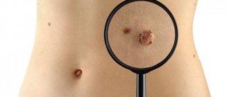
There are no specific rules for preventing the development of such neoplasms. Until now, the etiology of such moles is not known. To avoid the development of negative consequences, you should consult a doctor in a timely manner.
Timely diagnosis will help determine the presence of such neoplasms and help get rid of them, especially if the nevi are malignant. Therefore, every person should visit an ophthalmologist 1-2 times a year.
In addition, do not forget to monitor eye and eyelid hygiene. If suspicious symptoms develop, you should consult a doctor. Progressive nevi can affect visual acuity. If the patient observes clouding or distortion of the image, then it is necessary to contact an ophthalmologist.
After a full diagnosis, the doctor will be able to determine the classification of formations and the method of eliminating them. As an additional prevention, you should adhere to a healthy lifestyle. Sleep should be complete, nutrition balanced and rich in vitamins. It is necessary to give up bad habits.
Prognosis for life with choroidal melanoma
Life expectancy for this type of cancer depends on the location and size of the tumor, the patient’s age, the morphology of the tumor, the treatment performed and other features. The five-year survival rate in the initial stages of choroidal melanoma after the use of organ-preserving radical methods is 93%, and the ten-year survival rate is 89%. In later stages, when liver metastases are detected, the median survival is only 4-6 months. For patients with metastatic disease to other organs, the one-year survival rate is 76%.
Book a consultation 24 hours a day
+7+7+78