Absolutely all new parents get scared when they discover any spot or bulge on the body of a newborn child that, in their opinion, is not the norm. And since mom and dad's baby's skin is examined frequently, while simultaneously caring for and cleaning it, discovering an infant hemangioma is only a matter of a short time. Then worries, suspicions and questions begin, most often ending in the pediatrician's office. And that’s right – you never know what’s wrong with the child. But, despite all fears, hemangioma, although it does not look very aesthetically pleasing, is not a dangerous formation. What is it and why does it occur? Answers from experts.
Hemangioma in newborns
What is infantile hemangioma?
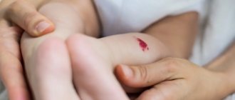
This is a tumor-like formation of a qualitative nature, which is formed by fragments of blood vessels of varying thickness. The nature of the origin is as follows: during the process of intrauterine development, the formation of blood vessels is disrupted. As a result, almost immediately after the birth of the child, or some time (about a month) later, a hemangioma appears on his body.
Cavernous hemangioma
By the way. Most often, this formation appears on the skin, at any point. But in 20% of cases, the tumor can localize itself to internal organs, muscle and even bone tissue.
The neoplasm looks like this:
- a thickened tumor-like spot of various sizes and degrees of elevation above the surface of the skin or depression;
- the color is either pale burgundy, closer to pink, or bright burgundy;
- the edges are uneven;
- the structure is dense;
- composition – atypical and normal cells.
New growth on a child's cheek
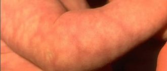
Important! The key point is that although this is a full-fledged tumor, it is always benign. Therefore, there is no need to do anything. Over time, in most cases, the hemangioma subsequently resolves on its own, without a trace.
In children, quite often hemangioma simply resolves over the years.
Of course, exceptions, deviations and anomalies occur, since each person’s body is individual, including a newborn baby. The most important thing for parents when a neoplasm is detected is not to panic and carefully study the issue of how the local pediatrician and this material can help them.
Clinical picture
The Mongolian spot usually has certain characteristics that help distinguish it from other marks on the skin:
- Present on the skin from birth.
- It is distinguished by a gray-blue or cyanotic color. Sometimes there are bluish-brown markings.
- Has an uneven color over its surface.
- Most often it has an irregular shape, but there are round and oval spots.
- It can have different sizes - from a small coin to 10 cm or even more.
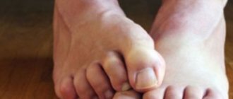
- Most often it is localized on the lower back, sacrum or butt. However, sometimes spots appear on the back, legs (their back side), etc. Some sources contain data on the possible migration of the spot when it moves over time.
- Most often it is present on the skin in a single copy, but sometimes several spots are observed on the body at once.
After the baby is born, the spot usually has a rich color. Over time, the trail gradually becomes smaller and loses brightness. By the age of 5, Mongolian spots usually disappear, although in some cases they can be present on the child’s body until adolescence and even adulthood. Such a prolonged presence on the skin is most often recorded if the spots are multiple in nature and are located in rather atypical places.
Fortunately, dermatologists have never encountered a Mongolian spot malingering, which leads to the development of skin cancer. Therefore, such a tumor on the body is considered safe.
How does a tumor occur?
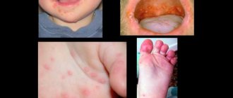
Statistically, hemangioma appears in ten percent of infants. The percentage is not so small if you think about it. Most often, formations found on the baby’s skin are located at such points that the tumor does not pose a danger to vital organs and does not cause damage to the mucous membranes. The only problems that education can cause are aesthetic ones. But they are precisely the most significant. Agree, a rough red “blot” in the middle of the forehead or on the child’s face, reminiscent of an exaggerated wart or depression, will bring few parents into a relaxed state.
Hemangioma on the forehead. Unsightly stain
By the way. Not every formation found on the skin of a baby is a hemangioma. Therefore, those parents who, at the sight of a spot, begin to sound the alarm and go to the doctor are right.
Signs of hemangioma.
- The most common type of tumor among newborns.
- Benign education.
- Appears only in children from age zero (immediately after birth) to the age of 60 days.
- Affects every tenth child on the planet.
- It is three times more common in girls (for every three newborn girls with this type of tumor, there is one male child).
- May appear as a flat spot.
- May resemble a depression (grow deep).
- Forms a bulge (most often).
- It has any size - from a “grain” to a large spot.
- There may be several or even many formations.
- If more than three hemangiomas are found on the skin, they are also present inside the body.
- Goes away on its own (in the vast majority of episodes).
Hemangiomas
Combined hemangioma in the breast area
Important! The most important sign that this is not a malignant formation, but a “harmless” benign hemangioma is that it contains elements of endothelial cells (the inner surface of blood vessels).
Of course, we are talking about “harmlessness” only when the formation is located outside, does not block physiological openings, does not damage the mucous membranes and does not increase in size.
There are several types of hemangiomas based on their location and structure.
Table. Types of hemangiomas.
| Variety | Description |
| It is formed from dilated vessels and cavities filled with venous or arterial blood. Does not penetrate deep into the skin, is located on the surface. It has the softest structure of all varieties. Amenable to basic therapy. |
| Located in internal organs, the blood supply to which is enhanced. These are the spleen and liver, adrenal glands, kidneys and brain. Diagnosed using ultrasound. It is considered dangerous, especially if localized in the liver, spleen and brain. Education is asymptomatic, and this poses the greatest danger. In case of injury, the spleen and liver rupture, filling the abdominal cavity or liver capsule with blood, which in more than 80% can be fatal. Rupture of a cerebral hemangioma leads to hemorrhage in the brain, which plunges the patient into a coma or causes death. |
| It consists of a combination of simple capillary and cavernous parts. And this is the greatest danger. The formation can go deep into the skin, and it is often mistaken for capillary, while the hidden cavernous part is capable of ruptures and hemorrhages. |
| Mixed | This is no longer quite a hemangioma, since, along with the vascular formation, in parallel with it, a tumor of other tissues develops - nervous, connective, and so on. |
| Capillary | It is considered the simplest formation consisting of blood vessels of the dermis. Does not enter the lower skin layers, usually has a convex shape. These are capillary vessels intertwined with each other, which in very rare cases are capable of hemorrhage. As a rule, its size is no more than a centimeter, and its color is light, which indicates its involutionary development. |
Distribution of the Mongoloid spot
This dermatological pathology most often occurs in children whose parents are ethnic natives of Western or Central Asian countries. According to medical statistics, at least 90% of Mongolian children who have not yet reached the age of one have pigmented neoplasms of this type. That is why the benign formation was named after the state in which the largest number of children with this congenital pathology of the skin is concentrated.
in the photo there is a Mongolian spot on a newborn
By the age of 10, most children completely get rid of excess skin pigmentation and are no different from their peers. In European countries, regardless of the location of the state, children with Mongolian spots are born extremely rarely. According to statistics, only 6% of newborns have such neoplasms in the lumbar and sacrum areas.
Along with representatives of the Asian race living on the Eurasian continent, Indonesians, Indians, Japanese, Koreans, Eskimos and indigenous Indians of North America are susceptible to the formation of a Mongoloid spot.
Education mechanism
Medicine has not found any exact reasons why this type of tumor forms on a baby’s body. There are more hypotheses than facts, despite the fact that many years of research into infant hemangiomas have been conducted.
Doctors are convinced of only one thing - the reasons are not genetic, therefore heredity in the formation mechanism is excluded by almost all specialists.
Development of hemangioma
However, there are aspects whose participation in the formation of hemangiomas is assumed with a high degree of probability.
- ARVI suffered by a pregnant woman in the early stages, between the third and sixth week. It is during this time period that the formation of the cardiovascular system of the embryo occurs.
- Rhesus conflict, when positive red blood cells from the fetus enter the maternal bloodstream.
- Pregnant women taking medications not authorized by a doctor, smoking during pregnancy, drinking alcohol.
- The presence of hormonal imbalances and disorders in both the fetus and its mother.
- Low level of ecology where the pregnant woman lives.
- Heredity aggravated by any vascular disease.
- Multiple pregnancy (starting with twins).
- The age of the woman giving birth is over 38 years.
- Birth of a child prematurely.
- Convulsive seizures in a pregnant woman.
In multiple pregnancies, there is a risk of developing hemangiomas in children
Bad habits increase the risk of developing fetal pathologies
If we briefly characterize the hypothesis of the mechanism of formation, it will sound like this. At the stage of bearing a child, when its cardiovascular system is formed, endothelial cells, under the influence of some (not precisely established) factors, are sent to the “wrong address”. Instead of forming the inner surface of the blood vessels, they form a tumor.
Bandages for pregnant women
If you want to find out in more detail what symptoms spinal hemangioma has, and also consider alternative treatment methods, you can read an article about this on our portal.
The significance of the Mongoloid spot on the butt in newborns
In dermatology, the birth of a child with a pigment spot of this type is not a special event. Doctors are always prepared for the fact that a certain number of children are born every year who have certain features of intrauterine development. In folk beliefs and mythology, the meaning of the Mongoloid spot in newborns on the butt and lumbar region has a broader interpretation.
It is believed that this new formation is the marks of the famous Mongol commander Genghis Khan. In Buryatia, a gray or bluish pigment spot is called “menge” in the local dialect. Representatives of these nationalities are sure that the larger the spot on the surface of the newborn’s skin, the happier his fate will be, because he is marked by the creator of the universe himself and will always be protected by higher powers.
In Kyrgyzstan, which is a fairly developed state in Central Asia, they still believe that the Mongolian spot on the body of a newborn child is a kind of natural talisman and a talisman against the forces of evil. The deep meaning of this type of pigment spot is reflected in the mythology of almost all the peoples of Western and Central Asia. Similar beliefs are held in Turkey, Kazakhstan, and Turkmenistan. In the countries of Western and Eastern Europe, as well as in most countries with developed medicine, the significance of the Mongoloid spot has a purely scientific basis, which consists in the oversaturation of epithelial cells with the pigment melanin and nothing more.
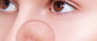
Melanoma-dangerous nevi
Phenotypically, these nevi do not reveal clinical signs of malignancy, but are distinguished by melanocyte dysplasia and a tendency to malignancy, thus giving reason to consider them as premalignant or borderline formations. These are the only tumors that require mandatory prophylactic removal with morphological verification of the pathological process. If it is multiple in nature, it is advisable to classify this group of patients as a risk group and subject them to mandatory clinical examination with dynamic monitoring by an oncologist.
Dysplastic nevus
Elements of the rash. A spot with a separate slightly raised area, usually in the center, above the skin level. The shape is round, oval or irregular with “ragged” edges. The boundaries are irregular and blurred.
Color. Various shades of black, brown, reddish, light red.
Localization. The most common are the torso, arms, buttocks, dorsum of the feet, and less commonly, the face.
Course and prognosis. A dysplastic nevus may not undergo any transformations throughout a person’s life, but may transform into a superficial melanoma; Complete regression of education is also possible. Against the background of a dysplastic nevus, melanoma develops in 9% of cases. It differs in clinical, histological, and biological manifestations from the known forms of melanoma, and is considered by some authors as “minimal melanoma.” The appearance of these nevi in young patients is not an alarming syndrome; they, as a rule, do not transform into melanoma.
With dysplastic nevus syndrome, the prognosis is unfavorable, and the risk of developing malignant melanoma increases significantly.
Treatment. Not all dysplastic nevi require immediate removal. Dynamic observation with photography and measurement of the size of the formation is necessary. Changing, suspicious, and traumatizing nevi are subject to excision.
Rice. 11. Dysplastic nevus
Localization Features
Mongoloid spots are also called coccygeal spots. The second name is associated with their frequent location. Often the pigment island in a newborn is located on the butt, in the tailbone area and on the buttocks, as well as on the lower back and hips. With extensive damage, pigmentation can occupy the entire back, including the neck and head of the child. Often the site of localization is the perineum and lower extremities, especially the back of the legs. Less commonly, islands are found on the arms, abdomen, and face.