Why is HPV genotyping needed?
HPV is a heterogeneous virus.
This means that there are many forms of it.
In fact, this is a whole family of viruses, varying in their level of pathogenicity.
When infected with different types of HPV, the risk of cancer increases to varying degrees and different symptoms occur.
About 40 types of HPV are transmitted sexually.
Fourteen of them are viruses of high oncogenic risk.
Thus, genotyping is used to find out what type of virus persists in the patient.
This is important for assessing oncogenic risk.
Patients with different types of HPV are managed completely differently.
If with low-oncogenic viruses a person can have warts removed and sent home, then detection of highly oncogenic types requires dynamic monitoring.
The role of drug therapy is increasing.
It is important for a person to get rid of carriers of the virus as soon as possible in order to prevent precancerous processes and cancer.
PCR diagnostics of papillomaviruses KVANT 21
Detection of the causative agent of papillomavirus infection (Human Papillomavirus), during which the DNA of viruses in a sample of biomaterial is examined using the polymerase chain reaction method in real time.
In this case, the presence and quantity of HPV is determined:
- low oncogenic risk (types 6, 11, 44);
- high oncogenic risk (16, 18, 26, 31, 33, 35, 39, 45, 51, 52, 53, 56, 58, 59, 66, 68, 73, 82 types).
Research method
Polymerase chain reaction (PCR).
Result format:
HPV type; HPV number: Lg copies per 100,000 epithelial cells, Lg copies/sample.
What biomaterial can be used for research?
Urogenital smear, urogenital smear (with prostate secretions), rectal smear, oropharyngeal smear.
How to properly prepare for research?
- Women are advised to take a urogenital smear or urine test before menstruation or 2 days after it ends.
- Men should not urinate for 3 hours before submitting a urogenital smear or urine test.
General information about the study
Human papillomaviruses (HPV) are widespread, infect the epithelium of the skin and mucous membranes and have oncogenic potential. HPV is transmitted through close contact with infected epithelium, so the main routes of infection are sexual and household contact. It is possible to transmit HPV from an infected mother to her fetus. A person can be infected with several types of HPV.
Factors that provoke the development of HPV infection include: early onset of sexual activity, a large number of sexual partners, reduced immunity, the use of oral contraceptives, vitamin deficiencies, sexually transmitted infections, smoking and living in large cities.
The incubation period can last from 2 months to 2-10 years. A latent course of the disease is characteristic, in which there are no clinical manifestations, and colposcopic, cytological and histological examination reveals the norm. In 30% of cases, the virus can be cleared within 6-12 months. Diagnosis of latent HPV infection is carried out only by PCR.
During acute infection, benign formations occur, such as papillomas, warts, and condylomas. In children, papilloma viruses can lead to laryngeal papillomatosis. Damage to trophoblast cells by papilloma viruses is fraught with spontaneous abortion.
The combination of papillomavirus DNA with the cell gene leads to dysplasia/neoplasia (most often in the transition zone of the cervix).
What is the research used for?
- To detect papillomavirus infection of high oncogenic risk.
- To confirm/exclude persistence of a certain type of HPV.
- To assess the degree of carcinogenic risk in women with cervical epithelial dysplasia.
What do the results mean?
Reference values: negative.
Reasons for a positive result
- Infection with papilloma virus.
Reasons for a negative result
- Absence of papillomavirus infection.
- Low levels of HPV in the blood.
Detection of HPV DNA in the test material indicates the presence of papillomavirus infection. If HPV DNA is not detected in the biomaterial under study, then the presence of papillomavirus infection is unlikely.
- from 3 Lg to 5 Lg (HPV/10 5 cells) - clinically significant viral load, dysplasia cannot be excluded, there is a risk of developing dysplasia
- > 5 Lg (HPV/105 cells) - clinically highly significant viral load, high probability of dysplasia
Do HPVs cause cancer in everyone?
Of course, viruses do not cause cancer in every person.
Even those that have a high oncogenic risk.
In 90% of cases, a person becomes infected and recovers on his own within 1-2 years.
Often he has no symptoms at all.
In other cases, condylomas or changes in the epithelium appear.
But pathological foci disappear after the virus is eliminated.
However, in 10% of cases, a chronic persistent infection develops.
She is the one who poses the greatest threat.
HPV most often causes cervical cancer in women.
Other types of malignant tumors, including among men, are very rare.
These include neoplasms:
- skin;
- member;
- anus;
- rectum;
- larynx;
- esophagus;
- Bladder.
Observation is required primarily for women.
In them, the virus causes cervical cancer, and this is the most common cause of death from cancer among patients of reproductive age.
Most often during this period only breast cancer occurs.
But they die from it less often.
The insidiousness of the disease is that it is detected late, and the effectiveness of treatment is low already from stage 2.
However, cervical cancer is common only in countries with poor medical standards.
This is a completely controllable disease.
Cancer does not develop out of the blue.
There are many ways to prevent it.
To do this, it is possible to identify HPV in time, carry out genotyping, and establish surveillance if a virus of high oncogenic risk is detected.
Already at this stage, drug treatment often allows you to get rid of the infection and prevent precancerous processes.
If precancerous diseases are identified, they can be cured.
And for all this, a woman has not even years – decades.
But if she never goes to the gynecologist and does not get tested, then cervical cancer appears and develops.
Although this happens slowly, there is no chance of identifying the disease in time.
Because there are no symptoms until the moment when the pathology becomes incurable.
Human papillomavirus infection
HPV is a group of common and genetically diverse viruses that infect and damage the epithelium of the skin (skin types of HPV) and the mucous membranes of the oral cavity and anogenital area (genital types of HPV). Genital types of HPV are transmitted primarily through sexual contact and through the birth canal from mother to child. The main clinical forms of human papillomavirus infection of the genitals are genital warts, as well as malignant forms of changes in epithelial cells, leading to cervical cancer.
The outcome of the infection depends on the type of virus. Low carcinogenic risk (LCR, low-tumorigenic) HPV types are associated with genital warts and mild dysplasias. HPV types of high carcinogenic risk (HCR, highly oncogenic) - 16, 18, 31, 33, 35, 39, 45, 51, 52, 56, 58, 59, 66, 68 - along with genital warts and mild dysplasia, also induce malignant transformation of the epithelium leading to invasive cancer.
Differential diagnosis.
Lesions of the cervix, vagina, vulva, penis, anus of non-papillomavirus etiology; in the case of genital warts - with manifestations associated with the herpes simplex virus, as well as with true papillomas.
Laboratory diagnosis of human papillomavirus infection includes
detection and quantification of HCR HPV DNA, HCR HPV genotyping, detection of NCR HPV DNA.
Material for research
- Scrapings of the epithelium of the cervical canal, vagina, vulva, anus - for detection, quantification and genotyping of HPV;
- smears from the oral cavity and larynx - detection of HPV NKR in children.
Detection and determination of the concentration of human papillomavirus of high carcinogenic risk
Indications for examination
- Determination of the risk group for the development of cervical cancer and anal cancer;
- screening programs with cytological examination simultaneously or at the first stage of screening (before cytological examination) for women over 30 years of age;
- resolution of uncertain and questionable results of cytological studies (presence of ASCUS - atypical flat cells of unknown significance);
- monitoring the effectiveness of therapy for severe dysplasia (CIN2+) 6 months after removal of the affected epithelium;
- carrying out differential diagnosis with diseases of non-papillomavirus etiology.
Comparative characteristics of laboratory diagnostic methods.
To detect HCV HPV DNA, NAAT (PCR, NASBA, TMA, etc.) and signal amplification methods (hybrid capture) are used. The virus cannot be cultured.
Hybrid capture is the first and best characterized method for detecting HPV DNA. The use of the method makes it possible to determine the concentration of the virus in arbitrary units of optical density; the linear range is small (3–4 orders of magnitude). Samples containing virus at concentrations below the threshold for clinical significance are counted as negative. The clinical significance limit is determined by testing a reference sample (HPV DNA concentration 1 pg/ml). All signals whose values are lower than the reference value are considered negative. Methods based on ANC are more modern, faster and easier to use. NAATs operating in a non-quantitative format detect either viral DNA (PCR, HDA, etc.) or mRNA transcripts of HPV oncogenes E6 and E7 (NASBA, TMA). To detect only clinically significant concentrations (according to different accounting methods - more than 1 pg/ml or 1000 copies of viral DNA per 100,000 human cells), methods with sensitivity are used, in which samples with low concentrations are counted as negative. It is most advisable to use real-time PCR, which allows you to accurately determine the concentration of viral DNA. Based on the data obtained, not only the concentration of the virus is assessed, but, importantly, the result is graded according to the clinical significance of the obtained concentration by:
- positive – above the threshold of clinical significance;
- positive – insignificant (below the significance threshold);
- negative.
Different sets of reagents may differ in the spectrum of detected genotypes. The HCR HPV group includes 16, 18, 31, 33, 35, 39, 45, 51, 52, 56, 58, 59, 68 genotypes. Types 16 and 18 are considered the most carcinogenic, the least – 51, 56, 68. For the Russian Federation and CIS countries, the clinical meaning of the study is retained only if at least 10 genotypes from those listed above are determined. Identification of low or unknown risk genotypes in a mixture significantly reduces the specificity of identifying a risk group for cancer pathology and cannot be justified.
Indications for the use of various laboratory tests.
To conduct a screening examination for the presence of precancer and cervical cancer, it is preferable to use smears/scrapings of the epithelium of the cervical canal and transformation zone, performed with cervical cytological brushes. It is permissible to use vaginal smears for these purposes. For cervical cancer screening, all tests, regardless of the technology chosen, must have balanced analytical and diagnostic performance that meets the following conditions: at least 98% sensitivity for CIN2+ and at least 90% specificity relative to a widely validated screening test (e.g. Hybrid Capture II). To achieve optimal analytical sensitivity, the approach of cutting off samples with HPV DNA concentrations below the threshold of clinical significance is chosen. To do this, either an artificial reduction in analytical sensitivity is carried out, or a control sample with the required threshold concentration is selected as a reference. The preferred method is to measure HPV DNA concentration by real-time PCR.
Features of interpretation of laboratory research results.
Detection of HPV DNA in any concentration indicates the presence of the pathogen, however, when interpreting test results, it should be taken into account that more than 80% of infected people are spontaneously cured within 12–24 months. The danger is represented by a persistent form of infection (lasting more than 12–24 months). A single positive result reliably indicates membership in a risk group for developing cervical cancer if the study is carried out in a group of women over 30 years of age or who entered into sexual activity more than 6-7 years ago. Particular caution should be exercised in relation to patients in whom persistence of the virus is confirmed when retested after 1 year. When screening using quantitative tests, only the clinically significant result is taken into account. For differential diagnosis and monitoring of patients after treatment, the additional value of a single quantitative determination is not shown. A promising way to predict the risk of relapse of CIN2+ is to assess the dynamics of the viral load after treatment (decrease or increase in virus concentration).
Genotyping of human papillomaviruses.
The purpose of HPV genotyping is to distinguish persistent infection from cases of re-infection (preservation of the genotype in repeated tests); clarification of patient management tactics depending on the oncogenicity of the identified types of virus.
Indications for examination.
Patients with detected HCV infection
Comparative characteristics of laboratory diagnostic methods.
NAAT is used for HPV genotyping. Techniques involving the detection of amplification products by electrophoresis (genotyping based on the size of the amplified fragment), hybridization on plates and strip strips, and real-time PCR have become widespread.
Classical methods using the principle of hybridization with specific probes make it possible to determine the widest range of genotypes (20–40). However, the use of universal primers for amplification leads to significant differences in the analytical sensitivity of detecting different HPV genotypes, leading to noticeable distortions in the results. In addition, the wide range of detectable genotypes is only necessary for scientific research. In clinical practice, HCR HPV genotyping (10–13 types) may be considered sufficient. Methods based on the use of type-specific oligonucleotides allow typing of a spectrum of clinically significant genotypes and are significantly free from the problem of differences in analytical sensitivity in relation to different HPV genotypes. Preference should be given to multiplex methods using real-time PCR as the fastest, contamination-safe, and with the possibility of automation. Abbreviated genotyping can be performed to identify only the most oncogenic HPV types 16 and 18.
Features of interpretation of laboratory research results.
To establish the persistence of the virus, a repeated genotyping approach is used after 12 months. Persistence is confirmed when the genotype/genotypes are preserved during repeated testing; a complete change in the spectrum of genotypes indicates cure and re-infection with alternative HPVs. Identification of the most oncogenic HPV types (16 and 18) may indicate the need for more aggressive patient management tactics (more frequent follow-up visits, more thorough examination to identify precancerous changes, etc.).
Detection of human papillomaviruses of low carcinogenic risk
Indications for examination
- Determining the risk group for developing laryngeal papillomatosis in a child (pregnant women and newborns);
- differential diagnosis with diseases of non-papillomavirus etiology.
Comparative characteristics of laboratory diagnostic methods.
To detect NCR HPV DNA, NAAT (PCR, NASBA, TMA, etc.) and signal amplification methods (hybrid capture) are used. Preference should be given to fast and contamination-safe methods, such as real-time PCR.
Clinically justified is the determination of two genotypes 6 and 11 (more than 90% of cases of laryngeal papillomatosis in children). The identification of genotypes 42, 43 and 44 has some additional value. Quantification is not justified.
Features of interpretation of laboratory research results.
Identification of 6 and/or 11 genotypes indicates a possible risk of developing laryngeal papillomatosis in a child (the risk increases if the virus is detected in the larynx). When planning pregnancy, expectant management may be recommended. If there is a concomitant presence of genital warts, they are treated by chemical and/or physical destruction. If there is pregnancy, its termination or delivery by cesarean section is not justified, since the risk of developing laryngeal papillomatosis is lower than the risk of possible complications of these operations.
HPV genotyping: what does this analysis provide?
This is the process of determining the type of virus.
It is based on determining a section of DNA, that is, the genotype of the pathogen.
Research objectives:
- determination of oncogenic types of virus;
- establishing the degree of oncogenic risk for a particular patient (the virus is determined in combination with other research methods);
- determination of therapeutic tactics.
As a result of the test, the doctor receives the following information:
- whether the virus that persists in the human body is highly oncogenic;
- what type of virus it is (even within the group of oncogenic HPVs there are viruses that, to a greater or lesser extent, increase the likelihood of cancer);
- in what concentration it is contained (if there are few viruses, it is likely that the body will soon get rid of them on its own).
HPV 16 and 18 analysis
Indications for the study
- Determining the risk group for developing cervical and anal cancer
- Cervical screening for women over 30 years of age
- Examination for questionable oncocytology results
- Monitoring the effectiveness of therapy for severe dysplasia
- Preventive diagnosis of HPV when changing partners, contact with carriers, practice of anal sex, etc.
Testing for HPV type 16 in the clinic is possible for men, women, virgin girls and teenage girls. Next, we will tell you in more detail about the main types of this research.
Places where analysis was taken:
- vulva,
- vagina,
- Cervix,
- anus,
- rectum,
- urethra (m+f),
- oro-nasopharynx (m+f),
- foreskin (m).
Qualitative analysis of HPV 16/18
A qualitative type of study allows you to determine only the presence of a specific strain of the virus in the body. The transcript for HPV 16 and HPV 18 in women will contain one of the words: “Detected” or “Not Detected.” For example, HPV 16 18 is positive (+) if viral DNA fragments were detected in the biomaterial, or HPV 16 18 is negative (-) if none were detected. Those women and men who are found to have oncogenic types 16 and 18 in a human papillomavirus test need to do additional clarifying tests to find out in what concentration (quantity) they are present.
Example of decoding HPV 16/18 (qualitative type of analysis)
| Index | The result is normal | Unit change |
| HPV DNA types 16 and 18 (qualitative research) | Not detected |
Quantitative HPV 16/18
The study allows you to determine the concentration in the body of a man or woman (viral load) of HPV type 16/18. The detection of a minimal amount of human papillomavirus DNA in itself indicates good immunity. But we cannot exclude the fact that the infection could have occurred quite recently (in this case, the result of a quantitative analysis for HPV 16/18 DNA will be doubtful). Therefore, after a while it is worth taking a second test.
Example of decoding HPV 16/18 (quantitative analysis)
| Indicators | The result is normal | Unit change |
| HPV type 16 DNA (quantitative study) | <3.0 (insignificant quantity) | lg DNA per 10^5 cells |
| HPV type 18 DNA (quantitative study) | <3.0 (insignificant quantity) | lg DNA per 10^5 cells |
Advanced analysis of HPV 16, 18 (DNA and count)
A test that determines the quantity and separate detection of 16 and 18 types of the virus allows you to identify the total number of high-oncology risk HPV (type 16, 18, 31, 33, 35, 39, 45, 51, 52, 56, 58, 59, 68) + number of HPV 16 and 18 separately.
In accordance with the international requirement for HPV tests intended for cervical screening, they must detect only types of high oncogenic risk. According to the International Agency Against Cancer (IARC), these include no more than 14 types: 16, 18, 31, 33, 35, 39, 45, 51, 52, 56, 58, 59, 66, 68. Among these types, it is important to identify the most carcinogenic types 16 and 18, since they more often and faster lead to the development of precancerous pathology of the cervix (cervical intraepithelial neoplasia - CIN). HPV types 16 and 18 together account for more than 70% of cervical cancer cases. Modern HPV tests based on PCR with real-time detection (Real-time PCR) can detect types 16 and 18 of the virus quite accurately.
Decoding HPV 16, 18 - result “Not detected”
| Index | Result | Norm | Unit change |
| Number of cells in sample | >500 | >500 | |
| HPV test (quantity and separate detection of types 16 and 18) | Ready | ||
| HPV DNA types 16, 18, 31, 33, 35, 39, 45, 51, 52, 56, 58, 59, 68 types | Not detected | Not detected |
Decoding HPV 16, 18 - result “Detected”
| Index | Result | Norm | Unit change |
| Number of cells in sample | >500 | >500 | |
| HPV test (quantity and separate detection of types 16 and 18) | Ready | ||
| HPV DNA 16, 18, 31, 33, 35, 39, 45, 51, 52, 56, 58, 59, 68 | Detection | ||
| HPV DNA/A9 (16, 31, 33, 35, 52, 58) | 4,9 | <3.0 | lg DNA per 10^5 cells |
| HPV DNA/A7 (18, 39, 45, 59, 68) | 0,0 | <3.0 | lg DNA per 10^5 cells |
| HPV DNA/A5/A6 (51, 56) | 0,0 | <3.0 | lg DNA per 10^5 cells |
| HPV 16 DNA | 6,0 | <3.0 | lg DNA per 10^5 cells |
| HPV 18 DNA | 0,0 | <3.0 | lg DNA per 10^5 cells |
Importance of the study
According to the guidelines for screening and prevention of cervical cancer adopted in the USA “American Cancer Society, American Society for Colposcopy and Cervical Pathology and American Society for Clinical Pathology Screening Guidelines for the Prevention and Early Detection of Cervical Cancer (2012)”, HPV- The test, together with a liquid PAP test, is recognized as the preferred method of screening for cervical pathology in women after 27-30 years of age.
For everyone, in our medical clinic on Kutuzovsky Prospekt you can take any tests for HPV, including types 16 and 18. Prices for services are presented in the table below.
HPV HCR genotyping
HCR stands for high carcinogenic risk.
Thus, this study differs in that only highly oncogenic viruses are detected.
For example, you will not receive a conclusion: 6, 11 or another type with low oncogenicity has been detected.
Testing may not be carried out on them.
Situations when typing specifically for BCR viruses is required:
- reduced immunity (including due to HIV);
- “bad” results of oncocytology analysis;
- clinical signs of cervical dysplasia.
That is, the analysis is done when the risk of cancer in a particular patient is assessed as high.
It may not matter whether other viruses are present among those that almost never cause cancer.
general information
These pathogens are transmitted through sexual contact (vaginal, anal, oral), through contact and household contact and cause damage to the mucous membranes of the vagina and cervix in women. The most harmless of these diseases are benign genital warts, the most dangerous is cervical cancer.
It is known that within 6-16 months, in 50-70% of infected women, the human papillomavirus leaves the body on its own, without any treatment, without causing disease (so-called spontaneous elimination). Only a few percent of women with chronic (persistent) HPV infection type 16 or 18 may develop a malignant process in the cervix after 10-15-20 years.
Oncology can only be caused by DNA types of human papillomavirus that belong to the group of high carcinogenic risk. Most often, HPV types 16 and 18 can lead to CIN grades 1, 2, 3 and cancer.
Scientists, after conducting a series of studies, came to the conclusion that the culprit in the development of invasive adenocarcinomas (cervical and rectal cancer) in most cases is still HPV-18, and in cases where both strains of the virus are detected, type 18 contributes to rapid progression of the disease. In the case of non-invasive types of glandular cancer, including the penis in men, the leading role is played by HPV-16.
HPV 16 HPV 18
According to the recommendations of gynecologists, a complete test for the virus (Digen test for oncogenic types of HPV) must be performed regularly - once a year. And as an initial screening test, a simple high-quality PCR test is suitable, which determines the DNA of all types of HPV of high carcinogenic risk, and one of them is tests for DNA of HPV 16 and HPV 18 types in women of all ages and men.
HPV genotyping: quantitative analysis
It is not enough for a doctor to know only the type of virus.
Information about its quantity is also needed.
The unit of measurement is the amount of DNA in 100 thousand epithelial cells.
There should be less than 10 to the 5th power.
If this threshold is reached or exceeded, this concentration of virus is considered clinically significant.
Oncogenic viruses in such quantities provoke intraepithelial neoplasia.
And then cancer develops against its background.
Why does a doctor need to know the amount of virus:
- to understand whether he is dangerous;
- give a prognosis for the further development of the infection;
- evaluate the effectiveness of treatment;
- plan further management of the patient (including the observation method, when periodic examinations and tests are carried out).
The analysis is carried out for 10-14 highly oncogenic types.
To determine quantitative indicators, real-time PCR is performed.
It makes it possible to differentiate clinically significant concentrations of the virus from resolving or transient forms of infection.
Human Papillomavirus, DNA quantitative [real-time PCR]
A molecular genetic study that detects the DNA of the human papillomavirus and quantifies the viral load in the material.
The quantity of only HPV of high carcinogenic risk (16, 18, 31, 33, 35, 39, 45, 51, 52, 56, 58, 59 types) is determined - without specifying the type.
Synonyms Russian
Human papillomavirus (HPV), human papilloma virus.
English synonyms
Human Papillomavirus, DNA, Quantitative; HPV, Viral Load.
Research method
Real-time polymerase chain reaction.
What biomaterial can be used for research?
Urogenital smear (with prostate secretions), rectal smear.
How to properly prepare for research?
- Women are recommended to take a urogenital smear or urine before menstruation or 2 days after its end.
- Men should not urinate for 3 hours before submitting a urogenital smear or urine test.
General information about the study
Human papillomavirus is a DNA virus from the papovavirus family, associated with the development of condylomas, warts, precancerous changes in the anogenital area, and cervical cancer. There are more than 100 types of HPV, more than 30 of which can infect the genital tract, and about 14 genotypes are associated with the development of cancer of the cervix, rectum, penis and certain other tumors (for example, oropharyngeal carcinoma).
Oncogenic papilloma viruses contain E6/E7 proteins in their DNA, which are capable of suppressing the processes of apoptosis (programmed death) in cells with altered genetic material. Genotypes 1, 2, 3, 5 are considered non-oncogenic, and genotypes 6, 11, 42, 43, 44 are considered papillomaviruses of low oncogenic risk. High oncogenic risk HPVs include genotypes 16, 18, 31, 33, 35, 39, 45, 51, 52, 56, 58, 59 and 68.
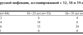
The main route of spread of infection is sexual, including oral-genital and anal contact, vertical (from mother to child) and contact-household transmission of infection is also possible. Several types of HPV can enter the human body at the same time. Infection usually occurs after the onset of sexual activity between the ages of 16 and 25. When infected with oncogenic genotypes of the virus between 25 and 35 years of age, intraepithelial lesions and, as a consequence, cancer after a few years are likely. In 70% of cases during the first year and in 90% of cases 2 years after infection, self-healing is possible.
Cervical cancer (CC) ranks third in prevalence among all malignant tumors in women (after breast cancer and colon cancer). The incidence of invasive cervical cancer in the world is 15-25 per 100,000 women. Cervical neoplasms occur mainly in middle-aged women (35-55 years), are rarely diagnosed in women under 20 years of age, and in 20% of cases are detected over the age of 65 years. The 5-year survival rate for localized (local, in situ) cervical cancer is 88%, while the survival rate for advanced cancer does not exceed 13%. In addition to infection with oncogenic HPV genotypes, the risk of developing cervical cancer is increased by smoking, chlamydial or herpes infection, chronic inflammatory gynecological diseases, long-term use of contraceptives, multiple births, cases of cervical cancer in the family, early onset of sexual activity, frequent change of sexual partners, insufficient dietary intake of vitamins A and C, immunodeficiencies and HIV infection. Despite the fact that the human papillomavirus does not always lead to neoplasms, more than 93% of cases of cervical cancer are associated with it.
Polymerase chain reaction (PCR) detects human papillomavirus (HPV) DNA with high sensitivity and specificity in more than 90% of cancers and 75-85% of intraepithelial neoplasia with severe dysplasia. According to research, the quantitative content of the virus in the material correlates with the degree of neoplasia: the higher the viral load, the more pronounced the cytological changes in the epithelium. During the examination, it is necessary to take into account the genotype of the virus, the presence and degree of cytological and histological changes in tissues, an increase or decrease in the viral load several months (6 months or more) after the previous examination.
What is the research used for?
- To assess the risk of developing tumors associated with HPV (cervical cancer, rectal cancer, cancer of the anogenital area and other localizations).
- To monitor the effectiveness of treatment of precancerous diseases associated with HPV.
- To monitor papillomavirus infection and predict its course.
When is the study scheduled?
- When detecting cytological changes in a smear for atypia, in a Papanicolaou smear, in a histological specimen.
- For condylomas and other morphological changes in the genital tract.
- When screening for cervical cancer in women over 25-30 years of age (as an additional study).
- When monitoring people infected with HPV.
- In the treatment of cancer and precancerous conditions associated with HPV.
What do the results mean?
Reference values: negative.
The level of viral load is interpreted taking into account the results of a cytological examination of smears, histological changes in the biopsy specimen and the genotype of the virus, changes in its quantity over time.
- The amount of HPV DNA is not determined if there is no virus in the test sample or its minimal amount (below the detectable level) - the risk of developing a pathological process associated with HPV is minimal.
- A clinically insignificant concentration of the virus (less than 103 copies of HPV DNA per 105 cells) means a minimal risk of developing dysplasia, a transient course of the viral process.
- A clinically significant concentration of the virus (more than 103 copies of HPV DNA per 105 cells) is a chronic infection with a high risk of developing dysplasia and cervical cancer.
- More than 105 copies of HPV DNA per 105 cells with an established fact of persistent infection (HPV has been detected for more than 1 year) - an increased viral load, associated with an increased risk of severe dysplasia, is often found in CC.
- A 10-fold decrease in viral load over 6 months is a transient infection.
- An increase in viral load 6 or more months after treatment indicates the possibility of relapse.
What can influence the result?
An unreliable result can be obtained when:
- improper handling and storage of material;
- contamination of the test material.
Important Notes
- HPV infection does not always lead to cervical cancer.
- Simultaneous infection with several HPV genotypes is possible.
- The result of the analysis should be interpreted taking into account the conclusions of cytological and histological studies.
Also recommended
- Human Papillomavirus 16/18 (HPV 16/18), DNA [real-time PCR]
- Human Papillomavirus 6/11 (HPV 6/11), DNA [real-time PCR]
- Human Papillomavirus of high carcinogenic risk (types 16, 18, 31, 33, 35, 39, 45, 51, 52, 56, 58, 59), DNA without type determination [PCR]
- Human Papillomavirus of high carcinogenic risk (types 16, 18, 31, 33, 35, 39, 45, 51, 52, 56, 58, 59), DNA genotyping [real-time PCR]
- Cytological examination of smears (scrapings) from the surface of the cervix (external uterine pharynx) and cervical canal - Papanicolaou staining (Pap test)
- Cytological examination of smears (scrapings) from the surface of the cervix (external uterine pharynx) and cervical canal for atypia
- Squamous cell carcinoma antigen (SCCA)
Who orders the study?
Gynecologist, oncologist.
Literature
- Arbyn M. et al. (2010). “European Guidelines for Quality Assurance in Cervical Cancer Screening. Second Edition–Summary Document.” Annals of Oncology 21(3):448–458.
- Hsiu-Ting Tsai, Ching-Hu Wu, Hsiao-Ling Lai, et al. Association between Quantitative High-Risk Human Papillomavirus DNA Load and Cervical Intraepithelial Neoplasm Risk Cancer Epidemiol Biomarkers Prev 2005;14:2544-2549.
- Saslow D, Solomon D, Lawson HW, et al. American Cancer Society, American Society for Colposcopy and Cervical Pathology, and American Society for Clinical Pathology Screening Guidelines for the Prevention and Early Detection of Cervical Cancer. Am J Clin Pathol. 2012;137:516-542.
- "Genital HPV Infection - CDC Fact Sheet." Centers for Disease Control and Prevention (CDC). April 10, 2008. Retrieved November 13, 2009.
- Materials and recommendations of the Anticancer Society of Russia. Access mode: /%d0%b0%d1%83%d1%82%d0%be%d1%81%d0%be%d0%bc%d1%8b
Who is recommended for HPV genotyping?
In our country, typing is prescribed not for preventive purposes, as in the West, but for medical reasons.
Many women do not get screened.
Therefore, the incidence of cervical cancer and mortality from it in the country remains high.
Typically, women come to the doctor if they have warts on the genitals or have received abnormal results from a cytological examination of a cervical smear.
But most international organizations recommend testing for HPV even without any symptoms or preliminary research.
That is, carry it out to everyone without exception.
According to FIGO recommendations, HPV testing is carried out once every 5 years until the age of 65 years.
WHO recommends testing at least once for all women aged 30-49 years.
The need for HPV testing is also provided for by Russian Ministry of Health Order 572n dated November 1, 2012.
Direct genotyping (determining the exact type of HPV) is not mandatory.
But if possible, it is recommended to carry out typing for at least types 16 and 18 as the most dangerous among all existing ones.
The use of alternative studies is also permitted.
The Digene test is widely used.
It is based on the hybridization of DNA sections with RNA probes.
They are subsequently captured by antibodies with a fluorescent label.
If the results are positive, quantitative indicators are given.
There are also tests based on the detection of HPV RNA.
In particular, the Aptima test is capable of detecting 14 highly oncogenic types with partial genotyping.
Post-rest health check (HPV test, cervical cancer screening)
Vacation is a time for yourself, when you want to fall in love. Time for holiday romances... It doesn’t matter whether it will continue or not, today we will talk about the most important thing - women’s health, namely, HPV. Why do we remember human papillomaviruses?
Human papillomaviruses are widespread and cause various diseases and lesions of the skin, larynx, male and female genital organs. Most of them are harmless. The most insidious types of HPV are called highly oncogenic, that is, capable of causing cancer. There is a clear connection between certain types of the virus and cervical cancer. The source of infection is exclusively human, the predominant route of transmission is sexual. But a household mechanism is also possible, when infection occurs in saunas, swimming pools, and spa salons. According to recent data, up to 90% of the population is infected with HPV and continues to spread it.
Highly oncogenic HPV types include 16, 18, 31, 33, 35, 39, 45, 50, 51, 56, 58, 64, 68. Types 16 and 18 are considered the most dangerous.
However, HPV infection does not always lead to cancer. If, in addition to HPV, there are risk factors, the likelihood of starting a tumor process is much higher. These include chlamydial infection, having multiple sexual partners, chronic inflammation of the cervix, smoking, and stress. A healthy and strong immune system usually copes with HPV and the viruses disappear from the body after 6-15 months.
For the discovery of the connection between HPV and cervical cancer, the German scientist Harald zur Hausen received the Nobel Prize. The peculiarity of the course of human papillomavirus infection is that it has no obvious manifestations. The woman is not worried or alarmed by anything. And at this time, HPV penetrates into the deep layers and integrates into the DNA, changing the structure of the cell, its ability to fully develop and reproduce. Dysplasia, a precancerous condition, gradually forms. If the process is not stopped at an early stage, the cells will turn into tumor cells. Cervical cancer is very insidious and can spread and metastasize, which without treatment leads to mortality.
Now do you understand the importance of timely testing for HPV and cytology? And this needs to be done regularly, and when changing sexual partners - it is mandatory! Even if a man considers himself healthy.
In order to be examined correctly, it is important to remember several important points:
- If a quantitative HPV test is done, the doctor looks at the concentration of the virus. There are studies in which the lowest concentration of the virus is determined, and in the absence of other signs, treatment is not required, only observation.
- There are many tests for HPV, and it is wrong to compare the results of studies performed by different methods.
- Consult a gynecologist. To make a decision, it is necessary to take into account all the data: complaints, the nature of the discharge, which is visible in the mirrors and during colposcopy, the results of the HPV test and cytology, and other laboratory findings.
- Situations often arise when one type of HPV is first detected, and after some time another, or several, are detected. This situation is possible. HPV rarely works alone and penetrates deeply, “hiding” in cells.
- It is important to properly prepare for the study: exclude sexual intercourse, local procedures, colposcopy and transvaginal ultrasound 48 hours before; Do not be examined during menstruation.
To establish the correct diagnosis, HPV tests and cytology smears are performed; each test has a specific purpose.:
- HPV tests - find oncogenic types, provide the woman with observation and treatment to prevent dysplasia
- Cytological examination - do not miss dysplasia (cell changes)
It is not always the case that each individual study allows us to understand the full picture of what is happening.
- Simultaneous HPV testing and cytology from the same biomaterial is the best solution, allowing the doctor to develop competent treatment tactics. It becomes clear that the cell is changing under the influence of HPV.
Benefits of KDL laboratory tests
HPV test for high carcinogenic risk (ROCHE COBAS).
- Detects the concentration of the virus that requires monitoring and treatment (the response “detected” will be received). Lower levels, which are not clinically significant but worry and frighten women, are not detected.
- The highest risk HPV types 16 and 18 are assessed separately.
- Other types of viruses are statistically much less likely to cause cancer. They are presented as a summary answer.
Cervical Cancer Screening (BD ShurePath Liquid Cytology) with HPV Test (ROCHE COBAS4800)
- Performs two tasks simultaneously
- Liquid-based cytology is an advanced method for cell monitoring that is accurate and reliable. The biomaterial is taken in a special container, and not on glass; the analyzer performs a high-quality and thin smear, allowing you to examine each cell individually; if necessary, you can perform several strokes; the test is carried out in PAP format (Papanicolaou staining), which complies with international recommendations.
- In countries where this test is mandatory, the mortality rate from cervical cancer is low.
Getting tested for HPV after active recreation is a chance not to miss the virus and prevent precancerous diseases and cancer. Get examined in a timely manner with KDL and be healthy!
What to do if dangerous types of HPV are identified?
The risk of detecting dangerous types of HPV in an individual person is low.
In most people, no variant of this virus is detected at all.
If the patient is infected, then with a high probability it is a harmless type of HPV.
Low-oncogenic varieties are much more common.
If you are unlucky and have been diagnosed with highly oncogenic HPV, you need to look at its quantity.
If it is small, no treatment is required.
Most likely, your body has already begun to get rid of the virus, and soon it will no longer be detected.
It's worse if its concentration is high.
In this case, the following is necessary:
- additional tests;
- treatment;
- dynamic observation.
A smear is taken from the patient for oncocytology.
Most often, this study is carried out not after HPV genotyping, but simultaneously with this test.
Based on the results of these two tests, a decision can be made about the need for colposcopy.
It is needed to identify areas of altered epithelium (precancerous processes).
In turn, the results of colposcopy (examination of the cervix) may require a biopsy.
This is a procedure in which a piece of tissue is taken and sent for histological examination.
Based on its results, both a malignant tumor and a precancerous process of stages 1-3 can be diagnosed.
Stage 1 is reversible, while stages 2-3 require cauterization to prevent cancer.
But precancerous processes develop very slowly.
If antiviral treatment is carried out in time, the virus disappears from the body and the cervical epithelium is restored.
Patients must be monitored.
They are tested every six months to assess whether the concentration of the virus is increasing or decreasing.
To eliminate it, antiviral and immunomodulating drugs are used.
Is it possible to do HPV without genotyping?
Of course, such an analysis can be done.
It is also quite informative.
HPV genotyping costs are higher, and such costs are not always necessary.
In any case, as a result of the test, the type will be determined or not, the doctor will find out whether the HCV papillomavirus is present in the body.
If it is present, and if it is present in high concentrations, in any case the patient must:
- treatment;
- dynamic observation.
And this is necessary, regardless of the specific type of virus detected.