What is herpes zoster?
Herpes zoster, also called shingles, is an infectious disease caused by the chickenpox virus. This is the same virus that causes chickenpox. Herpes zoster occurs only in those people who have previously had chickenpox. When chickenpox goes away, the varicella zoster virus remains in the body as inactive. This means you may not feel symptoms, but the virus is still present in your body. When the varicella zoster virus becomes active again, it causes herpes zoster.
to come back to the beginning
Herpes zoster, symptoms and treatment of the disease
09.09.2021
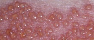
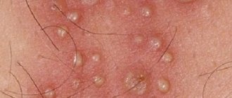
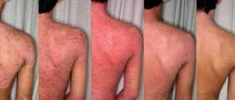
Herpes develops quickly and appears on the skin as small blisters. After the rash ends, separate scars form,
which are quite noticeable on the skin. Bubbles develop in groups, and the degree of appearance appears to be at one stage, parallel to
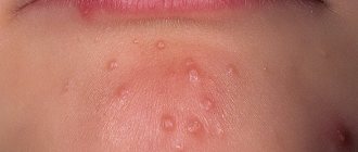
This other group of bubbles has its own special development. Where blisters with the disease form: head; body; face ; back .
Is herpes zoster contagious?
Cases of infection of an adult from a patient have not been recorded, but illnesses occur immune systems
chicken pox. Doctors noticed transplantation of the virus from mother to fetus, which caused its own complications, and the fetus died as a result.
During pregnancy , herpes medications will lead to death!
Yes, herpes is quite contagious and is transmitted by airborne droplets, and it can even occur in an adult if
weakened immunity .
In children, the disease usually occurs when certain circumstances coincide: infection enters the body; weakening of immunity and its transmission.
The source of infection usually becomes: older children with lichen or smallpox; adults with illness; carriers.
The disease occurs in children under 10 years of age, but is quite rare.
Is it possible to wash yourself when you are sick?
During the onset and exacerbation of the disease, washing is strictly prohibited; it is better to do this when the disease has completely receded and does not manifest itself.
symptoms. Procedures involving the entry of water should be limited to only those necessary, and not to taking a bath or shower.
Herpes zoster, symptoms and treatment of the disease
Before the rash appears, the patient tends to feel highly sensitive skin and severe pain. The origin of the disease begins
from the formation of small blisters that appear on slightly pinkish skin. The bubble formation time is often 2-3 days.
As a result, the pimples are opened, releasing a transparent liquid that dries and becomes crusty, after which the wound heals.
Symptoms that often appear include: increased body temperature; strong tooth in the blistering area.
How does the treatment work?
In patients with severe manifestations, they must be treated in hospital . Eliminating the disease at home is contraindicated.
In an outpatient setting, treatment consists of: eliminating symptoms; removal of pain syndrome; prevention of re-localization;
taking antiviral drugs. Herpes medications are used throughout the entire period of the disease until the rash appears.
will break through and no crusts will appear on them.
How to prevent the manifestation of the disease?
As you know, it is better to prevent the development of a disease than to cure it, this also applies to herpes zoster, since the disease is quite
serious and difficult to treat. To prevent the onset of the disease, you need to boost your immune system , drink vitamin complexes
or eat right. In addition, it is worth stabbing the body; how exactly to do this is up to the individual to decide. In adulthood you need
Walk more, exercise and take care of your health.
Published in Dermatology Premium Clinic
How is herpes zoster spread?
Herpes zoster can be spread by touching the blisters of an infected person. Disseminated herpes zoster can spread through contact with droplets from the nose and throat of an infected person. Droplets containing the virus become airborne when an infected person coughs or sneezes. They are easy to inhale and become infected.
If you have had chickenpox before, your virus will not become active as a result of contact with someone who has herpes zoster. However, if you have not had chickenpox, you may get it after being exposed to someone who has herpes zoster.
to come back to the beginning
Prodromal period
During which pain and parasthesia appear in the area of the affected dermatome (less commonly, itching, tingling, burning). The pain can be periodic or constant and accompanied by skin hyperesthesia. The pain syndrome can simulate pleurisy, myocardial infarction, duodenal ulcers, cholecystitis, renal or hepatic colic, appendicitis, intervertebral disc prolapse, early stage glaucoma, which can lead to difficulties in diagnosis and treatment. Pain in the prodromal period may be absent in patients under 30 years of age with normal immunity.
Stages of development of the rash with herpes zoster
| Erythematous-papular stage | |
| The disease begins with the appearance of grouped erythematous papules, spreading along 1-2 dermatomes. Herpes zoster rashes have a short erythematous phase (often completely absent), after which papules quickly appear. | |
| Vesicular stage | |
| Within 1-2 days, papules turn into vesicles, which continue to appear for 3-4 days. The elements tend to merge. If the period of appearance of new vesicles lasts more than 1 week, this indicates the possibility of the patient having an immunodeficiency state. | |
| Pustular stage | |
| Pustulation of vesicles begins a week or earlier after the appearance of the primary rash. | |
| Regression stage | |
| Then, after 3-5 days, erosions appear in place of the vesicles and pustules and crusts form. The crusts usually disappear by the end of the 3rd or 4th week of the disease. However, scales and hypo- or hyperpigmentation may remain after the herpes zoster rash resolves. | |
Localization of herpes zoster by dermatome
A feature of rashes with herpes zoster is the location and distribution of the elements of the rash, which are observed on one side and limited to the area of innervation of one sensory ganglion.
The areas of innervation of the trigeminal nerve, especially the ophthalmic branch, as well as the skin of the trunk T3-L2 segments are most often affected. Skin lesions in the chest area are observed in more than 50% of cases; Most rarely, the rash appears on the skin of the distal extremities. The rash may be located in the tailbone area. In this case, a picture of a neurogenic bladder develops with urinary disorders and urinary retention (due to the migration of the virus to neighboring autonomic nerves); may be associated with herpes zoster of the sacral dermatomes S2, S3 or S4.
General symptoms
The clinical picture of herpes zoster includes skin manifestations and neurological disorders. In 20% of patients, general phenomena are observed - fever, headache, poor health, increased fatigue, enlarged regional lymph nodes, changes in the cerebrospinal fluid (in the form of lymphocytosis and monocytosis). Pain often precedes skin changes and, with appropriate localization, can simulate myocardial infarction, appendicitis , renal colic, etc. The most common localization of pain is along the intercostal nerves.
Abortive (erased) herpes zoster
| It is characterized by the appearance of erythematous plaques and papules in the foci of hyperemia, but vesicles do not develop. |
Bullous herpes zoster
| Occurs when vesicles merge. Characterized by blisters, erosions and crusts with finely scalloped outlines. |
Hemorrhagic herpes zoster
| In the hemorrhagic form of the disease, the vesicular rashes have bloody contents, the process spreads deep into the dermis, and the crusts acquire a dark brown color. |
Gangrenous-necrotizing herpes zoster
| In some cases, the bottom of the vesicles becomes necrotic and a gangrenous form develops, characterized by the formation of grouped small scabs or a solid black scab with finely scalloped outlines. After healing, scars of appropriate size remain. The duration of the course of ordinary shingles is 2-4 weeks, gangrenous shingles is 2-3 months. A particularly severe course is characterized by gangrenous herpes zoster, which develops in the area of the branch of the 1st branch of the trigeminal nerve - on the skin of the forehead, eyelids, nose and temporal region. |
Generalized herpes zoster
| A number of patients (2-4%) experience a generalized form of herpes zoster: in addition to the usual lesion, a more or less widespread rash appears, consisting of vesicles resembling elements of chickenpox throughout the skin along with rashes along the nerve trunk. Recurrence of infection in the form of generalized rashes is usually not observed. In the presence of immune deficiency (including HIV infection), skin manifestations may appear far from the affected dermatome - a disseminated form. The likelihood of the appearance and severity of dissemination of skin rashes increases with the age of the patient. They are distinguished:
|
Herpes zoster of the oral mucosa
| Damage to the second and third branches of the trigeminal nerve, as well as other cranial nerves, can lead to the development of rashes on the oral mucosa (tongue, hard palate, gums), pharynx and larynx. |
Hyperkeratic herpes zoster
| A rare form of the disease, manifested in the form of continuous layers of horny masses or in the form of focally located hyperkeratic plaques, papules, nodules, sometimes resembling cutaneous horns and warts. The rashes persist for a long time within one or more adjacent dermatomes. Almost always observed in HIV-infected patients, but Cases are also rare in HIV-negative people. Also called verrucous herpes zoster. |
Ophthalmoherpes
| Ophthalmoherpes is a herpetic lesion of any branch of the optic nerve. Damage to the ophthalmic branch of the trigeminal nerve is observed in 10-15% of patients; the rash can be located on the skin from the level of the eye to the parietal region, abruptly interrupted along the midline of the forehead. Damage to the nasociliary branch, which innervates the eye, tip and lateral parts of the nose, leads to the penetration of the virus into the structures of the organ of vision. In this case, the cornea is often affected, leading to the occurrence of keratitis. In addition, other parts of the eyeball are affected with the development of episcleritis, iridocyclitis, and inflammation of the iris. The retina is rarely involved in the pathological process (in the form of hemorrhages, embolisms), the optic nerve is more often affected, which leads to optic neuritis with outcome in atrophy (possibly due to the transition of the meningeal process to the optic nerve). With herpes zoster affecting the eyes, the rash spreads from eye level to the crown, but does not cross the midline. |
Hutchinson's symptom
| Grouped vesicles are observed, localized on the wings or tip of the nose. Associated with the most serious complications of ophthalmoherpes and is a poor prognostic sign. |
Hunt syndrome
| Ganglionitis of the geniculate ganglion (geniculate ganglion of the facial nerve), caused by the herpes zoster virus and manifested by pain in the node, often radiating to the face, back of the head, neck, rashes in the external auditory canal, eardrum, sometimes on the tongue and palate, paralysis of the facial muscles and vestibular-cochlear nerve with dizziness, noise in the ear and hearing loss on the affected side. It is characterized by: pain in the area of the external auditory canal, rashes in the area of the external auditory canal, paralysis of the facial muscles, decreased taste in the anterior two-thirds of the tongue. The pain syndrome has a pronounced vegetative coloring in the form of burning, paroxysmal, sharp pains that intensify at night. Subsequently, the pain recurs and bothers the patient for many months and years, causing loss of ability to work, disrupting sleep, changing his mental and emotional status, forming a permanent syndrome - postherpetic neuralgia. The protracted, severe nature of the disease with a long-term, pronounced algic syndrome contributes to the formation of personality mental disorders. |
Herpes zoster in children
| In newborns whose mothers suffered a primary infection with the herpes zoster virus during pregnancy, intrauterine infection is possible. In these children, shingles develops without a previous history of chickenpox. Infantile shingles often leads to temporary paresis of the affected limbs. There are isolated reports of herpes zoster in young children. Risk factors for the occurrence in children include: maternal chickenpox during pregnancy or primary VZV infection in the 1st year of life. The risk of the disease is increased in children who have had chickenpox before the age of 1 year. Herpes zoster in children is not as severe as in older patients, with less pain; Postherpetic neuralgia also develops rarely. |
Herpes zoster in patients with HIV infection
The risk of developing the disease in patients with HIV infection is higher, and they are more likely to develop relapses of the disease. Additional symptoms may appear due to the involvement of motor nerves (in 5-15% of cases). The course of OH is longer, gangrenous and disseminated forms often develop (25-50%), while in 10% of patients in this category severe damage to internal organs is detected ( lungs, liver, brain). In HIV infection, frequent relapses of OH are observed within both one and several adjacent dermatomes.
Herpes zoster in pregnant women
The disease in pregnant women can be complicated by the development of pneumonia and encephalitis. VZV infection in the first trimester of pregnancy leads to primary placental insufficiency and, as a rule, is accompanied by termination of pregnancy. The presence of infection should serve as the basis for intensive prevention of the consequences of hemodynamic disorders (placental insufficiency, intrauterine hypoxia, intrauterine growth retardation).
Pain syndrome with herpes zoster
Pain is the main symptom of herpes zoster. It often precedes the development of a skin rash and is observed after the rash resolves (postherpetic neuralgia). Pain from herpes zoster and postherpetic neuralgia are caused by different mechanisms. In the early stages of the course, anatomical and functional changes are formed, leading to the development of neuralgia, which explains the connection between the severity of primary pain and the subsequent development of postherpetic neuralgia, as well as the reasons for the failure of antiviral therapy in its prevention.
The pain syndrome associated with herpes zoster has three phases: acute, subacute and chronic (postherpetic neuralgia). The acute phase of the pain syndrome occurs during the prodromal period and lasts for 30 days. The subacute phase of the pain syndrome follows the acute phase and lasts no more than 120 days. Pain lasting more than 120 days is defined as postherpetic neuralgia, which can last for months or years, causing physical suffering and significantly reducing the quality of life of patients.
The immediate causes of prodromal pain are subclinical reactivation and replication of VZV in neural tissue. Damage to peripheral nerves and neurons in the ganglia is a trigger for afferent pain signals. In a number of patients, the pain syndrome is accompanied by general systemic inflammatory manifestations: fever, malaise, myalgia, headache.
In most immunocompetent patients (60-90%), severe acute pain accompanies the appearance of a skin rash. A significant release of excitatory amino acids and neuropeptides caused by blockade of afferent impulses in the prodromal period and acute stage of OH can cause toxic damage and death of inhibitory interneurons of the dorsal horn of the spinal cord. The severity of acute pain syndrome increases with age. Excessive nociceptor activity and the generation of ectopic impulses can lead to an increase and prolongation of central responses to common stimuli - allodynia (pain and/or unpleasant sensation caused by stimuli that normally do not cause pain, such as the touch of clothing).
Predisposing factors to the development of ostherpetic neuralgia are: age over 50 years, female gender, the presence of a prodrome, massive skin rashes, localization of rashes in the area of innervation of the trigeminal nerve or brachial plexus, severe acute pain, the presence of immunodeficiency.
With postherpetic neuralgia, three types of pain can be distinguished:
- constant, deep, dull, pressing or burning pain;
- spontaneous, periodic, stabbing, shooting, similar to an electric shock;
- allodynia.
Pain syndrome is usually accompanied by sleep disturbances, loss of appetite and weight loss, chronic fatigue, depression, which leads to social maladaptation of patients.
Complications of herpes zoster
Complications of herpes zoster include: acute and chronic encephalitis, myelitis, retinitis, rapidly progressive herpetic necrosis of the retina, leading to blindness in 75-80% of cases, ophthalmic herpes with contralateral hemiparesis in the long term, as well as lesions of the gastrointestinal tract and cardiovascular systems, etc.
Wolf's isotopic response (phenomenon)
The development of a new skin disease directly at the site (anatomical area) of regressed herpes zoster. The mechanism is not fully understood, but it is assumed that herpes infection is a trigger for local immune regulation - an excessive or defective immune response. A new dermatosis can develop over varying periods of time from several days to several years. Described dermatoses with an isotopic response:
|
|
What precautions does the hospital take if I have herpes zoster?
Isolation measures are the measures we take to prevent the spread of infection among patients. If you were diagnosed with chickenpox during your hospital stay or were exposed to chickenpox:
- You will be placed in a separate room.
- The door to your room should always be closed.
- A sign will be posted on your door asking all staff and visitors to wash their hands with soap and water or use an alcohol-based hand rub before entering and after leaving your room.
- Isolation measures taken in the case of localized and disseminated herpes zoster are different. For localized herpes zoster, all visitors and staff should wear a yellow gown and gloves while in your room. They are distributed outside your room and can be disposed of in your room.
- For disseminated herpes zoster, visitors and staff should wear a yellow gown, gloves, and a face mask while in your room. While following these isolation measures, you are prohibited from walking around the unit.
- a food pantry in your department;
You can stop taking these precautions once all the blisters have dried out and crusted over.
to come back to the beginning
Shingles
Shingles (herpes zoster) is a common human disease that is characterized by general infectious symptoms, skin manifestations and neurological disorders of the central and peripheral nervous system.
The disease is caused by the Varicella zoster virus, which is also the causative agent of chickenpox. The virus contains DNA, being neurodermotropic, it affects the skin, cells of the central and peripheral nervous system. The virus is unstable in the external environment: it quickly dies when heated, under the influence of ultraviolet rays and disinfectants. It lasts for a long time at low temperatures.
Initially or after chickenpox, the virus penetrates through the skin and mucous membranes, then through the lymphogenous and hematogenous route into the intervertebral nodes and dorsal roots of the spinal cord, where it can persist for a long time in a latent state. With a decrease in immunological reactivity under the influence of various factors, such as exacerbation of chronic diseases, taking immunosuppressants, intoxication, latent infection, it can become more active. Herpes zoster is most severe in patients with cancer, HIV-infected people, as well as in people who have received corticosteroids or radiotherapy. Activation of the virus is accompanied by the development of ganglioneuritis with damage to the intervertebral ganglia or ganglia of the cranial nerves, as well as the dorsal roots (E. S. Belozerov, Yu. I. Bulankov, 2005). In severe cases, the anterior and posterior horns, white matter of the spinal cord, and brain may be involved in the process. The virus can also infect the autonomic ganglia, causing dysfunction of internal organs.
Pathomorphological changes in the brain with lesions of the central nervous system can be varied. In mild cases, changes occur only in the spinal cord and root ganglia; swelling is recorded in the brain. In severe cases, pronounced infiltration of the subarachnoid space, cerebral edema, hemorrhage in the white matter, basal ganglia and brain stem are noted.
The incubation period for herpes zoster can be several years from the moment of infection. In the clinical course, the main ones are: the prodromal period, the period of clinical manifestations and the period of residual effects. It all starts with an increase in temperature, a tingling sensation, burning, itching at the site of the rash, and a headache. Along the nerve trunks of the trunk, limbs or head, limited pink spots up to five centimeters in diameter appear. On the second day, bubbles 2–3 mm in diameter appear, filled with transparent contents. The number of lesions can vary from one to several, closely adjacent to each other, forming a continuous line. Over time, the contents of the bubbles become cloudy. At about 8–10 days, the blisters dry out and crusts form, which disappear after 3–4 weeks. In many patients, neurological manifestations can last for several months (up to a year).
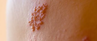
Typical clinical manifestations of herpes zoster are characterized by a certain sequence of skin rashes. The rashes are segmental, unilateral and do not spread to the other side of the body, unlike chickenpox.
Lesions of the nervous system with herpes zoster occupy first place among the complications of this disease. In the structure of neurological disorders, the leading place is occupied by lesions of the peripheral nervous system [10]. The most common disorders are neuralgia, neuropathy of the cranial and peripheral nerves, serous meningitis, etc. The most common manifestation is pain in the area of skin rashes. The pain is paroxysmal in nature, intensifying at night. In the future, the pain may intensify and bother you for several months and even years. Herpes zoster can also occur only with the symptoms of radicular pain, which is not preceded by a period of rash.
Most often, the rashes are located on the skin of the torso and limbs. The localization of pain and the appearance of a specific rash correspond to the affected nerves, most often intercostal nerves and are encircling in nature. The intensity of the pain increases with the slightest touch to the skin, with movement, or temperature changes. After the blisters disappear, the erosions epithelialize, and temporary red or red-brown pigmentation may remain on the skin. Some patients may have no pain. And sometimes herpes zoster can manifest itself only as neurological symptoms without the absence of skin manifestations.
Very often the localization of the disease is the skin of the face and head, especially the branches of the trigeminal nerve. Manifestations of the disease begin acutely, with general symptoms of intoxication and fever. Some patients may experience facial paralysis and trigeminal neuralgia lasting up to several weeks.
There may be manifestations of motor functions that occur not only when herpes zoster is localized in areas of the skin innervated by cranial nerves, but also when the cervical, thoracic and lumbar spinal cord, roots and ganglia are involved. Almost 5% of patients with rashes of various locations experience paresis of the upper and, more often, lower extremities, which indicate the phenomenon of focal myelitis.
To understand the pathogenesis of herpes zoster, data from pathological studies are important, indicating a connection between areas of the rash and damage to the corresponding ganglia. Head and Campbell (1900), based on pathohistological studies, came to the conclusion that both the neurological phenomena of herpes zoster and the areas of skin rashes that characterize them arise as a result of the development of a pathological process in the spinal nodes and their homologues (Gasserian node, etc.). But Volville (1924), having studied the nervous system of patients who died from a generalized form of herpes zoster, came to the conclusion that damage to the intervertebral ganglia in herpes zoster is not necessary. The inflammatory process often involves the spinal cord, and not only the posterior horns are affected, but also the anterior ones. Volville and Shubak (1924) described cases in which herpetic rashes were the first manifestations of a polyneurotic process occurring like Landry's paralysis. Volville believes that the inflammatory process first affected sensory neurons, and then spread to the spinal segments and peripheral nerves. In the case described by Shubak, a pathological examination revealed nests of inflammatory infiltration in the sciatic nerves, cervical sympathetic ganglia and the corresponding spinal ganglia, and the dorsal horns of the spinal cord.
Thus, the process involves not only the spinal and cerebral ganglia, which are most often affected, but also the substance of the spinal (anterior and especially posterior horns, white matter) and brain (oblongata, pons, hypothalamic region) brain, as well as the meninges.
Pathological and virological studies indicate that the herpes zoster virus widely disseminates throughout the body. During illness, it can be isolated from the contents of vesicles, saliva, tear fluid, etc. This gives reason to believe that herpetic eruptions can be caused not only by the settling of the virus in the sensory ganglia and damage to the parasympathetic effector cells located in them, but also by direct penetration it into the skin. Penetrating into the nervous system, it is localized not only within the peripheral sensory neuron (spinal ganglia, etc.), but also spreads to other parts of the central nervous system. When it is introduced into motor cells and roots, a picture of amyotrophic radiculoplexitis arises; in the gray matter of the spinal cord - myelitic syndrome; into the cerebrospinal fluid system - meningoradiculoneuritis or serous meningitis, etc.
The clinical picture of herpes zoster consists of skin manifestations and neurological disorders. Along with this, most patients experience general infectious symptoms: fever, enlargement of hormonal lymph nodes, changes (in the form of lymphocytosis and monocytosis) and cerebrospinal fluid. Typically, erythematous spots of round or irregular shape, raised, swollen, are found on the skin; when running a finger over them, a certain shagreen-like appearance of the skin is felt (tiny papules). Then, in these areas, groups of bubbles appear successively, often of varying sizes. The bubbles can merge with each other, but most often they are located in isolation, although close to one another - the vesicular form of herpes zoster. Sometimes they look like a small bubble surrounded on the periphery by a red rim. Since the rash occurs simultaneously, the elements of the rash are at the same stage of their development. However, the rash may appear in separate clusters over 1 to 2 weeks. In the latter case, when examining the patient, rashes can be detected at various stages of evolution. In typical cases, the bubbles at first have transparent contents, which soon turn cloudy, and then dry out in the form of a crust. A deviation from the described type is a milder abortive form of herpes zoster. With this form, papules also develop in the foci of hyperemia, which, however, do not transform into vesicles, which is how this form differs from the vesicular one. Another type is the hemorrhagic form of herpes zoster. The blisters have bloody contents, the process spreads deeply into the dermis, the crusts become dark brown. In severe cases, the bottom of the vesicles becomes necrotic - a gangrenous form of herpes zoster, after which scarring changes remain. The intensity of the rashes in this disease is very variable: from confluent forms, leaving almost no healthy skin on the affected side, to individual blisters, although in the latter case the pain can be sharply expressed. Such cases have given rise to the assumption that herpes zoster may exist without a skin rash.
Skin manifestations correspond to the level of damage to certain vegetative formations. By localization, lesions of the following ganglia are distinguished: gasserian, geniculate, cervical, thoracic, lumbosacral. One of the leading symptoms of the disease are neurological disorders, usually in the form of pain. Most often it occurs 1–2 days before the rash appears. The pain, as a rule, is intense, burning in nature, and the area of its distribution corresponds to the roots of the affected ganglion. It should be noted that the pain syndrome intensifies at night and under the influence of a variety of stimuli (cold, tactile, kinesthetic, barometric) and is often accompanied by vegetative-vascular dystonia of the hypertensive type. In addition, patients experience objective sensitivity disorders: hyperesthesia - the patient can hardly tolerate the touch of linen, hypoesthesia and anesthesia, and hyperalgesia may exist simultaneously with tactile anesthesia. Objective sensitivity disorders vary in form and intensity, they are usually limited to temporary sensitivity disorders in the area of rash or scars. Anesthesia affects all types of sensitivity, but in some cases a dissociated type of disorder is observed; sometimes within the same type of sensitivity, such as hot and cold. Occasionally, hyperesthesia takes on the character of irritation in the form of causalgia. Not in all cases, the intensity of the pain syndrome corresponds to the severity of skin manifestations. In some patients, despite the severe gangrenous form of the disease, the pain is minor and short-lived. In contrast, a number of patients experience prolonged intense pain with minimal skin manifestations.
Some patients in the acute phase have diffuse cephalgia, which intensifies with changes in head position, which may be associated with a meningeal reaction to herpes zoster infection. According to a number of authors [11, 12], herpetic ganglionitis of the Gasserian ganglion is more common than ganglionitis of the intervertebral ganglion. Most patients with this localization of the process experience increased temperature and swelling of the face on the affected side, as well as pain at the exit points of the trigeminal nerve.
The cornea is often affected in the form of keratitis of various types. In addition, other parts of the eyeball are affected - episcleritis, iridocyclitis, iris zoster. The retina is very rarely involved (hemorrhages, embolisms), more often the changes affect the optic nerve - optic neuritis resulting in atrophy, possibly due to the transition of the meningeal process to the optic nerve. With ophthalmoherpes (iritis), glaucoma may develop; Usually, with zoster, hypotension of the eyeball is observed, which is apparently caused by damage to the ciliary nerves. Complications of zoster from the motor nerves are quite common and are located in the following order: III, IV, VI nerves. Of the branches of the oculomotor nerve, both external and internal branches are affected. Ptosis is often observed. Skin rashes with ophthalmic zoster are often more severe than on other parts of the body, possibly depending on the structure of the skin in the eye area. Quite often, necrosis of the vesicles and severe neuralgia, accompanied by lacrimation, are observed. Bubbles appear not only on the skin, but also on the mucous membranes of the eye. As a result of the process in the cornea with ophthalmic zoster, optic nerve atrophy and complete blindness can develop. In addition, some patients experience loss of eyebrows and eyelashes on the affected side. The maxillary branches of the trigeminal nerve are affected both in the skin and in the mucous membranes (half of the hard and soft palate, velum, upper gum, inner surface of the buccal mucosa, while the nasal mucosa may remain unaffected). The branches supplying the mucous membranes may be more affected than the cutaneous branches, and vice versa. Lesions of the nerves of the upper and lower jaws do not always remain strictly localized, since pain sometimes radiates to the area of the ophthalmic and other branches.
Herpes zoster usually affects the autonomic nervous system. However, clinical observations have shown that the animal nervous system may also be involved in the pathological process. Evidence of this is that some patients, simultaneously with damage to the Gasserian ganglion, had peripheral paresis of the facial nerve on the side of the herpetic rash. With ophthalmic zoster, both the external and internal muscles of the eye are paralyzed. IV paralysis is rare. Oculomotor paralysis is more often partial than complete; More often than other muscles, m. is affected. levator palpebrae. There are cases of ophthalmic zoster with isolated changes in the shape and size of the pupil; unilateral Argil-Robertson (Guillén) sign. These paralysis sometimes partially or completely resolve spontaneously without special treatment.
Simultaneous damage to the facial, auditory and trigeminal nerves was first described by Frankl-Hochwart in 1895. Hunt (1907) described in detail the four clinical forms of this disease, which later became known as Hunt's syndrome, or herpes zoster oticus. The defeat of the geniculate ganglion in this form of herpes zoster was first pointed out by Nordahl (1969). Usually, herpetic rashes appear on or around the ear, and sometimes in the ear canal and even on the eardrum. There is sharp pain in the circumference of the auricle. Dysfunction of the facial, cochlear, and vestibular nerves occurs in the first days of the rash or precedes it. Pain in such cases is localized in the depths of the ear canal and the auricle with irradiation to the mastoid region, auricular and temporoparietal areas.
Objective sensitivity disorders are found behind the ear, in the fold between the auricle and the mastoid process. This skin area is supplied by the auricular branch of the X pair, which innervates the posterior walls of the auditory canal. Finally, in cases of very common ear zoster, the latter affects not only the external auditory canal, the auricle, the mastoid process, but also the eardrum, which is sometimes extremely seriously affected. In such cases, the area innervated by pairs V, VII and X is affected, and damage to these nerves is accompanied by damage to the ganglia, corresponding cranial nerves, or anastomoses connecting the terminal branches of all of the above nerves.
Often, simultaneously with paralysis of the VII pair, there is paralysis of the soft palate, anesthesia and paresthesia in the tongue, and often a taste disorder in the anterior two-thirds of the tongue due to the lesion. The defeat of the VIII pair usually begins with tinnitus, which sometimes lasts a long time after the disappearance of other phenomena. Hyperacusis with damage to the VIII pair is caused by paresis n. stapeblii, although this symptom can also occur with isolated and previous damage to the auditory nerve and in such cases represents a symptom of irritation. Hypoacusis can occur regardless of damage to the auditory nerve due to local lesions of the middle ear, eruption of bubbles on the eardrum, blockage of the external auditory canal, due to swelling of the mucous membrane due to eruption of zoster.
Vestibulatory phenomena, in contrast to cochlear phenomena, usually develop extremely slowly and are expressed differently: from mild subjective symptoms of dizziness to significant static disorders.
Neuralgia with auricular zoster, as opposed to ophthalmic zoster, is rare. Long-term results are not always favorable, as persistent paresis of the facial nerve and deafness may occur.
Volville emphasizes that the combination of paralysis of the VII and VIII pairs, although it occurs especially often with zoster, nevertheless the same combination occurs when the Gasserian node, II, III, cervical ganglia are affected, and, finally, all of the above areas can be affected simultaneously.
Zoster rashes are also described in the area of innervation of the IX pair; the posterior part of the soft palate, the arch, the posterolateral parts of the tongue, part of the posterior wall of the pharynx; In addition to IX, this same area is also innervated by branches of the X pair: the root of the tongue, larynx, epiglottis, basal and posterior parts of the pharyngeal wall. Although zoster predominantly and even selectively affects the sensory systems, movement disorders are sometimes observed with it, especially when the rash is localized in the head, neck, and limbs. Paralysis with zoster is radicular in nature, and damage to the posterior roots in these cases is accompanied by phenomena from the corresponding anterior roots.
Damage to the cervical sympathetic nodes is often accompanied by rashes on the skin of the neck and scalp. Pain in this case is observed not only in places of rashes, but also in the area of paravertebral points. Sometimes attacks that mimic facial sympathalgia may occur.
With ganglionitis of the lower cervical and upper thoracic localization, along with the usual symptoms of this disease, Steinbrocker syndrome can be observed. The dominant picture of this syndrome is pain of a sympathetic nature in the form of burning or pressure, occurring initially in the hand, and then in the entire arm. Soon swelling of the hand appears and quickly increases, spreading to the entire arm. Trophic disorders are added in the form of cyanosis and thinning of the skin, hyperhidrosis, and brittle nails. Movement of the fingers is limited and painful. Often pain and other autonomic disorders persist even after the rash disappears. Thoracic ganglionitis often simulates the clinical picture of myocardial infarction, which leads to diagnostic errors.
With herpetic lesions of the ganglia of the lumbosacral region, the rashes are most often localized on the skin of the lower back, buttocks and lower extremities; Along with pain in the areas of rashes, pain syndromes simulating pancreatitis, cholecystitis, renal colic, and appendicitis may occur. Herpetic lesions of the lumbosacral ganglia are sometimes accompanied by involvement of the animal nervous system in the process, giving a picture of ganglioradiculitis (radicular syndrome of Pori, Matskevich, Wasserman).
Sometimes, along with rashes along the nerve trunk, vesicular rashes appear throughout the skin - a generalized form of herpes zoster. Usually the disease does not recur. However, it is known from the literature that recurrent forms of the disease occur against the background of somatic complications: HIV infection, cancer, diabetes mellitus, lymphogranulomatosis, etc.
Treatment . When treating herpes zoster of varying localization and severity, early administration of antiviral drugs is necessary. It is known that the virus contains proteins that form its shell and carry enzymatic functions, as well as nucleic acid, the carrier of its genetic properties. Penetrating into cells, viruses are freed from their protective protein shell. It has been shown that at this moment it is possible to inhibit their reproduction using nucleases. These enzymes hydrolyze the nucleic acids of viruses without damaging the nucleic acids of the cell itself. It was found that pancreatic deoxyribonuclease sharply inhibits the synthesis of DNA-containing viruses, such as herpes virus, vaccinia, and adenoviruses. Considering the above, it is recommended that patients with herpes zoster be prescribed deoxyribonuclease intramuscularly 1-2 times a day, 30-50 mg for 7 days. In addition, in patients with rashes on the oral mucosa, conjunctiva and cornea, the drug is used topically in the form of an aqueous solution. The administration of deoxyribonuclease promotes rapid regression of skin rashes and a reduction in pain.
The drug Isoprinosine has a good effect in the treatment of herpes zoster. This is an immunostimulating agent with an antiviral effect. Isoprinosine blocks the reproduction of viral particles by damaging its genetic apparatus, stimulates the activity of macrophages, the proliferation of lymphocytes and the formation of cytokines. The second component increases the availability of Isoprinosine for lymphocytes. Reduces the clinical manifestations of viral diseases, accelerates convalescence, and increases the body's resistance.
Indications: viral infections in patients with normal and weakened immune systems (diseases caused by herpes simplex viruses types 1 and 2, Varicella zoster, including chickenpox, measles, mumps, cytomegalovirus (CMV), Epstein-Barr virus); viral bronchitis; acute and chronic viral hepatitis B and C; diseases caused by the human papillomavirus; subacute sclerosing panencephalitis. Chronic infectious diseases of the urinary and respiratory systems; prevention of infections in stressful situations; the period of convalescence in postoperative patients and persons who have suffered serious illnesses; immunodeficiency states. Isoprinosine is taken orally, for adults - 50 mg/kg/day in 3-4 doses; for children - 50–100 mg/kg/day in 3–4 doses. Duration of treatment is 5–10 days, in severe cases up to 15 days. For diseases caused by herpes simplex viruses types 1 and 2, treatment is continued until the symptoms of the disease disappear and for another two days. For subacute sclerosing panencephalitis for adults and children - 50–100 mg/kg/day in 6 divided doses. For acute viral encephalitis for adults and children - 100 mg/kg/day in 4–6 divided doses for 7–10 days. This is followed by a break for 8 days, then a repeat course for 7–10 days. If necessary, the dose and duration of the continuous course can be increased, subject to a mandatory break in taking the drug for 8 days. Long-term treatment is carried out under medical supervision. For genital warts in complex therapy with a CO2 laser - 50 mg/kg/day in 3 divided doses for 5 days, then with a 3-fold repetition of the specified course at intervals of one month.
In recent years, antiviral chemotherapy drugs from the group of synthetic acyclic nucleosides have been used to treat herpes zoster. The most well studied drug at present is acyclovir. The mechanism of action of acyclovir is based on the interaction of synthetic nucleosides with the replication enzymes of herpes viruses. Thymidine kinase of the herpes virus binds to acyclovir thousands of times faster than cellular ones, so the drug accumulates almost only in infected cells. This explains the complete absence of cytotoxic, teratogenic and mutagenic properties in acyclovir. The synthetic nucleoside is arranged in a chain of DNA being built for the “daughter” viral particles, and this process ends in this way: the reproduction of the virus stops. The daily dose of acyclovir for herpes zoster is 4 g, which should be divided into 5 single doses of 800 mg. The course of treatment is 7–10 days. The best therapeutic effect is achieved with early administration of the drug; The duration of rashes is reduced, crusts form quickly, intoxication and pain are reduced. Second generation acyclovir - valacyclovir, retaining all the positive aspects of acyclovir, due to increased bioavailability, allows you to reduce the dose to 3 g per day, and the number of doses - up to three times. The course of treatment is 7–10 days. Famciclovir has been used since 1994. The mechanism of action is the same as that of acyclovir. The high affinity of the virus thymidine kinase for famciclovir (100 times higher than the affinity for acyclovir) makes the drug more effective in the treatment of herpes zoster. The drug is prescribed 250 mg 3 times a day for 7 days.
Along with antiviral drugs, ganglion blockers, such as Gangleron, are used to reduce pain. Gangleron is used intramuscularly in the form of a 1.5% solution, 1 ml once a day for 10–12 days or 0.04 g in capsules 2 times a day for 10–15 days, depending on the severity of the pain syndrome. In addition, the use of carbamazepine gives good results, especially in cases of damage to the Gasserian node; the drug is prescribed with 0.1 g 2 times a day, increasing the dose by 0.1 g per day, if necessary, up to 0.6 g daily dose (in 3– 4 doses). After the pain decreases or disappears, the dose is gradually reduced. Usually the effect occurs 3-5 days after the start of treatment.
In cases of severe pain, analgesics and, in the form of injections, reflexology are prescribed. In reflexology, both points of general action and points corresponding to the affected ganglion are usually used. The course consists of 10–12 sessions. It is also recommended to prescribe multivitamins, in particular B vitamins. Local irrigation with interferon or ointments with interferon, aniline dyes, Eridin aerosol, Florenal, Helepin, Alpizarin ointments can be used. For gangrenous forms of herpes zoster, pastes and ointments containing an antibiotic, as well as Solcoseryl, are used.
Good results are obtained by irrigation with Epigen spray 4–5 times a day for 7–10 days from the first days of the disease. When combined with oral acyclovir therapy, a decrease in pain is noted.
After the skin rash resolves, treatment is carried out by neurologists until the neurological symptoms disappear.
Thus, treatment of herpes zoster should be comprehensive and include both etiological and pathogenetic agents.
Literature
- Batkaev E. A., Kitsak V. Ya., Korsunskaya I. M., Lipova E. V. Viral diseases of the skin and mucous membranes. Textbook manual, RMAPO. M.: Pulse, 2001.
- Butov Yu. S. Skin diseases and sexually transmitted infections.
- Kartamyshev A.I. Skin and venereal diseases. Medgiz, 1954.
- Skin and venereal diseases: Directory. Ed. O. L. Ivanova. M.: Medicine, 1997.
- Paltsev M. A., Potekaev N. N., Kazantseva I. A. et al. Clinical and morphological diagnosis of skin diseases (atlas). M.: Med., 2004.
- Pospelov A.I. A short textbook of skin diseases. M., 1907.
- Skripkin Yu. K., Kubanova A. A., Prokhorenkov V. I. et al. Dermatological syndromology. M. - Krasnoyarsk, 1998.
- Sukolin G.I. Clinical dermatology. St. Petersburg, 1997.
- Lezvinskaya E. M., Piven A. L. Laboratory diagnostics: skin diseases and sexually transmitted infections. M.: Practical Medicine, 2005
- Yushchuk N. D., Stepanchenko A. V., Dekonenko E. P. 2005.
- Kalamkaryan A. A., Kochetkov V. D. 1973.
- Zucker M. B. 1976.
I. M. Shakov, Candidate of Medical Sciences
GOU DPO RMAPO, Moscow
Contact information about the author for correspondence
gormed.su
Herpes zoster, or shingles, occurs against a background of weakened immunity as a result of activation of the Herpesviridae virus. Elderly people, as well as patients with congenital or acquired immunodeficiency, are most susceptible to infection. This disease is not uncommon among children. What typical and atypical symptoms are known to modern medicine?
What is herpes zoster
Several years may pass between the initial infection and the onset of symptoms, so the disease is characterized by a record incubation period. Shingles “moves” along the nerves, gradually reaching the skin, where an outwardly noticeable inflammatory process occurs. In the infected area, the pain intensifies and the lymph nodes become inflamed.
Typical manifestations
The disease usually begins with fever, headache, weakness, nausea or vomiting. Against the background of general malaise, the patient experiences itching, pain and paresthesia in the affected area - a sensitivity disorder. Swelling and erythema become noticeable after 3-5 days, and they are replaced by papules and blisters with serous contents. On days 7-10, the contents dry out and crusts form. Another development of events is that the bubbles burst and painful bright red erosions appear in their place.
Herpes zoster can affect the mucous membranes of the nose and mouth, conjunctiva, vagina, bladder and rectum. You cannot do without consulting a neurologist if the manifestations are accompanied by nerve damage, disorders of pain and tactile sensitivity, and paresis. In severe cases, the clinical picture includes damage to the heart, lungs and gastrointestinal tract.
Atypical forms of the disease include:
- Abortive. The rash appears only once, without repeated waves, and is not accompanied by unbearable itching and pain. After 3-4 days the disease subsides.
- Bullous. The rashes merge, forming one continuous bubble. The latter gradually dries out and leaves behind a scab in the form of a large spot.
- Hemorrhagic. The serous contents turn red-brown after 3-5 days.
- Gangrenous. It is characterized by severe and prolonged (more than three months) course, after which scars remain on the body.
What is the complication rate?
- If herpes zoster affects the trigeminal nerve, there is a 30-60% chance of developing conjunctivitis, keratitis and neuritis of the oculomotor and optic nerves. In this case, consulting a dermatologist is no longer enough: a comprehensive examination by a number of specialists is needed.
- Dislocation of skin inflammation in the area of the cervicothoracic segment in 40-50% of patients leads to acute myopathy and paresis of the arms.
- Lesions of the lumbosacral areas are accompanied in 10-15% of cases by leg paresis, urination disorders and intestinal obstruction.
- Sometimes the disease “reaches” the membranes of the brain, provoking the development of serous meningitis. Such forms occur without complications in only 20-25% of patients.
- In 0.1-1% of cases, encephalitis becomes a consequence of brain damage.
Most patients recover well, but in some, neurological symptoms persist for a long time. Therefore, it is better to entrust the treatment to specialists, especially if the development of the disease occurred against the background of a weakened immune system and it will be difficult for the body to cope on its own.
What is important to know about the causative agent of the disease and infection with it
Shingles is caused by the varicella zoster virus. It is also called herpes virus type 3. Initially, usually in childhood, this virus causes chickenpox (popularly called chickenpox), and only later, often years and even decades later, under certain conditions can lead to herpes zoster.
The photo below shows an example of chickenpox in a child:
When it enters an organism that has never encountered this infection before, the virus after some time penetrates the blood, with which it spreads throughout the body, primarily affecting skin cells. In these cells, viral particles replicate, the cells die, which is externally manifested in the appearance of multiple foci of inflammation - a rash of small itchy papules. In parallel, characteristic symptoms of intoxication of the body develop due to cell death, inflammatory and immune reactions.
Individual viral particles penetrate the processes of nerve cells and move along them to the nucleus of the neuron, located in the spinal cord. As a result, viral DNA penetrates into the nucleus of the affected neuron, which remains here until the end of the cell’s life. In fact, the virus remains in the body until the end of a person’s life.
Note: the chickenpox virus most often affects the processes of nerve cells innervating the sides of the body, although during the acute period of the disease, viral particles are also present in other parts of the body. Next we will see how this is related to the specifics of the rashes with herpes zoster.
As chickenpox develops, the body develops immunity to its pathogen and gradually destroys all viral particles located in the cells and intercellular space. The disease ends, but the DNA of the virus remains in the nuclei of the nerve cells of the spinal cord.
The infected cells will then continue to produce small amounts of virus particles throughout the person's life. These virions periodically leave nerve cells to infect surrounding tissues, but encounter antibodies, macrophages and other agents of the immune system in the intercellular space, which destroy them.
For humans, such activity of the virus is not noticeable, therefore the infection at this stage is called latent, hidden.
It is useful to consider the following nuances:
- The chickenpox virus is highly contagious (contagious). Its incidence among people without specific immunity (who have never encountered this particular virus before) is almost 100% - this is a unique indicator for viruses in general;
- When first infected with the Varicella zoster virus, only chickenpox develops;
- The older the infected person, the more severe the illness. Children suffer from chickenpox easily, but adults, on the contrary, are seriously ill with it, which, however, happens rarely, since most people get sick with this disease in childhood;
- If a person has had chickenpox once, then he will not get it a second time (thanks to lifelong immunity). The exception is severe immunodeficiency in the patient.
It is this specificity that determines why it is impossible to become infected with herpes zoster directly.
Ways to reduce the risk of infection
The already low risk of infection from a patient with herpes zoster can be further reduced if you follow basic infection safety rules:
- Do not contact the sick person. It is advisable not to be in the same room with him at all, and if necessary, physical contact should be avoided;
- Do not use the patient’s things (for example, a towel);
- Do not sit in the chair in which the patient was sitting, on the sofa or bed;
- It makes sense for the patient himself to cover the rash areas with clothing as much as possible.
At the same time, it is usually not worth worrying too much about protecting against such infection. For the vast majority of adults, a second encounter with the chickenpox virus is not scary, and for children it is sometimes even desirable. It is not for nothing that in France and Germany there is a widespread tradition of visiting friends or relatives who have chickenpox as a child - this guarantees that healthy children from the guest’s family will become infected, recover from the disease and be protected from this disease in adulthood, when it is much more severe.
To summarize: shingles is contagious, but mainly only for children, and chicken pox will develop when infected. And only then, after a very long period of time (usually decades), with a very low probability, herpes zoster can develop (if the immune system is weakened). If a person has previously had chickenpox and does not have an immunodeficiency, then he is not at risk of becoming infected from a person with herpes zoster.