Micropapillomatosis is a rather rare skin disease of the labia of women, the incidence of which is about five percent.
Micropapillomatosis in women is a dermatological disease that manifests itself in the form of multiple epithelial neoplasms of benign origin. In appearance, micropapillomatosis resembles a large accumulation of skin nodules.
The pathology in most cases does not cause serious discomfort or serious consequences, however, when diagnosed, treatment is recommended.
In addition, often in such cases there is a threat to the body associated with the immediate causes of tumors on the labia.
Description of the disease
Micropapillomatosis is a collection of rashes that form on the inner and outer areas of the labia minora of women. It practically does not occur on large lips, and is also not diagnosed in men.
There is also vestibular micropapillomatosis (micropapillomatosis of the vulva) located in the vestibule of the vagina. The rash is localized on the mucous membrane of the labia minora and may not be visible to the eye.
Most often, the occurrence of rashes does not cause serious discomfort, but the phenomenon is a cosmetic defect and in rare cases causes pain.
In addition, when such dermatological problems are detected, it is important to establish the cause of their occurrence and make a prognosis regarding the progression of micropapillomatosis and possible consequences.
Important! The disease has nothing to do with papillomavirus and is not transmitted through direct contact.
Malignant neoplasms
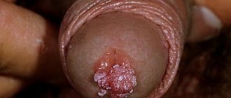
The external part of the female genital organs is susceptible to cancer. Young women are diagnosed with papilloma, which most often develops into a malignant neoplasm. Penetrating to the cellular level, the pathogen causes gene changes. This leads to abnormal protein release and accelerated cell proliferation.
Various factors for the development of vulvar cancer are known:
1. Age-related changes. More than half of women over 70 years of age have this diagnosis.
2. Bad habits. Smoking has a detrimental effect on blood vessels. Increases the risk of developing cancer.
3. HIV. The infection weakens the woman's immunodeficiency. The risk of contracting papillomavirus increases.
4. Vulvar neoplasia. Precancerous condition. An abnormal number of epithelial cells are found in the subcutaneous layers, then transform into cancer.
5. Cervical cancer. Oncology spreads to the vulva.
The appearance of malignant tumors is caused by the occurrence of adenoma-carcinomas. They are located in the thickness of the labia. Tumors are mistakenly confused with cysts. Adenocarcinoma can arise in the Bartholin glands and sweat gland cells of the vulva. Basal cell carcinoma sometimes occurs in this area. Melanomas arise from cells that produce melanin. It most often occurs in areas of the skin exposed to sunlight.
Vulvar cancer is divided into 4 stages:
1. The first stage of the tumor is caused by tumor growth within the vulva. It does not affect the lymph nodes and neighboring organs.
2. The second stage tumor grows into nearby organs. This can be the vagina, anus and urethra.
3. Neoplasms at the third stage affect the lymph nodes.
4. The last stage is characterized by damage to the lymph, vagina, bladder, and pelvic bones. Metastases penetrate to distant areas.
The stage of the tumor determines the further prognosis of the patient’s life activity and treatment. The growth of the tumor deeper causes manifestations; before this, no prerequisites may appear. The color of the skin differs significantly from healthy tissues; a pink or red tint predominates. A skin lump appears; it could be a wart, papilloma, or ulcer. The occurrence of pain, burning and itching.
Many women experience bloody discharge outside the menstrual cycle. These symptoms indicate the presence of a malignant neoplasm of the vulva. Adenocarcinoma is located deep in the labia majora and can be palpated, creating the sensation of a foreign body.
Types of micropapillomatosis
Rashes on a woman’s labia minora can occur after the incubation period or suddenly for no apparent reason.
Depending on the nature of the formation, the following types of micropapillomatosis are distinguished:
- Blisters (as a rule, do not require treatment and disappear on their own, do not cause discomfort);
- Plaques (rashes form in small clusters on the labia, the blisters are relatively small in size);
- Erythema (represents a small convex hyperemic area of skin, papule);
- Chancre (an abnormal red spot on the labia with an ulcerated nest and hard edges);
- Nodules (dense rashes forming in the layers of the dermis);
- Blisters (micropapillomatosis, forming singly or in groups, filled with fluid and eventually transforming into ulcers);
- Pustules (often occur as a complication of other types of rash and contain yellow or greenish purulent discharge);
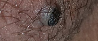
- Condylomas (brown or flesh-colored seals are an exception).
Each type of rash has its own nature, causes and possible consequences.
be careful
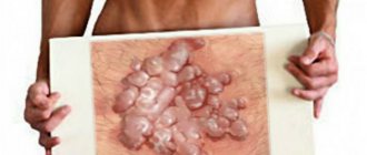
Papillomas and warts can at any time become melanoma - skin cancer, which leads to death in 89% of cases in the first 2 years!
Moreover, in most cases, it all starts with harmless papillomas and warts, which do not interfere with everyday life.
The more papillomas and warts on your body, the more likely it is that one of them will develop into a malignant tumor. The spread of papillomas in the armpits and groin area is especially dangerous.
Infections
Some types of sexually transmitted infections have the natural ability to masquerade as micropapillomatosis. In the first stages of the development of an infectious disease, only external symptoms of damage to the skin of the labia are observed.
Subsequently, the symptoms worsen and, in addition to small benign neoplasms, an acute inflammatory process with damage to the vaginal mucosa occurs.
Diagnostic procedures
The vulva is examined during a pelvic examination. A gynecologist performs vaginal examinations. A transvaginal ultrasound is prescribed. If necessary, vulvoscopy and colposcopy. A bacterial smear is examined to exclude infectious diseases. Clarification of the nature of the neoplasm using histological examination. For biological analysis, scrapings and punctures of tumors are taken.
Removing the tumor often causes a new growth to appear. Removing papillomas requires long-term treatment. Surgeries and the postoperative period may occur with complications. Hematomas may form, the urinary system may be disrupted, and severe bleeding may occur.
Causes
The following causes and risk factors can lead to the emergence and progression of micropapillomatosis in the vestibule of the vagina:
- Hormonal imbalance. First of all, this applies directly to sex hormones. A sharp change in their level leads to dermatological problems in the intimate area.
- Poor quality linen. Synthetic or too tight underwear irritates the skin of the labia and can lead to micropapillomatosis. In this case, therapy should begin with eliminating the main source of the problem.
- Taking anabolic steroids. Drugs of this type affect hormonal levels and health, and therefore can cause micropapillomatosis.
- Oral contraceptives. Most often, correctly selected contraceptives have a positive effect on the condition of the female body. If medications are selected incorrectly, a pathological rash may form.
- Infectious diseases. Some types of viral, fungal and bacterial infections can cause inflammatory processes on the mucous membrane and area of the labia minora.
- Mechanical tissue damage. It can occur during sexual intercourse due to a lack of natural lubrication.
Micropapillomatosis can develop during menopause or puberty.
Important! The progression of micropapillomatosis, changes in the appearance of the rash and the occurrence of pain may indicate that the phenomenon is pathological and requires medical correction.
2. Reasons
Etiopathogenesis, i.e. The causes and trigger factors, mechanisms and patterns of development of micropapillomatosis are currently unknown. The similarity with papillomas and genital warts, not to mention the typical localization for the latter, is so obvious that the viral hypothesis suggests itself and seems to be almost the only reasonable explanation, especially since “papilloma” is clearly heard in the name. Nevertheless, there are serious histological and physical differences on which differential diagnosis is based (see below).
Perhaps the diagnosis should be changed to micropapulosis or pseudopapulosis, from “papule” - nodule, pimples. However, there is clearly a venereal disease called “bovenoid papulosis”, for which papillomavirus etiology has been proven, and the clinical picture of this disease also does not coincide with the picture of micropapillomatosis.
A competing hypothesis is that micropapillomatosis is caused by fluctuations and imbalances in the hormonal formula. However, this hypothesis does not yet provide a satisfactory answer to the question of why exactly such pimples appear in this particular area and in the unlucky 1-5% of women, while in the rest, any rashes, polyps, neoplasia, vesicles occur with great frequency , papules, papillomas, condylomas (or nothing similar is observed), but not micropapillomatosis.
Perhaps this is not the main and most pressing question for modern gynecology, but for now it remains open, and research continues.
Visit our Gynecology page
Associated symptoms
The occurrence of small formations rarely causes discomfort. However, in some cases, irritation, pain, and itching may occur. In addition, patients suffering from micropapillomatosis often experience psychological problems and self-doubt.
The rashes themselves are characterized by the following symptoms and signs:
- Moderate density of formations on the labia;
- Absence of pain or its occurrence due to an additional irritant;
- Pale pink or flesh tone;
- Symmetrical formation of growths.
Even if the detected growths on the inner labia do not cause any discomfort and do not affect your well-being, it is recommended to undergo a diagnosis from a qualified doctor to rule out possible more serious illnesses
Types of surgery
Based on the characteristics of the tumor, its treatment is prescribed. Surgical interventions to eliminate this disease:
- Cryodisruption. Nitrogen exposure is carried out. Freezing tissue stops cell development. During the operation, low-temperature liquid nitrogen is applied to the condyloma. The operation is performed without anesthetic. Not painful. The operation was well tolerated. Removing tumors with liquid nitrogen is a special technology; not every clinic performs these manipulations. The operation has minimal complications. There may be a small scar or burn.
- Radio wave excision. A directed flow of low frequencies dissects the tumor. Radio wave is the most gentle method in relation to neighboring tissues. There is no pain. There is no risk of scarring, bleeding, suppuration, or necrosis. Healing occurs in the shortest possible time.
- Laser. A directed laser evaporates the tufted growths. Laser ablation is used to excise resistant condylomas. The skin is not injured, and under the influence of the laser, epithelial cells grow. The depth of laser exposure is controlled. Does not leave scars. No contact with tissue occurs.
- Electrical coagulation. Electric current is used to cauterize the damaged area. The removed material is sent for histological analysis. The process has a number of contraindications. For example, herpes, oncology, hemostasis, inflammation of neoplasms. There are cases of relapse. There may be a small scar on the surface of the vulva.
- Chemical exposure. The destruction method involves applying a special drug to the affected area. A special composition with organic and inorganic acids, nitrates causes burning and death of condyloma cells. It is not accompanied by pain and is used without anesthesia.
- Plasma coagulation. An arc discharge containing gas penetrates the tissue and vaporizes areas with growths. Cauterizes blood vessels. The method has a high level of efficiency. Thermal damage to tissue occurs at a minimum depth, which promotes rapid healing. No scars are formed and pain is minimal.
Micropapillomatosis and papilloma virus
Unlike papillomavirus (HPV), micropapillomatosis does not pose a health risk. However, in some cases, it can be difficult to distinguish between both abnormalities on the labia. The progression of HPV is accompanied by the formation of pointed anogenital warts.
It is possible to distinguish diseases by the following signs:
- Condylomas are located on the labia in a chaotic manner, while micropapillomas are formed symmetrically and in even rows;
- Condylomas of the labia minora are usually painted white or red, but during a test using acetic acid they turn pale;
- Papillomas have a denser structure than micropapillomatous growths;
- The rash is not transmitted through contact or sexual intercourse; it is possible to become infected with HIV.
Papillomas on the labia also often go away without any medical intervention. However, the likelihood of unpleasant consequences from the progression of papillomatosis is much higher than with other dermatological problems.
Vestibular micropapillomatosis should not be distinguished from anogenital warts, which are caused by papillomavirus.
They are also localized in the vestibule of the vagina on the mucous membrane, but are not dangerous and do not cause serious problems. Micropapillomatosis of the vulva most often has a pink tint and does not turn pale, and is located symmetrically.
Vulvar neoplasms
Vulvar neoplasms are tumors of benign and malignant nature in the area of the external female genitalia. A tumor occurs when the process of cell differentiation is disrupted. A healthy immune system identifies such cells and destroys them. With weakened immunity, they begin to grow and multiply without obeying the body.
The vulva includes:
- pubic area;
- clitoral area;
- external and labia minora;
- entrance to the vagina.
Vulvar tumors can spread:
- Posteriorly - to the perineum, posterior commissure of the labia, rectum.
- Anteriorly - on the opening of the urethra, bladder.
- Up to the vagina, cervix, uterus. The boundary between the external and internal genitalia in girls is the hymen.
The function of the vulva is to participate in sexual intercourse. With tactile stimulation of the receptors of the vulva, the production of secretion is stimulated, moisturizing the entrance to the vagina.
Symptoms of vulvar tumors
If a tumor process develops in the area of the external genitalia, a woman may be bothered by the following symptoms:
- pain;
- foreign body sensation;
- discomfort when moving, walking, during sexual relations;
- discharge mixed with blood and pus;
- menstrual irregularities;
- general weakness, temperature;
- enlarged inguinal lymph nodes;
- urinary disorders - frequent painful urge, pathological impurities in the urine;
- pain in the perineum, defecation disorders.
It is advisable to identify the disease as early as possible. This will facilitate the treatment process, relieve unpleasant symptoms, and reduce the risk of complications. Therefore, at the first suspicious sensations from the reproductive system, you should consult a gynecologist.
Among the complications that vulvar tumors can lead to:
- Anemia due to chronic blood loss.
- Problems with conception.
- Refusal to have sexual intercourse due to pain upon contact with the tissues of the external female genital organs.
- Degeneration of a benign tumor into a malignant one.
- Compression of neighboring organs - urethra, bladder, rectum.
Diagnostics
A woman is examined by a doctor on a gynecological chair. Evaluates the size, shape, symmetry, color of the external genitalia, then proceeds to examine the vagina and cervix. Small tumors deep in the tissue may not be visible upon examination. If they are 1–2 cm, the bulging of tissue in this place is noticeable. Skin color can be reddish, bluish, or normal.
A colposcope is used for a targeted examination. Applying a solution of acetic acid or toluidine blue to the skin of the external genitalia makes areas with neoplasms more visible against the background of healthy tissue.
Condylomas have the appearance of pointed pyramids of a dark color, they are painless, soft to the touch, located one at a time or in groups.
During palpation, the doctor specifies the consistency of the tumor (soft, dense), boundaries, whether the lesion is fused with the surrounding tissues, and pain. Also be sure to check regional lymph nodes. They can be enlarged, painful, and dense.
They take smears for cytology, this will allow you to examine the tissue under a microscope and make an accurate diagnosis. Sometimes a piece of tissue is plucked from a suspicious area for histological examination.
To assess the condition of the internal genital organs, a vaginal examination, colposcopy, and transvaginal ultrasound are performed.
If necessary, additional research methods are prescribed:
- MRI, CT, X-ray of the pelvic organs.
- General blood test, urine test, biochemical study.
Due to the fact that the female genital organs are located next to the rectum and bladder, with tumors of the vulva the process often spreads to neighboring organs. Disorders of urination, defecation, false and painful urges, and pain in the perineum may occur. To examine the bladder, the following is prescribed:
- general and special urine tests;
- Ultrasound;
- X-ray with contrast;
- endoscopic examination of the bladder.
To examine the intestine in detail, the woman is prescribed:
- Sigmoidoscopy – examination of the mucous membrane using an optical instrument.
- Consultation with a proctologist, digital examination.
- X-ray with contrast.
Lymph nodes are assessed by ultrasound, MRI, CT images, and palpation. The PET scanning method is good at detecting metastases. To detect metastases in the breast, it is enough to do a contrast mammography.
Types of benign diseases of the vulva
Benign tumors include neoplasms that are not aggressive towards neighboring cells and tissues. They grow by pushing away healthy tissue rather than invading it. They do not metastasize, do not affect the lymphatic system and other organs, and do not spread throughout the body. The cells that make up a benign tumor are not very different from normal ones.
However, this does not mean that they can be ignored and that they are not at all dangerous. Tumors can compress neighboring cells, causing ischemia, inflammation, and tissue necrosis. Some types of tumors can degenerate into malignant ones, so they need to be removed in a timely manner.
If the tumor does not cause discomfort, is accidentally discovered during examination, is small in size, grows slowly, it is monitored. If active growth begins and symptoms appear that bother the woman, it must be removed surgically.
Depending on what cells the tumor consists of, all benign neoplasms of the vulva are divided into:
- For fibroids, they develop from muscle tissue. If it is from smooth muscle, it is called leiomyoma, if from striated muscle, it is called rhabdomyoma. The tumor looks like a movable dense ball and is most often found on the surface of the labia majora.
- Fibroma - consists of connective tissue fibers. The tumor is elastic to the touch, with clear contours, may have a stalk or is located deep in the tissue. The most common localization is the labia majora, the entrance to the vagina.
- Condylomas, papillomas are superficial formations from a layer of epithelial cells. They have the appearance of a tubercle, often with papillary outgrowths on the surface. Human papillomatosis viruses (HPV) play an important role in their development. They have a high risk of malignancy, especially if the process spreads to the woman’s internal genital organs. The color often differs from the surrounding tissues - brown or whitish. Condyloma has a wide base, while papilloma has a narrow stalk that connects the tumor to the organ. Single formations are possible, but more often multiple. They are located on the labia, at the entrance to the vagina, and on the pubic area.
- Adenoma - consists of epithelial cells of the vulvar mucosa.
- Angioma is a tumor of vascular tissue. There is lymphangioma, it is soft, has a blue tint, and consists of lymphatic vessels filled with fluid. Hemangioma is the growth of small blood vessels. It can spread from the external genitalia to the vagina, to the cervix. Reddish or blue nodules protrude above the surface of the vulva and bleed heavily when injured.
- Lipoma, or wen. It is not dangerous, soft to the touch, round. Found on the pubis and outer labia.
- Myxoma - the formation has a capsule, filled with a yellowish liquid inside.
- Tumors from mixed tissues - fibrolipoma, fibromyoma (consists of modified cells of connective and muscle tissue).
- Hydroadenoma - from the sweat glands in the vulva area. The nodules are small, multiple, usually brown, pink or yellow. They are found on the pubis and in the labia area, often located symmetrically.
- Bartholin gland cyst - located in one of the labia, filled with fluid, painless.
Features of the treatment of benign tumors
The doctor decides whether it is necessary to remove a benign formation. It depends on the woman’s age, the growth rate and type of tumor, and the severity of symptoms. If it is necessary to remove a formation, this is done within healthy tissues:
- If the tumor is pedunculated, it is cut.
- If deep in the tissue, the skin or mucous membrane is cut, the tumor is bluntly removed, the bleeding is stopped, and the tissue is sutured.
After the operation, observation and change of dressings with antiseptic ointments and solutions are necessary. Medicines are prescribed locally to speed up healing. Sometimes antibiotics are given orally or by injection.
To remove superficial benign formations, not only a scalpel is used, but also more gentle methods:
- Cryodestruction - under the influence of nitrogen, tumor cells are frozen and die. The operation is used for condylomas and does not require anesthesia. It is easily tolerated by women.
- Radio wave therapy involves the separation of tissues with a directed flow of low-frequency radio waves. The procedure does not cause pain and is not accompanied by bleeding. The tissues heal with virtually no scars. The rehabilitation period is minimal, as is the risk of complications.
- Laser removal of tumors is also characterized by careful treatment of healthy tissues. The condyloma cells are vaporized due to the high temperature created by the directed beam of light.
- Electrocoagulation is a cruder method; the tumor is exposed to electric current. Subsequently, a scar may remain in the treatment area, deforming the vulva.
- Plasma coagulation is the evaporation of a tumor by exposure to a plasma discharge. As a rule, the scars are small, and the woman does not experience pain.
- Cauterization with chemically active substances, such as acids, solcoderm. The most commonly used is nitric acid; it causes coagulation of tumor cells. There is no need for anesthesia; the tissue heals well without large scars.
- Sclerotherapy is used in the treatment of hemangiomas - a substance is injected into the vessels, leading to tissue sclerosis, gluing and emptying of the vessels.
All these methods can be used if there are no signs of inflammation or malignancy in the area of tumor growth, the woman does not have a herpes virus infection of the genital organs, or problems with blood clotting. After removal, the tissue is sent for histological examination. If malignant cells are detected, treatment will be supplemented with chemotherapy or radiation exposure.
Papillomas tend to recur. Therefore, after surgical removal it is necessary to undergo a course of antiviral treatment.
Types of malignant neoplasms of the vulva
Malignant diseases are more dangerous, but they can also be successfully cured if detected in the early stages. About 80% of all cases of vulvar cancer are diagnosed in women over 55 years of age. Types of malignant tumors of the external genitalia:
- Squamous cell carcinoma (90% of all cancer cases) is a malignant formation of epithelial cells. It is often found in the thickness of the labia majora. The woman complains of a feeling of a foreign body, irregular discharge with blood impurities. Possible burning, itching, pain. The skin in the area of formation changes color to red or bluish, there are ulcers and seals. The ulcers do not heal for a long time, have an unpleasant odor, and bleeding occurs on contact. Carcinoma is often preceded by a benign papilloma, which grows from sweat or Bartholin gland cells.
- Melanoma is made up of pigment cells. Warning should arise if a mole begins to grow rapidly, its color and shape change, redness, swelling of the skin appears around it, itching and pain occur.
- Less common are sarcoma, Paget's cancer, basal cell carcinoma, and adenocarcinoma.
How to treat malignant tumors?
The main methods of treating malignant neoplasms:
- Surgical – the lesion and adjacent tissues are removed so that there are no tumor cells left in the body. If the lymph nodes are affected, they are also removed along with the fiber. In the presence of metastases to other organs, the scope of the operation increases even more. Therefore, everyone is interested in identifying the disease at the earliest possible stages.
- Chemotherapy - cytostatics are prescribed that inhibit the processes of growth and cell division in tumors. It is often prescribed before or after surgery, which makes it possible to reduce the primary lesion and reduce the risk of relapse.
- Radiation therapy – irradiation of tumor growth zones inhibits the development of the tumor and facilitates its surgical removal.
Depending on the histological structure of the tumor, these methods are combined to achieve the best effect.
In the development of malignant tumors of the external female genital organs, four stages are conventionally distinguished. This is necessary to select a treatment regimen:
- Precancer is a disease that often develops into a malignant tumor. Precancerous diseases include papilloma, genital warts (caused by HPV), leukoplakia (increased keratinization of the epidermis), kraurosis of the vulva (sclerosis and deformation, atrophic changes in the external female genitalia), intraepithelial neoplasia.
- The first is a lesion up to 2 cm, located within one anatomical zone.
- The second is that the tumor covers more than 2 cm, but does not extend beyond the vulva.
- The third is characterized by spread to the lymph nodes and nearby organs - anus, urethra, vagina.
- Fourth, distant organs are involved in the tumor process, such as bones, liver, lungs, and brain. Metastases may be indicated by bone pain, exhaustion, rapid weight loss, and unhealthy skin tone.
In the early stages, it is possible to perform organ-preserving operations, while the appearance and shape of the vulva is preserved. If the tumor has invaded more tissue, part or all of the vulva may need to be removed. In such cases, the woman may be offered reconstructive surgery, after which the external genitalia will be formed from the patient’s other soft tissues. This will allow her to lead a normal sex life and not feel psychological discomfort.
If distant organs are affected, surgery cannot always be performed. In advanced cases, palliative treatment is prescribed, aimed at alleviating the woman’s suffering. For pain, analgesics are prescribed orally or by injection. If the tumor spreads to the urethra, a urologist is involved in operations, and a proctologist is involved in the rectum.
Measures to prevent vulvar tumors
It is impossible to 100% prevent the appearance of formations. However, if you follow a number of rules, you can reduce the risk of developing a tumor:
- Treat acute and chronic diseases of the female reproductive system. Trichomonas, chlamydia, the causative agent of gonorrhea, Candida fungi, and the herpes virus cause vulvitis and vulvovaginitis. Despite the chronic course, complaints may be practically absent. Without treatment, fibrotic changes in organs increase, their shape and function are disrupted, and local immunity is weakened.
- Be sexually active with one partner. If you do not want to get pregnant, you should use contraceptive methods so as not to resort to abortion.
- Visit a gynecologist once a year for preventive purposes. Even in the absence of symptoms, a tumor can be detected.
- An active lifestyle, walks, and exercises will reduce blood stagnation in the pelvis.
- HPV infection can be prevented with the help of a vaccine, contact methods of contraception, and limiting the number of sexual partners. HIV weakens the body's immune defenses. Against the background of this disease, HPV infection occurs more easily. The virus penetrates the cell nucleus, leads to changes in the genetic apparatus, and causes the proliferation of atypical mutated cells. Thus, HPV leads to the development of not only vulvar cancer, but also cervical cancer - one of the most common pathologies in women.
- Refusal of bad habits - overeating, smoking, alcohol, sweets.
- Genital hygiene.
- Injury to the external genitalia should be avoided. More often this happens during rapid childbirth, during sports, during falls, during intimate relationships.
You can be examined by a gynecologist in our clinic. Reception is by appointment. On site you can take all the necessary tests, do ultrasound, x-rays and use other imaging methods. The older the woman, the higher the risk of developing tumor diseases.
Experienced doctors will help you identify an emerging problem in a timely manner. The staff treats each patient carefully. Treatment can be completed in full, including surgery, chemotherapy, and radiation if necessary. The clinic widely uses gentle treatment methods. After them, the woman’s recovery is faster and she is not bothered by pain. Whenever possible, organ-preserving operations are always chosen.
Author
Radlevich Natalya Vadimovna
obstetrician-gynecologist, ultrasound diagnostics
Doctor of the highest category, Candidate of Medical Sciences
25 years of experience
+7
Diagnosis of the disease
Any type of rash on the genitals should be a reason to visit a doctor, since their presence does not always mean the formation of a harmless rash.
To determine the type and causes of micropapillomatosis on the labia, a differential diagnosis is carried out by a gynecologist. An external examination is usually not enough to make an accurate diagnosis.
Therefore, during the examination, a smear and blood test are also performed. The process also determines:
- Cellular structure of formations;
- Presence of a pathogen;
- Type and nature of anomalous processes;
- Cell damage.
Also important in diagnosis are anamnesis and collection of information about the medical history. Additionally, tests for the presence of papillomatosis can be performed.
Diagnosis and differences from other diseases
Diagnosis of this condition is carried out in order to distinguish the signs of micropapillomatosis from condylomas or papillomas, the initial stages of syphilis, inflammation, malignant neoplasms and other dangerous diseases. The main differences between harmless rashes and HPV infection include the following:
- genital warts that occur with HPV are located chaotically over the entire surface of the vulva or on one side, while micropapillomas are arranged in symmetrical rows;
- condylomas are hard and dense in structure, the rash with micropapillomatosis is much softer and resembles skin elevations rather than warts;
- upon contact with acetic acid, HPV warts change color to a paler color, while micropapillomas do not react in any way to such exposure.
Despite the obvious differences, external examination is not enough to make an accurate diagnosis. Only a specialist can distinguish harmless neoplasms from dangerous ones. He must examine the patient and then take a swab from the affected area to analyze its cellular structure and determine whether the rash was caused by exposure to a virus.
HPV is characterized by fairly specific cell damage (koilocytosis), so it is usually not difficult to distinguish pathologies from each other.
If necessary, more accurate results will be shown by a method such as polymerase chain reaction. To clarify the diagnosis, the patient is also prescribed a serological blood test.
Treatment of micropapillomatosis
Despite the fact that micropapillomatosis type rarely poses a health hazard, it is recommended to eliminate it as a cosmetic defect to improve the psychological state of a woman.
In addition, micropapillomas may hide such serious causes of lesions as hormonal imbalance and infectious pathologies. The acquisition of a pustular form of the rash is also a prerequisite for mandatory therapy.
Important: Surgical removal of growths is the least effective treatment method. This is due to the small size of the lesions and the high likelihood of prolonged tissue healing and complications.
Therapy using a laser device
Without complications, the rash can be removed using a laser beam. Its main advantage is painless and quick excision of abnormal tissues, which does not require rehabilitation.
As a rule, to achieve the best effect, no more than two sessions are performed, during which the skin becomes as smooth as possible. Loose small structures are eliminated in one short session.
Cryodestruction
Growths in the area of the labia minora can be cauterized using liquid nitrogen. After applying the product to the affected skin, micropapillomas disappear without the possibility of relapse.
For complete and high-quality treatment of the skin, one procedure is usually sufficient.
If you follow all the doctor’s recommendations, cryodestruction leaves virtually no scars and does not cause discomfort afterwards.
Electrocoagulation
Cauterization of micropapillomatosis can also be carried out using electrocoagulation - a point effect of high-frequency electric current.
The procedure is performed with local anesthesia, as otherwise it may cause pain and discomfort.
Use of medications
There is no direct impact on the growths of the labia minora with medications for external and internal use.
However, medications are often necessary to restore the hormonal levels of patients, as well as eliminate the underlying causes of the disease.
If pustules occur, antiseptic treatment of areas of the skin of the labia affected by micropapillomatosis is also possible.
Traditional medicine
At home, it is possible to safely treat micropapillomatosis using medicinal plants.
In this case, the following folk remedies are effective for use:
- Celandine decoction . Pour a handful of dry or fresh herbs into a liter of water and boil the mixture for five minutes. Cool the broth and strain. Use the product for daily washing once or twice a day.
- Chamomile decoction . The composition is prepared more concentrated than a decoction of celandine. In this case, it is possible to use ordinary pharmaceutical chamomile. It is recommended to wash with the product for at least two weeks with regular use once or twice a day. The composition has anti-inflammatory and antiseptic effects. In the same way, you can also prepare a decoction of oak bark.
- Herbal collection . To prepare, you need to mix mint, linden, and chamomile in equal quantities. It is necessary to prepare a decoction from the collection in the same way as from chamomile.
Before using traditional medicine recipes, it is recommended to consult a gynecologist and therapist. The body's response to home treatment is not always positive and depends on the cause of the skin lesion.
Important: Decoctions of chamomile, mint and linden are also useful to take orally as anti-inflammatory and painkillers in the absence of contraindications.
4.Treatment
It should be repeated that micropapillomatosis, if all other symptomatically similar diseases are conclusively excluded, does not require treatment. This is objective.
Subjectively, such a rash can, as shown above, represent a serious problem that requires psychological correction and elimination of the causes.
Today, fortunately, this problem can be solved quite simply and safely, painlessly and bloodlessly, on an outpatient basis, without general anesthesia and long-term rehabilitation. Mainly three methods of removing micropapillomatous rashes have become widespread: cryodestruction, electrocoagulation and laser therapy. The last approach is the most popular.
Prognosis and possible complications
The disease rarely causes any complications and is easy to treat. At the same time, the likelihood of recurrence of formations is minimal and insignificant.
However, in most cases, women simply do not pay attention to the rash on the labia due to the fact that it does not cause any discomfort.
In some situations, rashes are considered as a variant of the norm that does not require treatment. However, the choice remains with the patients. If micropapillomatosis causes physical and psychological discomfort, the rash should be removed using one of the suggested methods.
Symptoms and signs
As a rule, rashes develop very quickly, without any previous signs or accompanying symptoms. They are usually discovered accidentally by the woman herself or by a gynecologist during a routine examination. Signs of inflammation in micropapillomatosis are as follows:
- the rashes are very small, located symmetrically relative to each other, lining up in rows or lines;
- to the touch, the affected areas are rough, soft or moderately dense;
- there are no inflammatory processes, discharge of pus, burning, itching or other unpleasant symptoms - the disease can only be identified by external signs;
- there is no pain either at rest or when touching or pressing on the rash;
- the rashes do not cause discomfort when sitting, walking, sexual intercourse, playing sports, etc.;
- the color of the neoplasms ranges from merging with the main skin tone to pale pink;
- Over time, the rashes do not change shape or grow, and do not degenerate into ulcers or cancer cells.
Micropapillomatosis does not cause physical discomfort to a woman and does not in any way affect her ability to work, engage in any activity, live a full sex life, or bear and give birth to children. But for many representatives of the fair sex, this pathology causes embarrassment, shame and disgust, so they strive to get rid of it by restoring the natural appearance of the genitals.
Preventive measures
In order to minimize the likelihood of micropapillomatosis on the labia, it is recommended to adhere to the basic rules of prevention:
- Carry out hygiene procedures daily;
- Wear only soft, breathable underwear with a minimum amount of synthetic fibers in the composition;
- Monitor the state of the endocrine system and promptly eliminate diseases associated with changes in the level of hormone production;
- Systematically visit the gynecologist’s office (at least twice a year);
- Use only pH-neutral mild agents for hygiene procedures;
- Eliminate infectious disorders in a timely manner;
- Use contraception to prevent sexually transmitted infections.
Diagnostics
Cancer is diagnosed using a biopsy. High accuracy of the study helps to distinguish a malignant tumor from a benign one. A gynecologist examines a woman using a colposcope. The skin is treated with a special solution, which makes the lesions appear more pronounced. If the tumor spreads beyond the vulva, perform the following steps:
- Research of the genitourinary system and rectum.
- Detection of tumor growth and penetration into lymph nodes and organs using CT or MRI.
- Search for metastases using radiography.