Traditional management of trophic ulcers
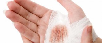
- Cleansing the wound should be done daily.
- The first step is to remove any dirt or dead tissue from the ulcer, the second is to apply an appropriate dressing. This provides the best conditions for healing.
- Use mild soap and water as a cleanser. The method of cleansing with saline solution has also proven itself well.
- Antiseptic solutions are also used to wash the ulcer, for example, chlorhexidine, a weak solution of furatsilin, a decoction of chamomile or string.
- Avoid using antiseptic cleansers such as iodine and hydrogen peroxide, which often damage sensitive skin and can interfere with healing.
- Some ulcers improve with the use of moist gauze dressings that dry after being applied to the wound. Dead tissue adheres to the gauze and is removed when you change the dressing.
- Daily hot tub or hydrotherapy may also be helpful as a method of cleaning the ulcer and reducing tissue that is dead or dirty.
- To better remove dead tissue, chymotrypsin is added to the wound after washing and covered with a napkin. The drug has anti-inflammatory, antiviral and healing effects. This dressing is done twice a day.
- To relieve inflammation, hormonal ointments are used (they are used for no more than 5 days and are not rubbed into the wound, but applied in a thin layer under a dry cloth).
Prevention of varicose ulcers after treatment
If the goal of treatment is achieved: blood flow is restored, adequate tissue nutrition is restored and the cause of the disease is eliminated, the risk of complications is minimal. However, the patient should remember that his future fate depends on himself. You need to lead a healthy lifestyle and constantly monitor the condition of your blood vessels. For this:
- take medications prescribed by a doctor,
- eat right (exclude fast food, alcohol, animal fats and simple carbohydrates),
- avoid excessive physical activity,
- perform a complex of physical therapy,
- wear compression stockings on the lower extremities,
- choose comfortable shoes,
- come regularly for medical examinations (twice a year),
- undergo specialized sanatorium-resort treatment.
Compliance with the doctor-prescribed regimen, nutrition and physical activity will help avoid relapse of a trophic ulcer. By contacting the A. Begma Medical Center regarding varicose veins, you can get a detailed consultation with a phlebologist, clarify the condition of the veins, get rid of trophic ulcers and make sure they never bother you.
Phases of the wound process
- Purification stage
If there is copious purulent discharge or the presence of necrosis or an unpleasant odor, the ulcer must be cleansed of infection and dead tissue. This is achieved by washing the ulcer with a sponge soaped with laundry soap. To separate dead tissue, chymotrypsin powder or mesh with enzymes (parapran) is used, which is placed into the ulcer after washing and covered with a napkin. Dressings are repeated 2 times a day before bedtime and after waking up. Before each dressing, the ulcer is washed with a soft sponge and laundry soap. In the morning, a compression stocking or golf stocking of 2-3 compression class is put on over the bandage; in extreme cases, a fresh elastic bandage is used. If the skin around the ulcer is eczematous, then it is necessary to reduce inflammation; if there is redness of the skin around the ulcer, hormonal ointments (Lorinden, fluorocort) are used, which are applied in a thin layer under a dry cloth at night and not rubbed in. Hormonal ointments are used for no more than 5 days.
- Healing stage (granulation)
When good granulations appear - the ulcer is bright red, relatively clean, and its depth is reduced, treatment is required to stimulate healing and protect the granulations from damage. We use special wax mesh (voskopran) on top of which we apply ointments that promote healing - olasol, curiosin, gel dressings. The compression rules remain the same. You can wash the ulcer at this time without a sponge and carefully. Dressings once a day. Improving blood flow is achieved by elevating the leg during sleep (15-20 degrees), and mandatory compression with a stocking or bandage while awake. Elastic compression is necessarily used for venous ulcers; for arterial ulcers it is contraindicated, since on the contrary it can cause harm.
- Epithelization stage (final covering with new skin)
After the ulcer begins to heal, only light protective coverings like the same mesh are used, and gel dressings such as “hydrocol” can be used. If a small dry crust forms, there is no need to remove it specifically. After the appearance of young skin, it will fall off by itself. For venous ulcers, after eliminating venous congestion (sclerotherapy, laser or surgery), the ulcerative surface closes in 2-6 weeks. For arterial ulcers, the situation depends on the degree of restoration of blood circulation. With good blood circulation, ulcers usually heal within 1-3 months.
Features of different types of ulcers
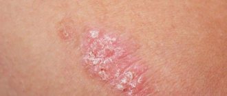
Venous ulcers
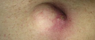
This type of trophic ulcer usually appears on the inner surface of the leg. Venous ulcers occur as a result of impaired blood flow in the lower extremities due to stasis dermatitis.
In this case, the lower part of the leg from the foot to the knee swells greatly, and cramps may occur at night. Then the lower leg begins to itch very much, and a mesh with enlarged veins appears on it. With further damage, red-violet spots and hyperpigmentation appear. The skin becomes denser, acquires shine and smoothness. Small ulcers quickly increase in size, the purulent process first affects the skin, then the Achilles tendon, calf muscle, and periosteum of the tibia. The ulcer first releases serous fluid, and when infected, pus with a nauseating odor.
If this disease is not treated or treated incorrectly, the lymph nodes may be affected and “elephantiasis” may develop (a type of swelling of the legs when the size and shape of the affected limb changes). And in some cases, even death is possible due to blood poisoning - sepsis.
Arterial ulcers
Arterial ulcers are initially, as a rule, small wounds with jagged edges filled with pus. They develop against the background of damage to the main arteries of the legs and form on the sole and outside of the foot, big toe and heel. This type of trophic ulcers occurs due to obliterating atherosclerosis. An arterial ulcer can occur due to hypothermia of the feet, wearing tight shoes, or damage to the skin. This disease most often affects smokers and older people.
Diabetic ulcer (Diabetic foot)
Diabetes mellitus causes various complications, one of which is a trophic ulcer. Although blood circulation in the legs may not be significantly impaired, due to damage to the nerve endings, patients complain of loss of sensation and coldness of the limb. Diabetic ulcers most often occur on the big toe. With further development of the disease, first the remaining toes are affected, and then the entire foot.
A diabetic ulcer is dangerous because it quickly becomes infected. This can lead to gangrene and leg amputation.
Hypertensive ulcers (Martorella)
Quite a rare type of ulcer. Hypertensive ulcers can appear as a result of constant high blood pressure (arterial hypertension), and this can provoke spasm of small vessels. Hypertensive ulcers most often affect women over 40 years of age. The peculiarity of these types of ulcers is the symmetry of the affected areas in the middle part of the legs.
Hypertensive ulcers are very painful and are most susceptible to bacterial infection.
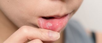
Neurotrophic ulcers
Occurs as a result of injury to the head or spine. A neurotrophic ulcer is located on the side of the heel or sole in the form of a small but deep hole, involving bone, tendon and muscle. The skin around the wound loses sensitivity.
Vacuum cleaning of wounds
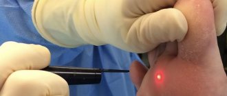
Vacuum therapy or negative pressure therapy is a method of removing serous fluid and dead tissue from a wound or surgical site. Currently, vacuum ulcer debridement can be used on all types of wounds: acute, subacute or chronic. It reduces swelling, promotes rapid healing and the formation of young connective tissue.
The essence of the method is that a piece of porous sponge with silver ions is inserted into the wound, then the whole thing is covered with a transparent membrane. A hole is made in it and a drainage tube is inserted, which is connected to a vacuum source. The fluid is drawn from the wound through the sponge into a reservoir for subsequent disposal.
The membrane prevents air from entering and allows a vacuum to form inside the wound, reducing its volume and making it easier to remove fluid. Before starting the procedure, the ulcer should be washed.
The duration of treatment depends on the size and depth of the wound. The dressing is changed every 24-48 hours.
Diagnostics
1 Consultation with a dermatologist in MedicCity
2 Consultation with a dermatologist in MedicCity
3 Consultation with a phlebologist in MedicCity
Our experienced surgeons and dermatologists can give you an accurate diagnosis based on the appearance of the wound.
But in order to find out the cause of the disease and propose the correct treatment program, it is necessary to undergo the following laboratory tests:
- clinical blood and urine tests;
- biochemical and immunological studies;
- ultrasound examination of the vessels of the lower extremities;
- vascular angiography.
1 Laboratory diagnostics in MedicCity
2 Vascular ultrasound in MedicCity
3 Vascular ultrasound in MedicCity
What to expect from treatment?
With proper care, the surface of the ulcer closes after 1.5 months. The use of vacuum therapy speeds up healing:
- Optimizes blood flow,
- Reduces swelling of local tissues,
- Removes excess fluid that can slow down cell growth,
- Reduces the number of bacteria.
- In addition, negative pressure changes the structure of cells in the wound layer, causing a cascade of intracellular signals that increase the rate of cell division and formation of granulation tissue.
There are a number of contraindications for vacuum therapy:
- malignant wounds;
- osteomyelitis without adequate treatment;
- necrosis with crust formation.
In such cases, cleansing is carried out using enzyme preparations and active surgical dressings. It is important to understand that cleansing trophic ulcers is an auxiliary method of treatment and in itself cannot lead to healing of the ulcer without eliminating the causes of its appearance.
Why is this complication dangerous?
If the patient ignores trophic ulcers on the legs and their treatment, then he will face a number of negative consequences:
- strong pain,
- development of dermatitis around the affected skin area,
- wound infection,
- progression of the disease to the chronic stage, which threatens more frequent relapses and longer therapy, which lasts from six months.
Treatment of trophic ulcers of the lower extremities includes 2 aspects: the work of the doctor and the care of the patient’s legs. As a rule, the doctor resorts to a conservative method of treatment, which is individual in each case:
- compression therapy – to return venous blood flow to normal, which leads to the closure of ulcers even without surgery:
- compression bandages made of breathable bandages and bandages with zinc oxide,
- special compression socks,
- washing and applying dressings (sorption, interactive, etc.),
- selection of drugs against inflammation, infections, to accelerate tissue healing.
Home treatment of trophic ulcers, in addition to following all the doctor’s instructions, involves
- cleansing the wound and treating it with antiseptic solutions,
- planned dressing, indicated by the attending physician,
- skin care around the ulcer,
- reducing the load on the legs,
- gentle hygiene procedures.