Angioma is a neoplasm that is classified by doctors as a benign tumor. This pathology is a collection of blood and lymphatic vessels. Outwardly, most neoplasms resemble moles, and therefore are popularly called “red moles” or “port-wine stains.” In most cases, angiomas occur during fetal development, but sometimes they appear in older people or can be an external manifestation of a number of diseases.
What is angioma
Venous angiomas are localized in organs and tissues and can be multiple or single.
They are located in most cases on the upper half of the body. Their morphological basis is highly dilated lymphatic/blood vessels. The shape and size of pathological areas can vary, color - from colorless to red-blue. The lesions tend to progress rapidly. There are angiomas of the brain, liver, external genitalia, and bones. Angiomas are classified according to their structure:
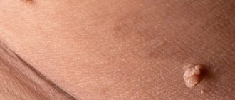
- Simple (hypertrophic, capillary). Small venous and arterial vessels grow. If you study a photo of such an angioma, it will become clear that it is a bright red or bluish-purple spot. Its sizes can be different, even gigantic. If you press on the lesion, it will immediately turn pale due to the outflow of blood. Capillary angiomas of the skin extremely rarely transform into malignant formations.
- Branched (racellose). They are represented by plexuses of tortuous dilated vascular trunks. They are characterized by the presence of constant pulsation. The limbs and face are most often affected. Traumatization of venous branched angiomas is fraught with severe bleeding, which cannot always be stopped on its own.
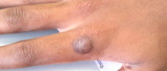
- Cavernous (cavernous). They are formed by wide spongy cavities that are filled with blood. Cavernous angioma looks like a purple-bluish bumpy spot. Most often located under the skin. It feels hotter than the surrounding tissue. When stressed, it increases in size. A complex form is cavernous angioma of the brain, the treatment of which is very long and involves restoration of impaired blood flow.
- Combined. Combine subcutaneous and superficial location. Formed by overgrown vessels and other tissues (depending on location on the body).
The shape of angiomas is:
- stellate (point angiomatous formations, from which dilated small blood vessels extend in all directions);
- flat (vascular spots of pink-blue color located in the upper layers of the skin);
- nodular (seals protruding above the surface of the skin);
- serpiginous (rashes on the skin in the form of small burgundy nodules).
Types of neoplasms
There are several classifications of angiomas. They are distinguished by shape, cause of appearance, tissue composition and type of vessels that provoked the problem.
Thus, according to their shape, neoplasms are divided into pineal, nodular, branched (arachnid) and flat. Pineal angiomas are characterized by a convex shape; they rise noticeably above the skin. Nodular angiomas are usually small in size. Outwardly, they resemble small red dots. However, they do not have branches from the capillaries - a characteristic vascular network. It is characteristic of branched (arachnid) angiomas, which in appearance can actually resemble a spider with many legs. Finally, flat angiomas resemble ordinary moles in appearance.
Content:
- Types of neoplasms
- Reasons for appearance
- Methods for removing angiomas
Based on the reasons for their appearance, formations are divided into hemangiomas and lymphangiomas. Hemangiomas are a manifestation of blood vessel pathology, and the cause of lymphangiomas is a disruption in the functioning of the lymphatic system. They are diagnosed less frequently than hemangiomas and are point formations.
As for hemangiomas, they are divided into capillary, which are bluish-purple or bright red spots, as well as cavernous (cavernous) and punctate. Cavernous hemangiomas can acquire very significant sizes. These are soft tubercles that change shape when pressed. As a rule, they appear on the skin of the face, in the groin, and in the armpit area. Punctate hemangiomas are characterized by miniature sizes, and in appearance they resemble tiny red dots, similar to an injection mark.
Causes of angioma formation
Angioma in newborns is a standard clinical picture, since this disease in most cases is congenital. The source of the development of neoplasms in adulthood are persistent fetal anastomoses located between the veins and arteries. The proliferation of blood vessels in a benign tumor leads to the growth of the tumor itself.
The causes of angioma are as follows:
- hereditary predisposition;
- changes in hormonal levels;
- diseases of the gastrointestinal tract;
- dysfunction of lipid metabolism;
- skin pigmentation disorders.
In rare cases, venous angiomas of the skin occur after trauma to the skin area (bruises, cuts). They can also accompany malignant neoplasms of internal organs and cirrhosis of the liver.
Causes of red moles
The exact reason why red moles appear on the body is unknown; it may be a predisposition due to a genetic factor. But there are other reasons:
- pregnancy;
- exposure to chemicals;
- some diseases, including infectious ones;
- unsuitable climate (too hot or cold)
- mechanical damage to the skin, such as a bite or cuts, in which pieces of skin are not completely torn off.
Their manifestations are also possible at a more mature age, this is due to a gradual decrease in immunity. Neoplasms occur in 75% of the population over the age of 60 years.
It is impossible to somehow influence the appearance or non-appearance of red moles on the body, since they are inherent in us at the genetic level; if one of the parents had a lot of them, it means that the children will have a similar situation, and even identical maps of the location of nevi are possible.
Symptoms of angioma
Symptoms of angioma depend on the type of vascular tumor, its size and location. In newborns, it is diagnosed immediately or several months after birth, and can grow very quickly, covering large areas of the skin.
Vascular angiomas can affect:
- Integumentary tissues - skin, mucous membranes of the genitals, subcutaneous tissue. Angiomas are also found in the mouth.
- Musculoskeletal system - muscles, bones. A severe form of the disease includes hemangioma of the vertebral body.
- Internal organs.
If integumentary hemangiomas cause cosmetic defects, then hemangiomas of internal organs provoke the occurrence of disturbances of important functions - urination, breathing, vision, defecation.
Angiomas of muscles and bones are accompanied by:
- skeletal deformation;
- severe pain in the joints;
- frequent fractures;
- radicular syndrome (compression of the spinal nerves).
Venous angiomas of the brain (cerebellum, left/right frontal lobe, etc.) are very dangerous. They lead to subarachnoid hemorrhage and epilepsy. Also, due to compression of the brain vessels by the tumor, the following symptoms may occur:
- convulsions;
- headache;
- nausea, vomiting;
- dizziness;
- noise in the head;
- taste disturbances;
- speech problems.
During the growth process, angiomas can become inflamed, causing phlebitis and thrombosis. A common complication is bleeding due to trauma.
If you notice similar symptoms, consult a doctor immediately. It is easier to prevent a disease than to deal with the consequences.
Spinal hemangioma: treatment with folk remedies
No matter how sad it may sound, any methods of alternative treatment for spinal hemangioma are ineffective. The only way “grandmother’s chest” recipes can help is to eliminate symptoms and alleviate the patient’s condition.
Today, many patients of the neurosurgical department note that baths with medicinal herbs are one of the most effective methods of relieving pain and fatigue.
If you are faced with a benign formation, and now you are interested in a spinal hemangioma, a photo of one can always be found on the Internet, and options for getting out of the situation can be found with an experienced specialist!
Diagnostics
Angiomas located on the surface of the skin are diagnosed during examination and palpation. The characteristic coloring, ability to contract and increase in size when pressed and strained allow the doctor to easily make the correct diagnosis.
If the localization of the lesion is more complex (internal organs, bones, brain), a number of diagnostic measures are required:
Skeletal hemangiomas are identified using x-rays of the spine, ribs, bones, skull and pelvis.
To identify angiomas of internal organs, antiography (contrast x-ray examination of blood vessels) of the kidneys, brain, lungs, etc. is performed.
Pharyngeal angiomas are examined by an otolaryngologist.
Ultrasound diagnostics allows you to determine the exact depth of growth of the angioma, its structure and location features. If a vascular angioma is suspected, the doctor may additionally prescribe a puncture. The resulting yellowish liquid is examined in the laboratory. This makes it possible to differentiate angioma from lymphadenitis, lipoma, cyst, hernia.
How dangerous is the disease?
A small hemangioma does not pose any danger. Such a benign tumor rarely manifests itself. About 10% of patients do not even suspect its presence. If the tumor is less than 1 centimeter, then it is only observed; if it begins to grow, it is removed.
Developing between the vertebrae, the tumor can invade several neighboring vertebrae, usually 2-3 vertebrae. Large tumors cause pain and can also cause vertebral collapse. In this case, compression of the nerve endings of the spine occurs, which leads to both pain and numbness in this area of the back. In addition, a vertebral hemangioma of significant size can cause its fracture.
A large tumor in the lumbar spine can cause:
- urinary disturbance;
- problems with bowel movements;
- numbness of limbs and so on.
Treatment of angioma
Treatment of angioma is aimed at stopping its growth, eliminating the pathological process and normalizing the functioning of the vascular network. Indications for urgent intervention are:
- extensive tissue damage;
- rapid growth of the tumor;
- localization in the neck, head;
- frequent bleeding;
- dysfunction of the organ in which the angioma is located.
Watchful waiting is justified only if regression of a vascular benign tumor is observed.
Among the most common treatments for angiomas:
- Diathermoelectrocoagulation. Cauterization with electric current is indicated if it is necessary to remove hard-to-reach punctate angiomas and angiofibromas. The method cannot be used if the angioma is deep and occupies a large area.
- Laser treatment. Using a laser, the doctor removes pathological tissue layer by layer. The advantage of laser treatment of angiomas is minimal bleeding.
- Radiation treatment. Allows you to achieve good results when removing angiomas of complex anatomical localization.
- Surgery. It is used if the vascular tumor is deep and it is not possible to remove it without affecting the surrounding healthy tissue.
- Cryotherapy. Makes it possible to remove simple small angiomas of any location. During the procedure, liquid nitrogen is applied to the tumor. The method is painless and does not cause bleeding.
- Sclerosing therapy. Seventy percent alcohol is injected into the tumor. The treatment is painful and is suitable if the angioma is located in the deep layers of the skin.
- Hormonal therapy. Relevant if the lesion is extensive, angiomas grow quickly.
- Excision of angiomatous areas followed by reconstruction of the vessel.
- Ligation of the arteries supplying the angioma. A ligature is applied to the end of the artery, as a result of which the neoplasm gradually dies.
It is possible to treat angioma with folk remedies:
- Apply the kombucha to the tumor site and fix it. After a day, replace the compress. The duration of such treatment is 2-3 weeks.
- Dilute a tablespoon of copper sulfate with 100 ml of clean water. Wipe the neoplasm with the resulting liquid 4-5 times a day for 10 days.
- Apply a compress of onion pulp to the affected area of skin for 10 days. Change the bandage every 12 hours.
- Cover the angioma with fresh grated carrots and tie gauze on top. Change 3 times a day.
- Mix fresh celandine juice in a ratio of 1:4 with petroleum jelly and add a drop of 0.25% carbolic acid. Use the ointment daily in the morning and evening.
- If the angioma has affected the internal organs, you can use the following recipe: pour 3 tablespoons of potato flowers into 300 ml of boiling water. Leave in a thermos. Drink half a glass 3 times a day half an hour before meals. The treatment course is 2 weeks.
It should be remembered that any folk recipe can be used only after consultation with your doctor. Some medicinal plants can provoke the growth of angiomas, so preparing decoctions and infusions from them yourself is not recommended.
Cavernous angiomas on CT
When using visualization methods, it is useful to separate the cavity into 3 components. These include (1) a peripheral pseudocapsule, consisting of glial tissue impregnated with hemosiderin, (2) an irregularly structured intermediate connective tissue separating the cavities, and (3) a central vascular part, consisting of vascular cavities with slow blood flow.
On CT images without contrast enhancement, a cavernoma appears as a focal formation of an oval or nodular shape, characterized by slightly or moderately increased X-ray density and does not have a volumetric effect on the surrounding parenchyma. Areas of calcification and hemosiderin deposition in the walls of fibrous septa, along with stagnation of blood in the cavities, contribute to increased x-ray density on non-contrast-enhanced images. On CT images, calcifications are detected in approximately 33% of all cavernomas. If the formation is old, then it may contain central non-contrasting areas of reduced density, which corresponds to cysts from resorbed hematomas.
Contrast enhancement can be minimal or maximal, although 70-94% of cavernous malformations are weakly or moderately enhanced after intravenous contrast. In most cases, good contrast is the result of increased blood flow in the vascular component of the mass. The heterogeneous “speckled” enhancement is caused by intravascular fibrous septa, and the low-density rim along the periphery is caused by a pseudocapsule of glial tissue surrounding the formation.
Mass effect is not typical for cavernomas, unless they are associated with recent hemorrhage. On CT images without contrast enhancement, cavernomas may not be detected at all. In hemorrhages and the formation of intracerebral hematoma, cavernomas are visualized as areas of focal signal enhancement in the area adjacent to the hematoma.
Any hemorrhage detected on CT in a relatively young patient should be carefully investigated, and cavernous angioma should always be considered as a possible cause. When evaluating a patient with a seizure disorder, cavernous angioma should also be considered as a likely etiological factor, especially if the patient is between 20 and 40 years of age.
Cavernous malformations identified by CT may also include other rare vascular malformations (thrombosis of arteriovenous malformation, capillary telangiectasia), glioma (poorly differentiated astrocytoma or oligodendroglioma), and metastatic melanoma.
Danger
A small spot formed on the skin can go unnoticed or be ignored by the patient for a long time. Since it can transform into a malignant formation, the lack of necessary treatment can lead to dire consequences.
Particular attention should be paid to angiomas located in places of increased friction with clothing (neck, chest, abdomen, shoulders), on the scalp. Frequent traumatization can lead to inflammation of a benign tumor and its rapid growth and bleeding.
The greatest danger to life is posed by angiomas of the brain and internal organs. As practice shows, if you do not treat a cerebral angioma, the prognosis is unfavorable - the area of accumulation of blood vessels will increase, which will lead to their rupture, hemorrhage in the brain and death.
Vascular malformations of the brain
Types of vascular malformations differ based on their macroscopic and microscopic characteristics. Typically, intracranial vascular malformations are divided into the following 4 groups:
- Capillary malformations (or telangiectasias)
- Cavernous malformations (cavernous angiomas/hemangiomas)
- Venous malformations
- Malformations with arteriovenous shunts
According to the newer classification, 2 more categories have been added: arterial malformations (without the formation of an arteriovenous shunt) and mixed malformations.
Cavernomas can be found in any area of the brain because they can arise anywhere along the vascular bed. Intracranial extracerebral cavernous angiomas sometimes occur, but they are quite rare. Cavernous angiomas are also sometimes found in the spinal cord, most often in association with multiple vascular lesions of the brain.
Causes of hemangioma
Spinal hemangioma occurs when improper (chaotic) division of vascular tissue cells occurs. What this depends on is still a matter of debate in scientific circles. However, it has been noted that the main factor in most cases is heredity. Studies have shown that such a tumor can also occur with increased levels of estrogen, so it is most often found in women. The disease also occurs when there is heavy load on the spine, for example, during pregnancy or when lifting heavy objects.