Ichthyosis - symptoms and treatment
The main symptom of ichthyosis is dry, scaly skin, usually over large areas of the body. The skin may also peel, itch, and become red.
Ichthyosis vulgaris
It usually proceeds quite easily. Symptoms develop from about 2 months of age. Layers of scales (keratoses) of various sizes and colors appear on the child’s skin: from whitish to gray-black. The skin becomes dry, rough to the touch and wrinkled. Skin changes most often appear on the elbows and knees. On the neck, in the elbow and popliteal folds, as well as in the armpits, the skin does not change [1][2][12].
In children, the facial skin, as a rule, is not affected or is slightly changed; in adults, the forehead and cheeks sometimes peel off. The skin of the palms and soles shows an altered network-like skin pattern and slight peeling. Follicular keratosis in the form of small dryish nodules is also typical [1][2].
The mildest form of ichthyosis vulgaris - xeroderma - is manifested by dryness, roughness, peeling of the skin of the elbows, knees and buttocks. Small non-inflammatory horny nodules are noticeable on the skin [1][2].
A more severe form of vulgar ichthyosis is simple ichthyosis (ichthyosis simplex). When it develops, the stratum corneum resembles cracked parchment. White grooves divide it into polygonal thin scales similar to mica. The color of the scales is grayish-white, sometimes they shine and resemble mother of pearl (ichthyosis nitida). Less common are people with large multifaceted horny scales similar to the skin of a snake (ichthyosis serpentina). Sometimes layers of thick, large horny plates resemble the skin of a lizard or crocodile (ichthyosis sauriasis), and the horny outgrowths resemble the skin of a porcupine (ichthyosis hystrix) [1].
X-linked recessive ichthyosis
This form occurs only in men. Symptoms appear soon after birth - usually in the first weeks of life. Dry skin is noted, and later light and dark brown dense scales appear on the elbows, knees and back of the neck. Unlike other forms, in X-linked recessive ichthyosis the skin of the face and hands is not affected [6].
Autosomal recessive ichthyosis
This type of ichthyosis is more severe, since the child inherits mutant genes from both parents.
In a milder form (lamellar ichthyosis), the child is born with reddening of the skin (erythroderma) and thickening in the area of the folds. Rarely do signs of the disease appear several days or weeks after birth. Sometimes shortening and deformation of the eyelids and ears are noticeable. The face is mask-like. The skin of the palms and soles is thickened. Increased sweating of the affected skin and other areas is noted [1][2][4]. With this form of congenital ichthyosis, children can survive, but their mental and physical development is sharply inhibited [1].
In a severe form (fetal ichthyosis), the child is born as if in a shell of thick horny plates that form in utero. Hyperkeratosis (thickening of the skin) is observed even in the groin folds, neck, elbow, popliteal folds and armpits. The eyelids and ears are either completely absent or severely deformed. Sometimes the upper lip becomes deformed and everted. The nasal passages, oral cavity, ears and external auditory canals are clogged with horny masses. The face is mask-like. This form of ichthyosis threatens the life of a newborn [1][2].
Hereditary ichthyosiform erythroderma
There are dry (inherited from both parents) and bullous forms of the disease (inherited from one parent). A characteristic feature of this form is erythroderma, in which the entire skin becomes red. Large natural folds, as well as the face and neck, are most affected. Hyperkeratosis and growths similar to papillae are noted not only on the trunk and limbs, but also in the armpits, inguinal and intergluteal folds, elbow and popliteal folds, as well as on the neck and face. The skin of the face is red, shiny, tense, covered with flaky scales.
In the bullous form, blisters periodically appear on the skin. When the bubbles burst, clear liquid flows out. Hyperkeratosis with increased sweating is also observed on the palms and soles. In addition, nails and hair grow faster and become thicker [1][2].
Syndromic ichthyosis
Sometimes ichthyosis is a component of hereditary syndromes and is combined with lesions of internal organs and body systems:
- Rood syndrome - ichthyosis in this case is combined with epilepsy, hypogenitalism (underdevelopment of the gonads), polyneuritis, atrophy of all muscles, arachnodactyly (spider finger syndrome) and Birmer's anemia.
- Sjogren-Larsson syndrome - ichthyosis is combined with mental retardation, symmetrical leg weakness, foot deformity and retinitis [1][2].
Acquired ichthyosis
Such ichthyosis can begin at any age, but most often develops after puberty. Symptoms of acquired ichthyosis are dry and flaky skin.
Ichthyosis in the photo with a description of 7 pieces
Ichthyosis on the hands of an adult
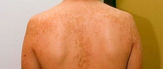
Ichthyosis on the skin of an adult
Ichthyosis on the face of a child
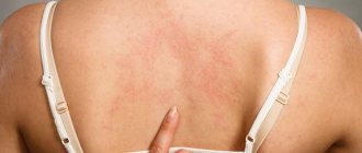
Ichthyosis on the body of a child
Ichthyosis on the body in an adult
Ichthyosis on the face of a child
Ichthyosis on the knees
The disease is life-threatening. Children are born with low birth weight. Modern medicine has achieved success in saving the lives of little sufferers.
If previously an infant could not survive a systemic infection because thermoregulation was impaired, now medicine is able to save their lives.
Prevention
Some patients are prone to recurrent fungal infections. These people may benefit from periodic oral courses of a systemic antifungal agent.
Prevention methods aimed at improving barrier function are very important. They are simple and clear:
- providing humidified , temperature-controlled environment;
- daily bathing using mild detergents;
- use of emollients (petroleum jelly-based products).
It should be remembered that exposure to sunlight worsens the condition by damaging dry skin. However, dead epidermal cells peel off much better when tanned while swimming.
Causes
Harlequin ichthyosis, also known as “harlequin fetus,” is an extremely rare form of congenital ichthyosis with a distinct and striking phenotype. It is inherited in an autosomal recessive manner and occurs secondary to mutations in the ABCA12 gene.
Babies born prematurely are encased in a tough, thick stratum corneum that is described as armour-like. The shell very soon cracks and dark red transverse and longitudinal cracks separate dense, yellow, geometric plates of skin.
Patients show ectropion and eclabium, underdevelopment of the ears and nose, swelling of the arms and legs. In the past, newborns with harlequin ichthyosis had no chance of survival. Currently, with intensive care combined with appropriate skin prophylaxis, many with such a complex diagnosis survive.
In some cases, harlequin ichthyosis presents with a milder phenotype, similar to a "colloid fetus", or with whitish-yellow condensed scales covering the body, resembling vernix.
Inherited types of ichthyosis are caused by mutations in genes passed on from one or both parents to the child. In most cases, parents carry the genetic mutation but do not have the condition themselves. They can occur spontaneously during the fusion of an egg with a sperm or shortly after conception.
Acquired ichthyosis is manifested by small whitish scales, concentrated mainly on the limbs. The form can be caused by diseases of the lymphoid tissue (Hodgkin's lymphoma, leukemia), systemic diseases (sarcoidosis, HIV infection, hypothyroidism, chronic hepatitis, malabsorption), bone marrow transplantation, or taking certain medications that disrupt the synthesis of sterols in epidermal cells.
Diagnostics
A doctor diagnoses ichthyosis based on characteristic skin features. In some cases, the diagnosis may be indicated before delivery, as is the case with X-linked ichthyosis.
The cause is low unconjugated estriol, the level of which is tested in the serum of a pregnant woman during the second three-month period. Harlequin ichthyosis is detected based on the results of ultrasound examination.
Other prenatal diagnostic methods include:
- examination of chorionic villi;
- biopsy ;
- preimplantation testing.
In the neonatal period, a probable diagnosis can only be made based on clinical reasons. It is important that the specific type of ichthyosis is not determined at this stage. During the first year of a child's life, many cases that are unclear at birth become more recognizable clinically when patients exhibit a characteristic skin phenotype.
Skin biopsy results are not effective in all cases, but the procedure is minimally invasive and sometimes provides useful information, for example in epidermolytic ichthyosis (characteristic histological findings). In the case of Netherton's syndrome, a hair cast is placed to examine abnormalities on the hair shaft.
When the diagnosis is unclear, genetic testing is performed by a dermatologist or geneticist.
Is it possible to cure ichthyosis of the skin?
Unfortunately, scientists have not yet fully understood the pathological processes that occur with this disease. It is incurable. There are a number of methods to alleviate the course of the disease.
Is it possible to cure mild ichthyosis? This is possible if the disease is acquired, not advanced, and treatment began immediately.
Only in very small areas of the stratum corneum can mealy, dry, brown formations remain.
Treatment of ichthyosis has many methods in its arsenal.
Statistics
Ichthyosis is a rare hereditary skin disease that occurs in newborns or in the first few months of life. Currently, in 100% of cases the disease cannot be completely cured.
The frequency of manifestation of various forms of pathology varies somewhat from each other.
Thus, the non-syndromic form of the disease is diagnosed in 1 child in 300 thousand. With the syndromic type, this figure is 1:200 thousand, and 1 child out of 250,000 suffers from the acquired form.
If we consider ichthyosis by type of inheritance, statistics present the following data.
In an autosomal recessive disease, the disease is transmitted to the baby only if both parents are carriers of the mutated gene, but in the absence of it on the X and Y chromosomes. In this situation, the probability of having a healthy child is 25%. Moreover, in 50% of cases the child will act as a carrier, and the probability of pathology in his children is 25%.
With the autosomal dominant type, when one of the parents is healthy and the defective gene is absent on the chromosomes, the probability of having a sick baby is 50%.
According to world statistics, the proportion of those born with ichthyosis is 1 child in 30,000. In Russia, the disease is diagnosed in approximately 3,000 people.
Clinical manifestations
The main symptom of ichthyosis - characteristic hard, scaly formations on dry skin - appears already in the first year of life, much less often - over the next few years. The child's skin becomes rough, and transparent, whitish or grayish scales appear on it, adjacent to each other. Only the elbow bends, the hollows under the knees and axillary areas, as well as the groin area remain clean. The palms and feet are peeling, and the skin pattern is clearly visible on them. In the mildest cases, the disease manifests itself only as slight peeling and dry skin.
Currently, there are more than fifty clinical and morphological forms of the disease, but in all cases the following manifestations are present:
- the stratum corneum of the epidermis is thickened, scaly formations and keratinization zones form on it;
- the skin is dry and flaky, the sebaceous glands practically do not work;
- the nail plates peel or break, in some cases they are deformed and resemble bird claws, peeling off from the soft tissues;
- the skin pattern becomes deeper and more expressive;
- There are rashes of different shapes and colors.
Skin ichthyosis usually worsens with the onset of cold weather. In warm weather, remission usually occurs. During puberty in adolescents, painful symptoms decrease, only to return in adulthood.
Answers to common questions
Does a solarium help with ichthyosis?
There is no complete cure for ichthyosis. But in the warm season, ichthyosis is not a concern. Many people resort to solariums in winter, and they really help.
Do they take you into the army with ichthyosis?
People with ichthyosis are not accepted into the army.
Is it possible to get tattoos?
During periods of exacerbation and rash, tattoos for ichthyosis are not recommended. It is better to wait for the remission stage.
Forms
To date, more than 25 genes have been identified that encode a wide range of epidermal proteins, including lipid metabolism and peptide cross-linking enzymes, proteases and their inhibitors, epidermal structural proteins and proteins involved in cell communication.
Why can our articles be trusted?
We make health information clear, accessible and relevant.
- All articles are checked by practicing doctors.
- We take scientific literature and the latest research as a basis.
- We publish detailed articles that answer all questions.
Abnormalities in any of these components result in a stereotypic epidermal response with epidermal hyperplasia and the formation of a redundant stratum corneum accompanied by abnormal desquamation, with visible accumulation of scales on the skin surface—the clinical feature of all ichthyoses. In some cases, the condition is characterized by increased skin fragility and blistering.
The most common or well-known types are the following:
- Ichthyosis vulgaris (common).
- Lamellar (lamellar).
- Epidermolytic hyperkeratosis.
- Ichthyosiform congenital erythroderma.
- X-linked ichthyosis is associated with Kallmann syndrome.